Case Report

Low Tracheostomy as a Potential Contributing Factor for Tracheo-Innominate Fistula: A Fatal Case and a Successfully Treated Case

Sukjune Byun1https://orcid.org/0009-0008-3955-439X, Chang Heum Han1https://orcid.org/0009-0003-2439-7356, Youna Chung1https://orcid.org/0009-0007-2525-441X, Yong Jin Song1,*https://orcid.org/0000-0003-1451-6676
Author Information & Copyright ▼
1Departments of Otorhinolaryngology, Gangneung Asan Hospital, University of Ulsan College of Medicine, Gangneung, Korea
*Corresponding author: Yong Jin Song, Department of Otorhinolaryngology, Gangneung Asan Hospital, University of Ulsan College of Medicine, Gangneung 25440, Korea, Tel: +82-33-610-3309, Fax: +82-33-642-3189, E-mail: Yjsong.yjs@gmail.com

© Copyright 2026. The Busan, Ulsan, Gyeoungnam Branch of Korean Society of Otolaryngology-Head and Neck Surgery. This is an Open-Access article distributed under the terms of the Creative Commons Attribution Non-Commercial License (http://creativecommons.org/licenses/by-nc/4.0/) which permits unrestricted non-commercial use, distribution, and reproduction in any medium, provided the original work is properly cited.

Received: Jun 11, 2026; Revised: Jul 03, 2026; Accepted: Sep 15, 2026

Published Online: Sep 30, 2026

ABSTRACT

Tracheo-innominate fistula (TIF) is a rare but frequently fatal complication of tracheostomy. We report two patients who developed TIF after low tracheostomy placement. A 47-year-old woman developed sentinel bleeding 16 days after tracheostomy. Imaging revealed a brachiocephalic artery pseudoaneurysm; despite endovascular stenting, fatal rebleeding occurred two months later. A 15-year-old male with long-term tracheostomy dependence presented with progressive bleeding. Angiography demonstrated active extravasation from the brachiocephalic artery, and covered stent grafting achieved sustained hemostasis. In both patients, the tracheostomy stoma was located 35–38 mm below the cricoid cartilage, below the third tracheal ring. Low tracheostomy may therefore represent a modifiable anatomical risk factor for TIF. Sentinel bleeding requires immediate evaluation, and long-term surveillance is necessary after endovascular treatment because delayed rebleeding may occur.

Keywords: Tracheostomy; Fistula; Hemorrhage; Endovascular procedures

Introduction

Tracheo-innominate fistula (TIF) is a rare but catastrophic complication of tracheostomy, occurring in fewer than 1% of patients and often proving fatal without prompt intervention.1–3) It results from erosion of the anterior tracheal wall into the brachiocephalic artery; sentinel bleeding should therefore prompt immediate evaluation.1,4,5) Reported risk factors include high cuff pressure, tube movement, infection, radiotherapy, corticosteroid use, and tracheostomy below the third tracheal ring, which increases tube-artery contact.1,3,6,7) Although endovascular treatment can rapidly control hemorrhage, recurrent bleeding remains a concern.8,9) We report two patients with radiologically documented low tracheostomy who had contrasting outcomes after endovascular treatment.

Case 1

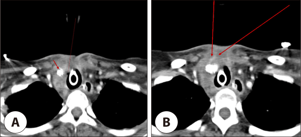
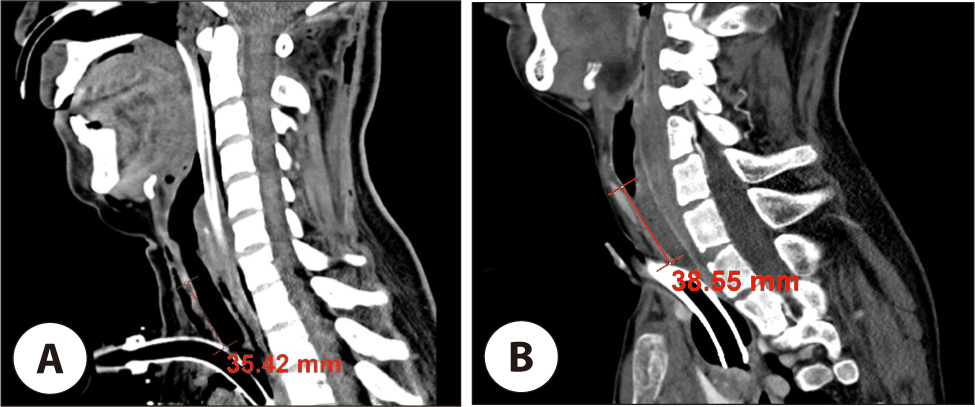
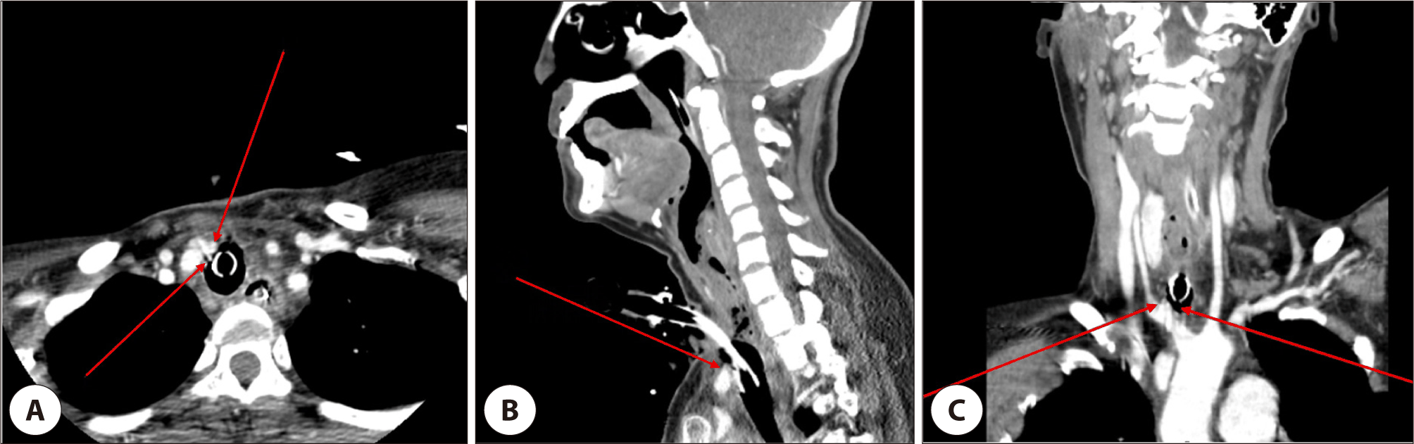
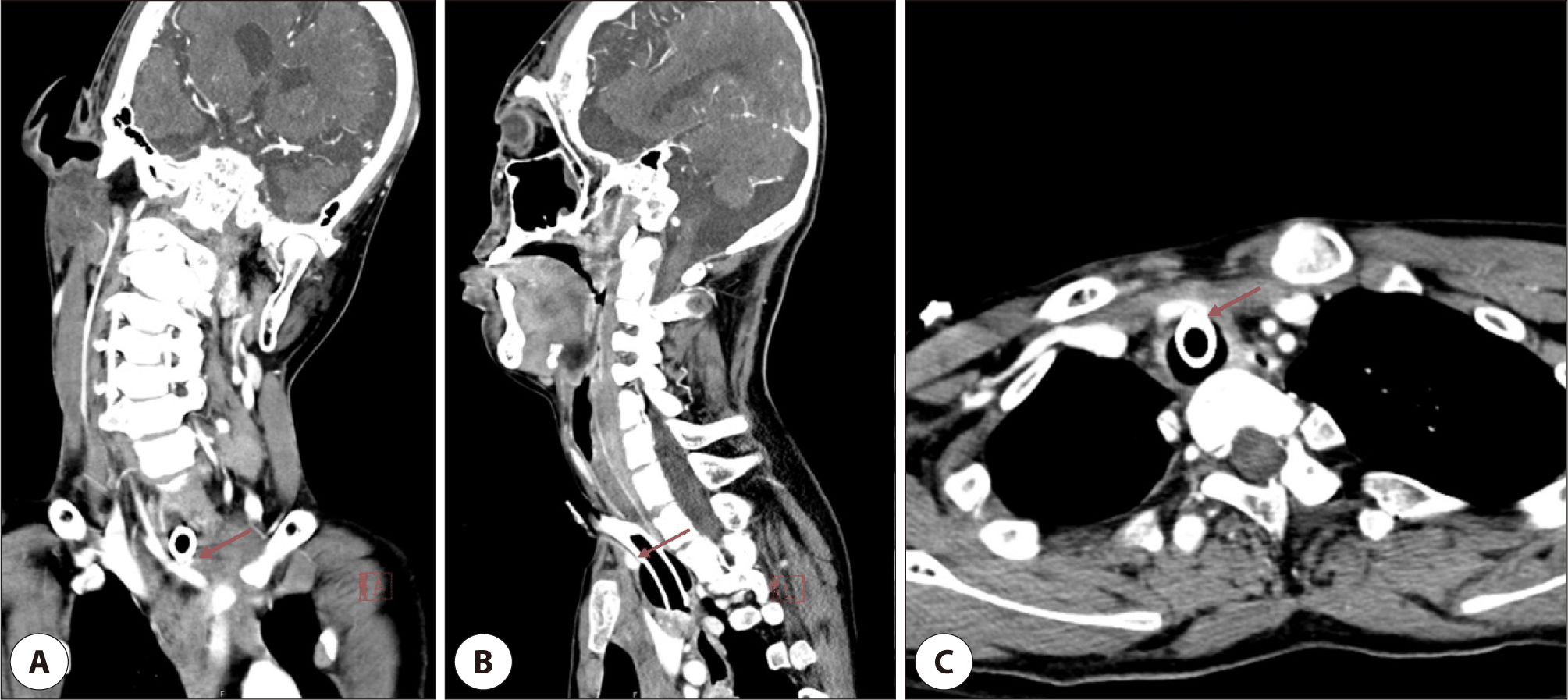
A 47-year-old woman with moyamoya disease, prior cardiac valve surgery, and hyperthyroidism presented with a massive right subcortical intracerebral hemorrhage. Surgical tracheostomy was performed on May 30, 2025, using a 7.5-mm Portex Blue Line Ultra® tube (Smiths Medical, Minneapolis, MN, USA); cuff pressure was maintained at approximately 28 cmH2O during mechanical ventilation. She was not receiving anticoagulant or antiplatelet therapy when TIF developed. Low tracheostomy was defined as a stoma below the third tracheal ring. On sagittal reconstructed CT, the distance from the inferior cricoid border to the stoma center was measured with a PACS electronic caliper as a supplementary quantitative measure. The stoma was 35.4 mm below the cricoid and below the third ring (Fig. 1A). Sixteen days after tracheostomy, sentinel bleeding progressed to active hemorrhage. CT showed a brachiocephalic artery pseudoaneurysm consistent with TIF (Fig. 2). Endovascular stenting achieved temporary stabilization. Follow-up CT showed exclusion of the pseudoaneurysm but a tubular enhancing structure anterior to the stent, raising concern for persistent or recurrent fistulous bleeding (Fig. 3). Approximately two months later, massive rebleeding occurred. Despite emergency embolization, the patient deteriorated and died; no surgical or pathologic confirmation of the rebleeding mechanism was available.
jcohns-37-3-124-g1
Fig. 1. Sagittal computed tomography images demonstrating low tracheostomy level in both cases. (A) Case 1. The hyoid bone, thyroid cartilage, and cricoid cartilage are indicated to illustrate the cervical anatomical landmarks. The distance from the inferior border of the cricoid cartilage to the tracheostomy stoma was 35.4 mm. (B) Case 2. Sagittal computed tomography image demonstrates a similarly low tracheostomy. The distance from the inferior border of the cricoid cartilage to the tracheostomy stoma was 38.6 mm. In both cases, the tracheostomy was created at a relatively low cervical level, illustrating the similarly low tracheostomy level observed in both patients.
Download Original Figure
jcohns-37-3-124-g2
Fig. 2. Contrast-enhanced CT demonstrating a pseudoaneurysm (arrows) arising from the right brachiocephalic artery adjacent to the tracheostomy site, consistent with tracheo-innominate artery fistula (Case 1). The lesion is located along the anterior wall of the trachea, supporting the diagnosis of vascular erosion secondary to tracheostomy (A: axial, B: sagittal, C: coronal).
Download Original Figure
jcohns-37-3-124-g3
Fig. 3. Follow-up contrast-enhanced CT after endovascular stent placement in the brachiocephalic artery (Case 1). (A) The endovascular stent is visualized within the vessel (arrow), and the previously seen pseudoaneurysm is no longer visualized. (B) Anterior to the stent, a tubular enhancing structure (long arrows) is noted, raising suspicion for persistent or recurrent bleeding.
Download Original Figure

Case 2

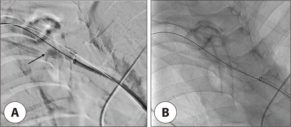
A 15-year-old boy with intractable epilepsy, severe scoliosis, and continuous home mechanical ventilation underwent surgical tracheostomy on August 24, 2023, using a 6.0-mm Portex Blue Line Ultra® tube with a cuff pressure of approximately 28 cmH2O. About 30 months later, minor stomal bleeding rapidly progressed to massive hemorrhage during tube exchange. The tube was reinserted and the cuff hyperinflated for temporary hemostasis. CT showed the stoma 38.6 mm below the cricoid, below the third tracheal ring, and in close proximity to the brachiocephalic artery (Fig. 1B and 4). Angiography demonstrated active extravasation from the brachiocephalic artery (Fig. 5A). Covered stent-graft placement achieved immediate hemostasis (Fig. 5B), and the patient recovered without recurrent bleeding before discharge.
jcohns-37-3-124-g4
Fig. 4. Anatomical relationship between the tracheostomy site and the brachiocephalic artery in Case 2. Imaging demonstrates the proximity between the tracheostomy tube and the brachiocephalic artery (arrows), illustrating the close anatomical relationship between the tracheostomy tube and the brachiocephalic artery, which may predispose to chronic vascular erosion (A: coronal, B: sagittal, C: axial).
Download Original Figure
jcohns-37-3-124-g5
Fig. 5. Angiographic findings in Case 2. (A) Digital subtraction angiography demonstrating active contrast extravasation above the overinflated cuff (arrow) from the right brachiocephalic artery, consistent with tracheo-innominate fistula. (B) Post-procedural angiography after deployment of a covered stent graft, showing complete cessation of extravasation and restoration of vascular continuity.
Download Original Figure

Discussion

TIF is attributed to pressure necrosis from repeated contact between the tracheostomy tube or cuff and the brachiocephalic artery.1,3) In both patients, the stoma was below the third ring and 35.4–38.6 mm below the cricoid (Fig. 1). These similar distances should not be interpreted as a disease-specific threshold because cervical dimensions vary with age and body habitus and no control group was available. They only support the shared radiologic finding of a low stoma. In Case 2, severe scoliosis and postural deformity may have altered cervical and mediastinal alignment, increased tube-artery contact, and made identification of tracheal landmarks difficult.10) Thus, low tracheostomy and scoliosis-related distortion likely acted together, and their relative contributions cannot be determined.

Endovascular treatment provides rapid, less invasive hemorrhage control in unstable or medically fragile patients.8,9,11,12) Both patients were considered poor candidates for emergent open repair because of severe underlying conditions and the need for immediate hemostasis. However, among 27 endovascularly treated patients in a systematic review, 18.5% required repeat intervention for rebleeding, 11.1% required rescue sternotomy, and mortality was 29.6%.13) Endovascular treatment may therefore serve as a bridge rather than definitive therapy because recurrent bleeding, infection, stent migration, occlusion, or a persistent fistula may occur.8,9)

The delayed fatal rebleeding in Case 1 may have resulted from persistent tube pressure, incomplete stent endothelialization, recurrent pseudoaneurysm, infection, or migration, although the exact mechanism remains speculative.8,9) Long-term clinical and imaging surveillance is therefore warranted even after technical success. In both patients, sentinel bleeding was the earliest clue. Even minor stomal bleeding should prompt airway stabilization, cuff hyperinflation, angiographic or CT evaluation, and definitive intervention.1,4,5)

Prevention requires direct identification of the cricoid cartilage and tracheal rings and creation of the stoma at the second or third ring whenever possible.4) In patients with distorted anatomy, preoperative CT review and preprocedural ultrasonography may delineate the brachiocephalic artery and aberrant pretracheal vessels, while bronchoscopic guidance may confirm the intraluminal level and tube/cuff position.10,14) Routine fluoroscopy is not established for standard tracheostomy.

This report is limited by its two-case design, the marked heterogeneity in age, underlying disease, ventilation duration, interval to TIF, and spinal anatomy, and the absence of pathologic confirmation of rebleeding in Case 1. These factors preclude causal inference or separation of the independent effect of low tracheostomy. Nevertheless, the cases support careful control of tracheostomy level, urgent evaluation of sentinel bleeding, and prolonged surveillance after endovascular treatment

Acknowledgements

Not applicable.

Funding Information

Not applicable.

Conflicts of Interest

No potential conflict of interest relevant to this article was reported.

Author Contribution

Conceptualization: Byun S.

Data curation: Byun S.

Formal analysis: Byun S, Han CH.

Methodology: Byun S.

Software: Byun S.

Validation: Song YJ.

Investigation: Byun S.

Writing - original draft: Byun S, Chung Y.

Writing - review & editing: Byun S, Han CH, Chung Y, Song YJ.

Ethics Approval

Research was conducted according to all ethical standards. This study was approved by the Institutional Review Board of Gangneung Asan Hospital (IRB No. 2026-05-009). The requirement for informed consent to participate in the study was waived by the Institutional Review Board due to the retrospective nature of this case report. Written informed consent for publication of the clinical information and images of Case 2 was obtained from the patient’s legal guardian.

References

1.

Grant CA, Dempsey G, Harrison J, Jones T. Tracheo-innominate artery fistula after percutaneous tracheostomy: three case reports and a clinical review. Br J Anaesth 2006;96(1):127-31.

2.

Jones JW, Reynolds M, Hewitt RL, Drapanas T. Tracheo-innominate artery erosion: successful surgical management of a devastating complication. Ann Surg 1976;184(2):194-204.

3.

Schaefer OP, Irwin RS. Tracheoarterial fistula: an unusual complication of tracheostomy. J Intensive Care Med 1995;10(2):64-75.

4.

Tarver EM, Lerant AA, Orledge JD, Stevens BP, Jefferson GD. Tracheoinnominate artery fistula. J Educ Teach Emerg Med 2021;6(3):S62-86.

5.

Joshi KD, Singh A, Singh DK, Gupta V, Saxena S, Hota A, et al. Tracheo-innominate artery fistula: a systematic review of diagnostic and management strategies. Otolaryngol Head Neck Surg 2025;173(4):824-39.

6.

Praveen CV, Martin A. A rare case of fatal haemorrhage after tracheostomy. Ann R Coll Surg Engl 2007;89(8):W6-8.

7.

Gelman JJ, Aro M, Weiss SM. Tracheo-innominate artery fistula. J Am Coll Surg 1994;179(5):626-34.

8.

Khanafer A, Hellstern V, Meißner H, Harmening C, Schneider K, Henkes H. Tracheoinnominate fistula: acute bleeding and hypovolemic shock due to a trachea-innominate artery fistula after long-term tracheostomy, treated with a stent-graft. CVIR Endovasc 2021;4:30.

9.

Taechariyakul T, Keller FS, Jahangiri Y. Endovascular treatment of tracheoinnominate artery fistula: case report and literature review with pooled cohort analysis. Semin Thorac Cardiovasc Surg. 2020;32(1):77-84.

10.

Bae MH, Lee YJ, Nam SO, Kim HY, Kim CW, Kim YM. Endovascular stenting of tracheoinnominate fistula after tracheostomy in a 14-year-old boy. Korean J Pediatr. 2016;59(Suppl 1):S76-9.

11.

Chien YS, Chao YC, Lee KS, Hsu KH. Successful rescue of a ruptured tracheoinnominate fistula with extracorporeal membrane oxygenation, endovascular stents, and debranching surgical bypass. Ann Thorac Cardiovasc Surg 2020;26(3):166-9.

12.

Hamaguchi S, Nakajima Y. Two cases of tracheoinnominate artery fistula following tracheostomy treated successfully by endovascular embolization of the innominate artery. J Vasc Surg. 2012;55(2):545-7.

13.

O’Malley TJ, Jordan AM, Prochno KW, Saxena A, Maynes EJ, Ferrell B, et al. Evaluation of endovascular intervention for tracheo-innominate artery fistula: a systematic review. Vasc Endovascular Surg. 2021;55(4):317-24.

14.

Barash M, Kurman JS. Patient selection and preoperative evaluation of percutaneous dilation tracheostomy in the intensive care unit. J Thorac Dis 2021;13(8):5251-60.