
 PDF Download
 PubReader
 Export Citation
 Email the Author
 Share Facebook
 Share Twitter
 Cited by 0 Article
 Metrics (View:1,005)








Introduction
Inverted papilloma (IP) is a benign tumor originating from the Schneiderian mucosa of the nasal cavity and paranasal sinuses. Despite its benign nature, IP demonstrates locally aggressive behavior with a high rate of recurrence and the potential for malignant transformation in approximately 5%–15% of cases.1,2) Preoperative imaging, particularly CT and MRI, plays a critical role in the diagnosis, localization, and surgical planning of IP.3–7)
On CT, the site of origin can often be inferred from focal hyperostosis, believed to result from chronic irritation and associated bone remodeling.5,8,9) However, this finding is not present in all patients (about 63.2%), which may lead to incomplete resection if the origin site is overlooked.6)
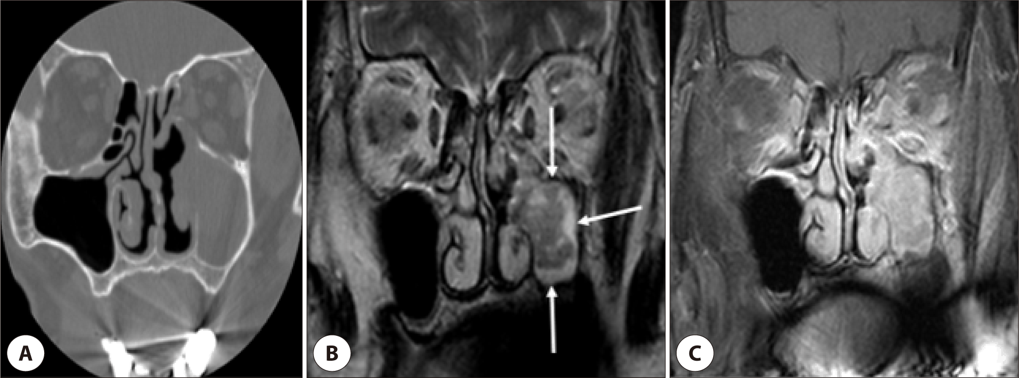
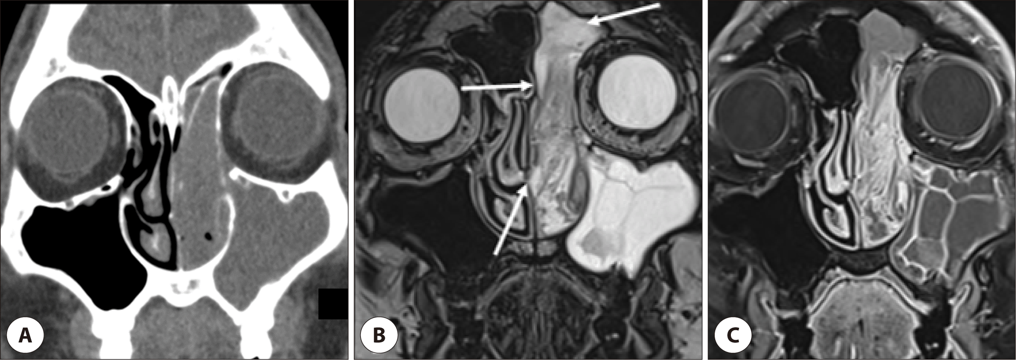
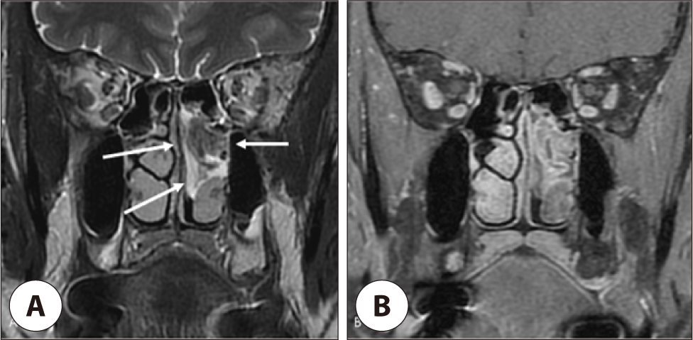
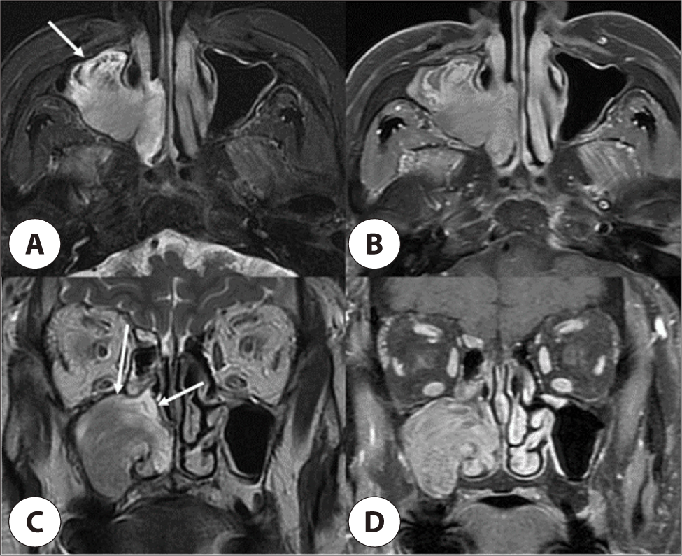
MRI is more useful for characterizing the lesion itself. The most characteristic finding on MRI is a cerebriform pattern of contrast enhancement, which is often not appreciable on CT.10,11) Conversely, hyperostosis is difficult to detect using MRI.7) In cases lacking hyperostosis on CT, careful evaluation using T2-weighted images (T2WIs) may help identify the site of origin. A peritumoral hyperintense layer, representing peritumoral mucosal edema or inflammation, can often be observed surrounding the lesion.12,13) This T2 peritumoral hyperintense layer contrasts with the intermediate signal intensity of the tumor itself and may serve as an indirect marker of the tumor origin.
The purpose of this study was to evaluate the role of T2WI in identifying the site of origin of IP and to assess the diagnostic utility of the T2 peritumoral hyperintense layer on MRI in cases without focal hyperostosis on CT, which is otherwise a well-established marker for tumor origin.
Materials and Methods
All patients underwent both CT and MRI before surgery between May 2007 – June 2025. The patients showing hyperostosis on CT were excluded. All patients underwent endoscopic sinus surgery (ESS), and the site of tumor origin was confirmed intraoperatively. 51 patients (39 males and 12 females: age range, 27-87 years; mean age, 57.0 years) with pathologically proven IP in the nasal cavity or paranasal sinus by surgery, were included. This study was approved by the Institutional Review Board of our hospital (IRB No. 2506-015-152).
The paranasal sinus CT scans were obtained preoperatively in axial and coronal planes in helical CT scanners (Cti, lightSpeed Plus, Discovery, Revolution, Revolution FT, Revolution RC, Revolution Apex, Revolution Max; GE Medical System, Milwaukee, WI, USA; Sensation 16, Definition, Somatom Plus; Siemens, Erlangen, Germany). CT parameters were shown on Table 1. MRI was performed on 1.5 and 3.0 T systems (SMS Skyra, Avanto, Vida, TrioTim and Symphony; Siemens, Erlangen, Germany, GMS Architect, GMS Premier; GE medical system, Milwaukee, WI, USA). The MRI protocols were shown on Table 2.
The image analysis of all CT and MRI was performed by two radiologists who have been practicing for 30 years and 23 years. All CT and MR images were reviewed retrospectively by the consensus of two experienced neuroradiologists (H.J.K. and Y.W.K.). Assessment of the hyperostosis of the nasal and maxillary bone was performed on CT for exclusion of the present study. When evaluating hyperostosis in the paranasal sinuses, we considered only focal hyperostosis to be a positive finding. Focal hyperostosis was defined as eccentric bone thickening and sclerosis that involved only a limited portion of the wall of a given paranasal sinus. The patients showing no hyperostosis were included in the present study.
In all MR images, a convoluted cerebriform pattern (CCP) was defined as a mix of linear or curvilinear hyperintense and hypointense striations partially observed on the Gd-enhanced T1-weighted images (Gd-T1WI) and analyzed on MRI as an IP itself. On T2WI, the solid components of the tumor with or without partially or diffusely distributed hypointense striations, were considered as IP itself. The origin of IP was suspected at the sites where the enhancing mass was adherent on Gd-T1WI and at sites lacking a preserved peritumoral hyperintense layer adjacent to the tumor on T2WI. The preserved peritumoral hyperintense layer adjacent to the IP was interpreted as non-adherent sites on T2WI.
We analyzed the origin site of IP on Gd-T1WI and T2WI and defined positive as possibly distinguishing the origin and negative as impossibly distinguishing the origin of IP. The radiologic predictions were compared to the surgically confirmed origin sites. We analyzed the predictive accuracy of Gd-T1WI and T2WI separately and discussed causes of mis-localization.
Statistical analyses were performed to evaluate the association between MRI sequences the ability to predict the origin of inverted papilloma and surgically confirmed origin. Categorical variables were compared using the Pearson’s chi-square test. A p-value of less than 0.05 was considered statistically significant. All statistical analyses were conducted using IBM SPSS Statistics (version 26.0; IBM, Armonk, NY, USA).
Results
Of the 51 cases, IP masses were located in the nasal cavity commonly (n=14), followed by maxillary sinus (n=12). Multi-sinus involvement including the nasal cavity was 25 cases. Of the 51 cases, surgically confirmed IP origin was in the maxillary sinus (n=16), frontal sinus (n=4), ethmoid sinus (n=14), sphenoid sinus (n=2) and nasal cavity (n=15). Compared to the surgically confirmed origin sites, T2WI was able to predict IP origin in 43 cases (43/51, 84.3%). Positive prediction of IP origin was 21 cases on Gd-T1WI (21/51, 41.2%). For comparison of IP origin prediction, the predictive accuracy of T2WI was better in 22 cases (8 cases in the maxillary sinus, 6 cases in the ethmoid sinus and 8 cases in the nasal cavity) and similar in 21 cases (8 cases in the maxillary sinus, 4 cases in the frontal sinus, 6 cases in the ethmoid sinus and 3 cases in the nasal cavity) compared to that of Gd-T1WI. In 8 cases (2 cases in the sphenoid sinus, 2 cases in the ethmoid sinus and 4 cases in the nasal cavity), the prediction of IP origin was unable on Gd-T1WI nor T2WI (Table 3) (Figs. 1–4).
| Gd-T1WI | T2WI | p-value | |
|---|---|---|---|
| Correct* | 21 (41.2%) | 43 (84.3%) | |
| Incorrect† | 30 (58.8%) | 8 (15.7%) | <0.001 |
| Total | 51 | 51 |
The origin of inverted papilloma was correctly predicted in 21 cases (41.2%) using Gd-T1WI and in 43 cases (84.3%) using T2WI. Comparison using the Pearson’s chi-square test demonstrated a statistically significant difference between the two MRI sequences (p<0.001), indicating that T2WI showed superior performance in predicting the tumor origin. These findings indicate that T2WI provides significantly higher accuracy than Gd-T1WI in predicting the origin of inverted papilloma.
Discussion
Although CT remains valuable for evaluating osseous structures and guiding surgical access, the reliance on CT alone may result in missed or inaccurately localized tumor origins, particularly in the absence of definitive hyperostotic changes. Hyperostosis, traditionally considered a hallmark of tumor attachment, was absent or located at non-origin sites in some cases, likely due to prior inflammation or remodeling changes.5,6,8) In addition, diffuse sclerosis secondary to chronic sinusitis may obscure the site of origin, highlighting the limited specificity of CT findings within the complex sinonasal anatomy.9)
According to the previous study,8) CT-based detection of focal hyperostosis correlated with the surgically confirmed tumor origin in 89.1% of cases (49/55) among patients with focal hyperostosis on CT.
However, the overall incidence of focal hyperostosis in the patients with IP was only 63.2% and the most common site of tumor origin was the lateral nasal wall (52.6%). Therefore, not all patients with IP demonstrate focal hyperostosis on CT, making it difficult to determine the tumor origin in such cases. Furthermore, plaquelike or diffuse hyperostosis may occur irrespective of the tumor origin, and diffuse sclerosis related to chronic sinusitis may further obscure the origin site.8,9)
Preoperative imaging of the paranasal sinuses is essential for staging and treatment planning of sinonasal masses including the IP.10–12) The presence of CPP on MR imaging may facilitate preoperative diagnosis and help guide therapeutic strategies, particularly in cases without focal hyperostosis on CT. In this study, we evaluated two MR imaging sequences, Gd-T1WI and T2WI for predicting the origin site of IP. MRI-based predictions were compared with surgically confirmed tumor origins. T2WI achieved a positive prediction rate of 84.3% (43/51), markedly outperforming Gd-T1WI, which identified the origin in only 41.2% (21/51) of cases.
The convoluted cerebriform pattern was described by Barnes et al. as a distinctive gross mucosal morphology of IP,10,13) and on MRI it appears as characteristic alternating hypointense and hyperintense bands on T2WI and Gd-T1WI. Jeon et al.13) reported that the CPP was observed on MRI in all IPs cases (100%) and that Gd-T1WI demonstrated a higher detection rate (93%) than T2Wis In contrast, our study demonstrated superior visualization on T2WI. This discrepancy may be explained by the relatively frequent multisinus involvement in our cohort and the use of a 3T MRI scanner, which may enhance the visualization of signal intensity differences with the lesion. Additionally, the non-adherent portions of IP were more clearly demonstrated on T2WI, which helped to determine the tumor origin and guide surgical planning.
MRI also provided important diagnostic information in cases lacking hyperostosis on CT. In particular, the presence of a peripheral hyperintense layer on T2WIs, attributable to adjacent mucosal inflammation, often helped identify the tumor attachment site.10,12,14) The similar findings were reported by Mohamed Eid et al.,13) who demonstrated that T2WI was superior to Gd-T1WIs in 11 out of 16 cases. The difference may be explained by limited differential enhancement between epithelial and subepithelial tissues. Variations in cellularity and stromal composition within subepithelial tissues can result in variable contrast enhancement comparable to that of the epithelium. Furthermore, extrinsic factors, including variability in acquisition timing and dynamic contrast enhancement techniques, may further reduce the conspicuity of differential Gd enhancement.
Nevertheless, MRI was not infallible. In our study, eight cases were negative for the prediction of IP origin on T2WI, primarily due to the relatively large tumor size within the confined space of the nasal cavity or paranasal sinuses. In such cases, the intermediate signal intensity of the tumor filled the narrow sinonasal cavity and obscured the surrounding hyperintense peritumoral edema. In addition, 30 cases were negative for the prediction of IP origin on Gd-T1WI, largely due to difficulty in differentiating contrast enhancement of the mass itself from hypervascular enhancement of the surround mucosa. The most frequent factors limiting MRI localization included large tumor volume causing mass effect and multifocal tumor attachment, both of which have been previously reported as limitations of imaging-based localization.5–7,10) One case (2.4%) originating from the medial maxillary wall was particularly challenging because of the complex adjacent soft tissue anatomy, and another case (2.4%) showed partial discordance between imaging and intraoperative findings, highlighting the need for cautious interpretation even with high-resolution MRI.
Complete surgical resection of IP is crucial to reduce the recurrence rates, which have been reported to range from 15% to 78%.13) Preoperative evaluation with cross-sectional imaging, including CT and MRI, is therefore critical for determining the location and extent of the tumor and selecting the optimal surgical approach. In our study, MRI demonstrated superior performance compared with CT for identifying both the origin and extent of IP. The CPP on MRI and focal hyperostosis on CT remain important imaging findings for preoperative localization of tumor origin and extent. In particular, peritumoral high signal intensity on T2WI in maxillary sinus-reflecting non-adherent mucosal sites-helped guide surgical decision-making, such as choosing between ESS and Caldwell-Luc operation.
Although MRI serves as powerful adjunct in the preoperative evaluation of paranal sinus IP, particularly when CT findings are inconclusive without focal hyperostosis, integration of imaging findings with endoscopic evaluation and surgical experience remains essential for optimal patient outcomes.
The present study has several limitations. First, the relatively small sample size may have restricted the robustness of our findings. In particular, evaluating the tumor origin was challenging when the lesion was located in a small cavity such as the ethmoid sinus. A larger sample size would allow for more reliable assessment even in cases involving small anatomical spaces. Second, this study was conducted in a single institution, which may limit the generalizability of the results to broader populations. Third, the retrospective design inherently carries risks of selection and information bias. Future prospective studies are warranted to overcome these limitations and provide more definitive conclusions.
Conclusion
In cases where CT lacks definitive signs, such as focal hyperostosis, MRI provides critical complementary information for identifying the origin of IP with T2 hyperintense peritumoral changes. MR T2WI demonstrated significantly greater accuracy than Gd-T1WI in localizing the tumor origin of the sinonasal IP. The hyperintense peritumoral edema on MR T2WI can improve preoperative surgical planning, increase the likelihood of complete tumor resection, and potentially reduce recurrence rates following ESS.


