


Introduction
Toxoplasma gondii is a protozoan parasite first discovered in 1901 by Nicolle and Manceaux in North African rodents. The first human infection was reported in 1923 by J. Janju.1,2) Human infection occurs through various transmission routes, including ingestion of raw or undercooked meat from infected pigs, cattle, or birds, consumption of oocysts present in cat feces or contaminated soil, blood transfusion, organ transplantation, or congenital infection via the placenta.3–5)
Most infections in immunocompetent individuals are asymptomatic; however, when symptoms occur, cervical lymphadenitis is the most common clinical presentation. In contrast, immunocompromised individuals and congenitally infected neonates may develop severe complications such as encephalitis, chorioretinitis, pneumonia, or myocarditis.6)
From 2010 onward, we encountered five cases of cervical toxoplasmosis diagnosed through histopathological and serological examinations in patients presenting with cervical lymphadenopathy. Here, we report these cases along with a review of the literature.
Case Reports
A 49-year-old man presented with a left submandibular mass that had developed two months prior. He had no history of livestock or pet exposure, raw meat consumption, travel, or occupational risk factors. On physical examination, multiple firm, non-tender, mobile lymph nodes measuring 2×2 cm were palpated in the left level II and V regions, with no additional abnormalities. Ultrasonography showed multiple hypoechoic lymph nodes measuring up to 1.5×1.5 cm in the left level II and V regions, along with additional lymphadenopathy (>1 cm) in both sides of the neck. Fine-needle aspiration cytology (FNAC) revealed abundant lymphocytes without malignant features. Neck computed tomography (CT) identified multiple homogeneous hypodense masses in the left level II and V regions, with the largest measuring 1.7×1.5 cm (Fig. 1A). Excisional biopsy was performed, removing a well-circumscribed firm mass from level V. Pathological examination revealed chronic lymphadenitis with clusters of epithelioid cells, raising suspicion for toxoplasma lymphadenitis. Postoperative serological testing confirmed positive Toxoplasma IgG and IgM antibodies, leading to a definitive diagnosis. The patient remained asymptomatic, with no recurrence observed during six months of follow-up.
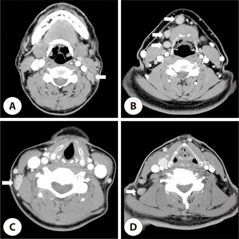
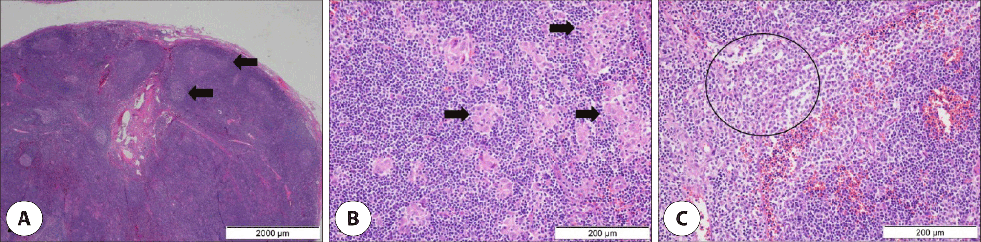
A 53-year-old man presented with a painless right cervical mass persisting for two months. His past medical history was unremarkable. Physical examination revealed a firm, mobile, 1.5×1.5 cm mass at level I of the right neck. FNAC did not reveal malignant features. Neck CT showed multiple homogeneous hypodense lymph nodes, with the largest measuring 1.7×1.5 cm at level I (Fig. 1B). Excisional biopsy was performed. Pathological findings included reactive follicular hyperplasia, epithelioid histiocytes, and monocytoid B-cell proliferation, confirming toxoplasma lymphadenitis (Fig. 2). Postoperatively, serological testing confirmed positive Toxoplasma IgG and IgM antibodies. The patient remained asymptomatic, with no recurrence noted at the two-month follow-up.
A 56-year-old woman presented with a right cervical mass that persisted despite a 10-day course of antibiotics. Physical examination revealed two firm, non-tender lymph nodes measuring 2.5×1 cm and 1×1 cm in level V of the right neck. No additional abnormalities were noted. FNAC showed no malignant features, and Mycobacterium Nested PCR was negative. Neck CT revealed two oval, contrast-enhanced masses measuring 2×1 cm and 1.5×1 cm in the right level V region (Fig. 1C). Excisional biopsy was performed and pathological examination revealed follicular hyperplasia with multiple small granulomas, suggesting toxoplasma lymphadenitis. Postoperative serological testing confirmed positive Toxoplasma IgG and negative IgM, indicating a chronic infection.
A 55-year-old man presented with bilateral cervical lymphadenopathy that had developed two weeks prior. His past medical history included raising poultry and dogs for over ten years, but he had no history of pulmonary tuberculosis or systemic symptoms. On examination, multiple lymph nodes (up to 1cm) were palpated in the posterior cervical region bilaterally. Laboratory tests showed normal findings, Interferon-gamma release assay (IGRA) negativity, and positive Toxoplasma IgG and IgM. To confirm the diagnosis, the excisional biopsy was done, and pathologic findings revealed chronic lymphadenitis with epithelioid cell clusters, leading to a diagnosis of toxoplasma lymphadenitis.
A 47-year-old man was referred for suspected T-cell lymphoma based on an excisional biopsy. On examination, multiple non-tender lymph nodes (–1.5×1.5 cm) were palpated in levels II and V of the right neck. No other abnormalities were noted, and laboratory tests were within normal limits. Serological testing showed positive Toxoplasma IgG and IgM. Neck CT revealed multiple homogeneous hypodense lymph nodes in levels II and V, with the largest measuring 1.5×1.3 cm at level V (Fig. 1D). Re-excisional biopsy was performed, and pathological findings included reactive follicular hyperplasia, epithelioid histiocytes, and monocytoid B-cell proliferation, confirming toxoplasma lymphadenitis. The patient remained asymptomatic, with no recurrence observed over one year of follow-up.
Discussion
Most patients infected with Toxoplasma gondii remain asymptomatic, with cervical lymphadenitis being the most common clinical manifestation. Involvement of posterior cervical lymph nodes is common, and some cases of intraparotid lymphadenopathy have also been reported in Korea.7) Other clinical symptoms may include maculopapular rash, arthralgia, myalgia, and hepatitis. In immunocompromised patients, toxoplasmosis may present as central nervous system infections, myocarditis, pneumonia, or chorioretinitis.6)
The diagnosis of toxoplasmosis requires both pathological and serological examinations. To obtain pathological specimens, fine needle aspiration (FNA), core needle biopsy (CNB), and excisional biopsy can be considered. Among these, FNA and CNB can be performed relatively easily under ultrasound guidance and are associated with a low risk of complications.8) However, FNA yields a smaller sample compared to CNB, and diagnosis may be challenging when based solely on aspirated cells.9) Although excisional biopsy provides the most accurate diagnosis, it often requires hospitalization and general anesthesia, leading to increased time and financial costs, as well as the potential for scarring.10) To avoid unnecessary excisional biopsies, it is preferable to first perform ultrasound-guided procedures and consider excisional biopsy only when a definitive diagnosis cannot be established.8, 9)
On contrast-enhanced CT, tuberculous lymphadenitis shows central low attenuation due to necrosis and peripheral enhancement whereas pre-treatment lymphoma presents as homogeneously enhancing nodes without necrosis or calcification.11) Toxoplasma lymphadenitis usually presents as multiple posterior cervical nodes with homogeneous or only mild enhancement, and otherwise nonspecific features, therefore CT scan alone is insufficient to reliably distinguish it from tuberculous lymphadenitis or lymphoma.12) Definitive diagnosis should rely on histopatholgy and serology (toxoplasma IgM/IgG).
Three key histopathological features of toxoplasma lymphadenitis include: (1) reactive follicular hyperplasia, (2) clusters of proliferating epithelioid histiocytes mixed with lymphocytes and immunoblasts, and (3) monocytoid B-cell hyperplasia.13) Although these three histological findings exhibit high specificity (96.9%), they have a relatively low sensitivity (44.4%). Eapen et al. proposed diagnostic criteria that include (1) the presence of microgranulomas, (2) the absence of multinucleated giant cells, (3) the predominance of stage II giant granulomas, and (4) follicular hyperplasia. This set of criteria was shown to diagnose toxoplasma lymphadenitis with 100% sensitivity and 96.6% specificity.13)
Serological tests, particularly enzyme-linked immunosorbent assay (ELISA), are widely used for detecting Toxoplasma antibodies. The presence of IgG and IgM antibodies is commonly used to diagnose acute toxoplasmosis. IgG antibodies typically appear 2–3 weeks after infection, peak within 1–2 months, and persist for life.14) In contrast, IgM antibodies become detectable around two weeks post-infection, peak at one month, and gradually decline, usually disappearing within 6–9 months.15)
In general, toxoplasmosis is self-limiting, and symptomatic treatment is sufficient for most immunocompetent patients. However, in cases of severe acute infection, systemic involvement, or immunocompromised patients (including pregnant women), treatment with macrolide monotherapy or a combination of pyrimethamine and sulfadiazine may be required.16)
In our study, all five patients were immunocompetent, and only one case had a history suggestive of potential toxoplasma exposure. None of the cases presented with clinical symptoms other than painless cervical lymphadenopathy. Three patients underwent IGRA testing for tuberculosis, with one showing a positive result. Given the relatively high prevalence of tuberculous lymphadenitis in Korea, careful differentiation is necessary. Serological testing revealed Toxoplasma IgG positivity in all five cases, with IgM positivity in four cases. All patients underwent excisional biopsy and improved without additional therapy. No recurrences were observed during the 6-month follow-up (Table 1).
Although the prevalence of toxoplasmosis in Korea remains relatively low, the increasing trends of pet ownership, meat-based diets, and international travel may contribute to a rising incidence. Therefore, clinicians should consider toxoplasma lymphadenitis as a differential diagnosis in patients presenting with cervical lymphadenopathy.