Introduction
Orbital apex syndrome (OAS) is characterized by the involvement of cranial nerves: optic nerve (CN II), oculomotor nerve (CN III), trochlear nerve (CN IV), abducens nerve (CN VI), The ophthalmic division of the trigeminal nerve (CN V). The clinical manifestations of orbital apex syndrome are highly variable, with the most common being vision loss, orbital pain, and limited eye movements.1) OAS is associated with diverse pathologies, including inflammatory, infectious, neoplastic, iatrogenic, traumatic, and vascular etiologies, all of which may lead to the aforementioned symptoms.2) A noteworthy anatomical consideration is the Onodi cell, which represents a variant of the posterior ethmoid air cells situated in close proximity to the optic nerve. Mucocele originating within the paranasal sinuses have the capacity to exert influence on the optic nerve through mechanisms of mechanical compression or direct extension of inflammation to the optic nerve, thereby precipitating ischemic injury.3) Nevertheless, the occurrence of mucocele arising from ethmoid cells especially Onodi cell is rare. In addition, cases in which symptoms improved after early surgical intervention have been well reported,4–6) but it is extremely rare for the main visual symptom to completely improve after more than 1 year.7) Therefore, we represent a successful treatment case in which normal vision was restored after surgery despite long-term visual impairment.
Case Report
A 62-year-old male patient with a medical history of myocardial infarction and stent insertion presented to the hospital with a chief complaint of decreasing visual acuity on the right side, which had been present for one year. The patient visited an ophthalmic hospital five months ago and was diagnosed with cataracts and subsequently underwent surgery; however, vision did not improve.
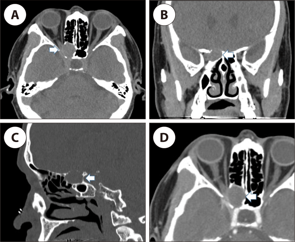
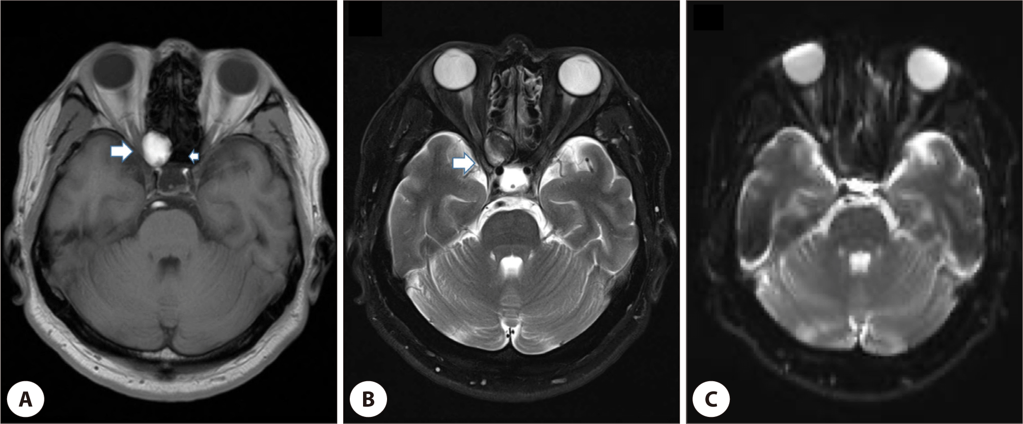
Based on the imaging findings of Brain CT and Brain MRI scans, a right ethmoid mass suspected to be mucocele was identified, and the patient was referred to our department for surgical treatment. A well-defined, non-enhancing mass measuring 2.0 cm in its largest diameter was identified in the right Onodi cell on paranasal CT. The mass had a mild expansile nature and caused narrowing of the right optic canal (Fig. 1). MRI finding of the mass showed no definite diffusion restriction in DWI image. Well marginated mass with contour bulging to optic canal showed hyper intensity in T1 weighted image (Fig. 2). The visual acuity measured at our medical center was 0.04 in the right eye and 0.8 in the left eye. Both uncorrected vision and corrected vision were same in right eye.


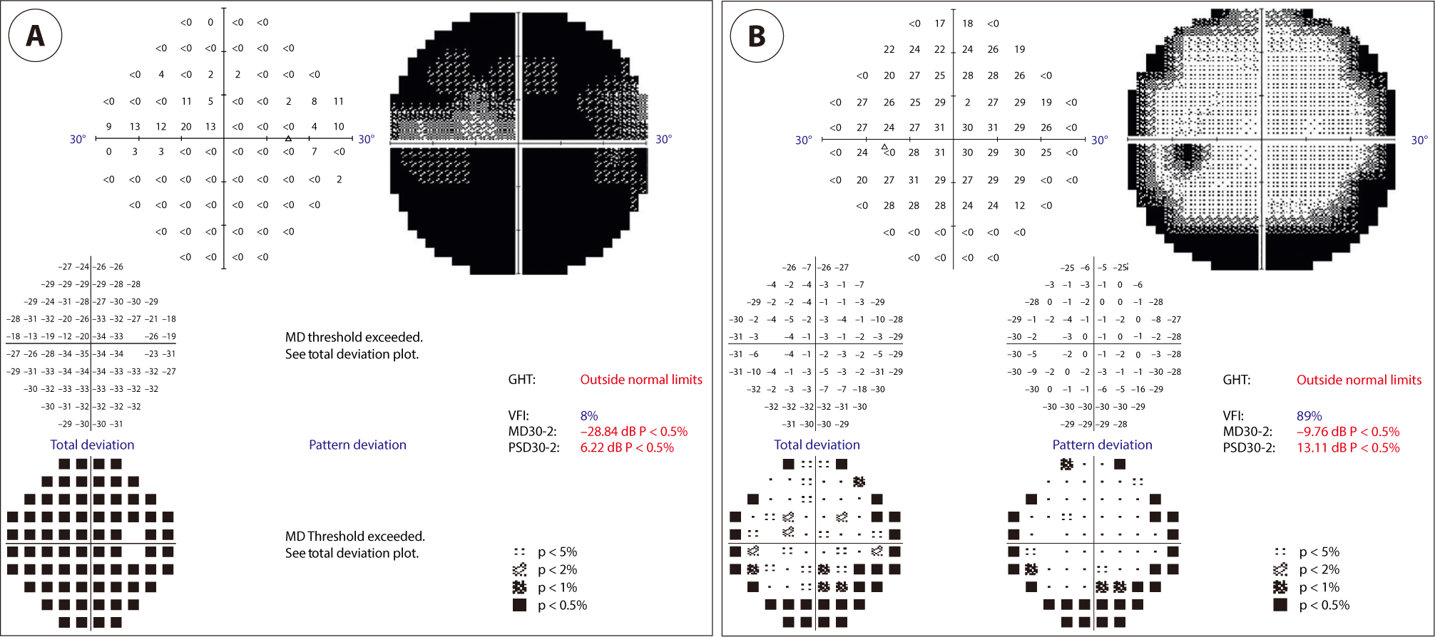
The intraocular pressure was in normal range with 10 mmHg in the right and 2 mmHg in the left, and fundoscopy showed a normal optic discs and macula in both eyes. Additionally, a visual field assessment was conducted, revealing a visual field index (VFI) of 8% in the right eye and 89% in the left eye (Fig. 3). This discrepancy indicates significant impairment of visual field function in the right eye. The light reflex in the right eye showed a sluggish response. The test showed an anisocoria of more than 1.8 unit (beyond the maximum limit of measurement) in the right eye, which suggests a possible optic nerve damage.

Endoscopic surgical intervention, including marsupilization of mucocele, was performed after the evaluation. The subjective visual acuity of the patient showed improvement immediately after surgery. After the surgery, he took a 3rd generation cephalosporin 200 mg bid and methylprednisolone 8 mg bid for one month. The visual acuity measured at 3 months after surgery was 0.8 in the right eye and 0.8 in the left eye, and was completely restored to the normal range.
Discussion
Visual loss and ophthalmoplegia are often the initial sign of an OAS. Therefore, most patients first visit an ophthalmologist to evaluate various tests because of uncomfortable ophthalmological symptoms. If optic nerve dysfunction is suspected after the examination such as best-corrected visual acuity, pupil size, and visual field test, additional radiologic examination such as CT or MRI should be considered.2) When a mass-like lesion is observed on an image, information can be obtained regarding the location, size, and type of the lesion, and assessment of intraorbital and intracranial extension.4) CT scan of a mucocele shows a homogeneous isodense lesion that is not enhanced by contrast agent and allows evaluation of the bone structure surrounding the lesion unless infected.
MRI of mucocele will show variable signal intensity in both the T1- and T2-weighted images based on the fluid content and viscosity of the mucocele or the degree of dehydration.8)
In this case, visual acuity gradually deteriorated with the presence of cataract. Consequently, given these clinical findings, additional imaging tests were not performed in the earlier stage, and the most important measure considered was the surgical intervention to resolve the cataract. Only after confirming the lack of improvement in visual field and acuity following cataract surgery, CT and MRI imaging were performed which led to a delay in diagnosis.
Mucocele in posterior ethmoid sinus can cause optic neuropathy through direct mechanical compression of the optic nerve, circulatory disturbance of the vasa nervorum, or optic neuritis due to inflammatory reaction. Although in cases of mechanical compression, symptoms worsen gradually, when mucoceles are accompanied by inflammation, the onset or progression of ophthalmologic signs and symptoms is rapid, potentially leading to a poor prognosis.7) A review study by Kim et al. also found that the presence of infection was a critical factor influencing postoperative visual outcomes.9) In our case, considering the gradual onset of symptoms, it is probable that the symptoms are caused by increased mechanical pressure that is transmitted to the optic nerve through the optic canal wall. Surgical intervention is the treatment of choice,10) and our patient showed immediate improvement in symptoms after surgery. It is remarkable that there was a complete improvement in visual symptoms, despite the one-year interval from symptom onset to surgery and the severity of symptoms before the surgery. The majority of the available literature indicate that initiating treatment within a few days after symptom onset has been associated with favorable outcomes.4-6,11) However, this case represents that even though more than a year has passed since the onset of symptoms, visual improvement can be achieved through surgical decompression in patients with orbital apex syndrome caused by direct compression of mucocele.
Therefore, even in patients whose vision loss persist for a long time, if mucocele is identified, surgical decompression must be performed immediately to expect restoration of vision.


