Case Report
내경동맥의 가성동맥류가 동반된 측두골 방사선골괴사 1례
A Case of Temporal Bone Osteoradionecrosis Accompanied by Internal Carotid Artery Pseudoaneurysm
Seunghwa Kim1
,
YoungJun Seo1,
Jai Ho Choi2,
Jae-Hyun Seo1,*
Author Information & Copyright ▼
2가톨릭대학교 의과대학 서울성모병원 신경외과학교실
1Department of Otorhinolaryngology-Head and Neck Surgery, College of Medicine, The Catholic University of Korea, Seoul, Korea
2Department of Neurosurgery, Seoul St. Mary's Hospital, College of Medicine, The Catholic University of Korea, Seoul, Korea
*Corresponding author: Jae-Hyun Seo, Department of Otorhinolaryngology-Head and Neck Surgery, College of Medicine, The Catholic University of Korea, Seoul 06591, Korea, Tel: +82-2-2258-6210, Fax: +82-2-2258-1354, E-mail:
revivalseo@catholic.ac.kr
© Copyright 2022 The Busan, Ulsan, Gyeoungnam Branch of Korean Society of Otolaryngology-Head and Neck Surgery. This is an Open-Access article distributed under the terms of the
Creative Commons Attribution Non-Commercial License (http://creativecommons.org/licenses/by-nc/4.0/) which permits
unrestricted non-commercial use, distribution, and reproduction in any
medium, provided the original work is properly cited.
Received: Oct 22, 2023; Revised: Dec 07, 2023; Accepted: Feb 01, 2024
Published Online: Mar 31, 2024
ABSTRACT
Radiation therapy plays a critical role in treating head and neck malignancies. However, it is associated with the potential for severe complications, including the development of osteoradionecrosis (ORN). An uncommon consequence of ORN is the emergence of pseudoaneurysms within the internal carotid artery (ICA). Pseudoaneurysms of the petrous ICA resulting from temporal ORN can lead to life-threatening bleeding, necessitating prompt interventions such as coiling, stenting, and carotid sacrifice. In instances where there exists a high risk of ICA rupture, a combination of these therapeutic approaches might be indispensable to prevent serious complications and enhance the well-being of the patients. We present a case involving a pseudoaneurysm within the petrous segment of the ICA caused by ORN. The treatment consisted of coil embolization coupled with subtotal petrosectomy and partial labyrinthectomy. Despite an initial treatment, recurrence occurred within two months, prompting the subsequent implementation of stent insertion with revisional subtotal petrosectomy.
Keywords: Osteoradionecrosis; Aneurysm, false; Carotid artery, internal; Temporal bone
서론
방사선 치료는 두경부 및 비인두의 악성 종양의 중요한 치료법이지만, 두개저 방사선골괴사(skull base osteoradionecrosis)와 같은 심각한 합병증을 초래할 수 있다.1) 두개저 방사선골괴사의 드문 후유증 중 하나는 내경동맥(internal carotid artery)의 파열 및 가성동맥류(pseudoaneurysm)이다. 내경동맥의 파열 및 가성동맥류의 치료로는 수술적 접근을 통한 주변 염증 부위의 절제와 동반하여 혈관 내 코일 색전술, 스텐트 삽입술 등이 보고되고 있다.2)
저자들은 최근 방사선골괴사의 내경동맥 침범으로 인해 발생한 가성동맥류 증례를 경험하였다. 활동성 출혈을 조절하기 위해 코일색전술을 시행한 후에 추체아전절제술과 부분 미로절제술을 통해 괴사된 골조직을 제거하였다. 그럼에도 불구하고 2달 후 가성동맥류의 재발이 확인되어 경관적 스텐트그래프트 설치술 후에 추가로 괴사 조직 제거 및 유리피판술을 통한 재건을 시행하였다. 이에 저자들의 경험을 문헌 고찰과 함께 보고하는 바이다.
증례
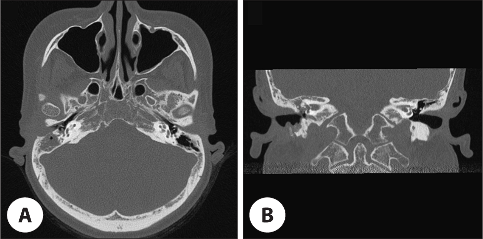
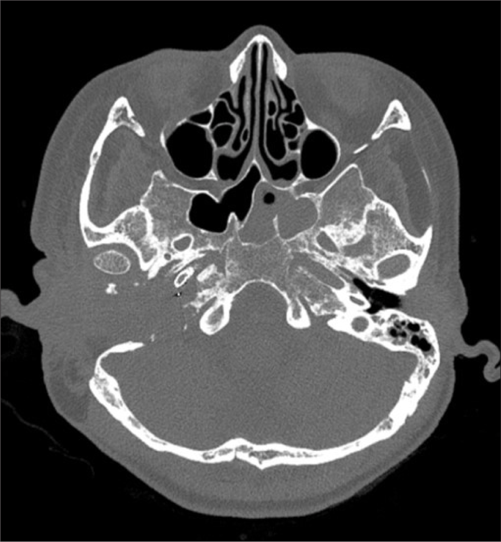
기저질환으로 26년 전 연수신경교종에 대한 방사선 치료력, 당뇨병성 만성 신부전 병력이 있는 41세 여자 환자가 1년 전부터 지속되는 우측 이루를 주소로 외래 내원하였다. 내원 당시 우측 농성 및 혈성 이루를 호소하였으며, 우측 외이도 부종 및 농성 이루, 외이도 전벽의 미란을 관찰할 수 있었다. 수술 전 시행한 순음청력 검사상 우측 기도 청력 35 dB, 골도 청력 23 dB였으며, 안면마비 증상은 없었다. 추가로 시행한 측두골 전산화단층촬영상 우측 외이도 후골벽에서부터 유양돌기뼈까지 불규칙한 골 미란 및 상고실과 유양돌기봉소을 가득 채우고 있는 연부조직 음영이 관찰되었다(Fig. 1). 영상의학적 소견과 방사선 치료 병력을 고려하였을 때 중이 및 외이도의 방사선골괴사의 가능성도 염두에 두고, 만성중이염을 치료하기 위하여 개방동 유양돌기절개술 및 고막성형술을 시행하였다. 수술 당시 유양동과 고실 내부는 육아조직으로 차 있는 양상이었으며, 조직병리검사상 만성 염증 소견을 보였다.
Fig. 1.
Preoperative temporal bone computed tomography scan of the patient showing Irregular bone erosions at right bony EAC and mastoid bone, due to expansile osteolytic lesions. A: Axial view. B: Coronal view. EAC: external auditory canal.
Download Original Figure
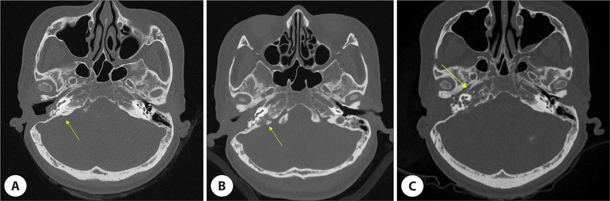
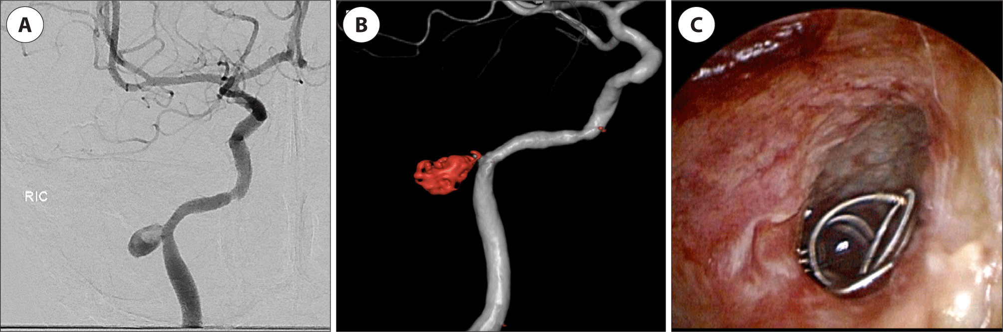
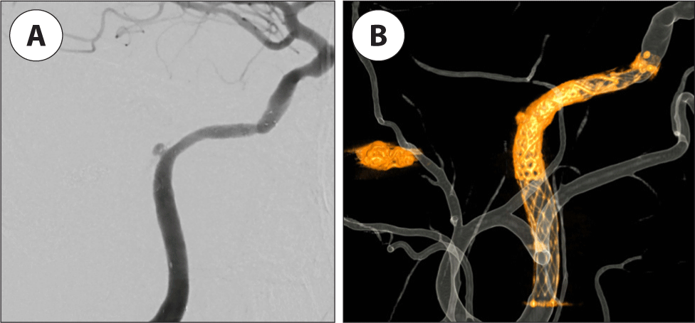
그 후 약 4년간 지속적으로 외래를 통하여 추적 관찰하였는데, 시간이 지날수록 측두골 전산화단층촬영에서 골 미란 병변은 추체첨부까지 점차 범위가 커지는 양상을 보였으며(Fig. 2), 우측 안면 마비는 수술 2개월 후 처음 발생하여 House Brackmann grade V까지 점진적으로 진행하였고 청력 손상도 점차 진행하여 심도 난청 소견을 보였다. 두개저 부위의 진행하는 골미란과 함께 주변부 연조직의 침범으로 악성 외이도염도 감별해야 하나 이통을 호소하지 않은 점, 녹농균이 배양되지 않은 점, 혈액 검사상 염증 수치가 정상 범위였으며, 환자의 방사선치료력을 고려하였을 때 방사선골괴사의 가능성이 높다고 판단하고, 내경동맥의 평가를 위한 추가 영상 검사를 권유하였으나, 환자 사정상 이를 거부하였고 간헐적으로 발생하는 농성 이루에 대해 항생제 치료를 하며 짧은 간격을 두고 외래를 통한 추적관찰을 진행하였다. 그러던 중 환자가 대량의 우측 혈성 이루를 주소로 응급실에 내원하였다. 전뇌동맥조영촬영상 우측 내경동맥의 추체분절에서 약 8 mm 크기의 가성동맥류가 발견되었고(Fig. 3A), 이에 신경외과에서 코일 색전술을 시행하였다(Fig. 3B). 시술 후에도 간헐적인 혈성 이루 증상을 호소하였으며, 시술 1주 뒤 우측 외이도에 코일이 빠져 나온 것이 귀내시경을 통하여 발견되었다(Fig. 3C). 이에 진행하는 중이 및 외이도의 방사선골괴사에 대해 추체아전절제술을 시행하였으며, 골괴사가 의심되는 측반고리관의 일부도 함께 제거하여 주었다. 그 후 성형외과 협진 하에 전외측대퇴 유리피판술을 이용하여 결손부분을 재건하였다. 수술 중 제거한 부골의 조직병리검사상 골의 괴사 소견이 확인되었다. 수술 직후 신경외과에서 남아있는 가성 동맥류 부위에 다시 코일 색전술을 시행하였다.
Fig. 2.
Follow-up images of post-operative temporal bone CT (computed tomography). A: 4 months after surgery, showing no definitive osteolytic lesion at petrosal bone (arrow). B: 3 years after surgery, showing osteolytic lesion at petrosal bone (arrow). C: 4 years after surgery, showing progressing osteolytic lesion at petrosal bone and internal carotid canal (arrow).
Download Original Figure
Fig. 3.
4-vessel angiography of the patient. A: A pseudoaneurysm approximately 8 mm in size in the petrous segment of right ICA (internal carotid artery). B: Status post coil embolization on aneurysm involving petrous segment of right ICA. C: Otoscopy of the patient. A coil was found in the right EAC, which is considered to be a part of previous coil embolization. EAC: external auditory canal.
Download Original Figure
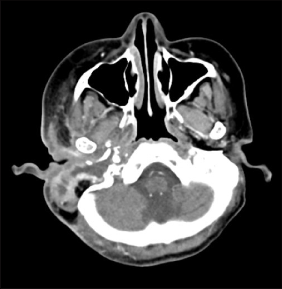
술 후 환자는 활동성 출혈은 없었으나 수술 부위에서 화농성 분비물이 배출되었고, 영상 검사에서 추체첨부 부위의 괴사 및 농양 형성이 확인되었다(Fig. 4). 이후 수차례의 절개배농 및 세척을 시행하며 경과 관찰하던 중, 수술 후 2개월에 진행한 전산화 단층촬영에서 이전 코일의 이탈 및 가성 동맥류의 재발이 확인되었다(Fig. 5A). 가성동맥류 파열에 의한 대량 출혈을 예방하기 위해 신경외과에서 우측 내경동맥에 스텐트 삽입술을 시행하였다(Fig. 5B). 스텐트 삽입 1주 후, 전신 마취 하에 수술 부위 탐색을 시행하였는데, 수술장 소견상 이전 추체아전절제술 부위 내부가 육아 조직으로 차 있고, 남은 골조직에도 부분적인 침범이 의심되었기에, 추체아전절제술을 재시행하여 창상세척, 육아 조직 및 괴사조직을 추가로 제거하였다. 그 후 성형외과 협진으로 대퇴근막장근 및 지방 이식술을 이용하여 결손부위 재건술을 시행하였다. 술 후 환자는 수술 부위의 피부 염증이 생겼으나 항생제 및 드레싱을 통해 조절되었으며, 이후 약 6개월간 특이 소견 없이 경과 관찰 중이다. 최근 시행한 전산화 단층촬영에서 추가적인 감염의 악화 및 골 미란은 확인되지 않았다(Fig. 6). 지금까지의 환자 임상증상 및 치료과정, 그리고 수술 부위 사진을 정리하였다(Fig. 7, 8).
Fig. 4.
Follow-up images of post-operative temporal bone CT (computed tomography). Abscess formation at right petrous apex.
Download Original Figure
Fig. 5.
Follow-up 4-vessel Angiography of the patient. A: Recurrent pseudoaneurysm in the petrous segment of right ICA (internal carotid artery). B: Stent insertion on right petrous ICA.
Download Original Figure
Fig. 6.
Recent follow-up image of temporal bone CT (computed tomography). Little interval change of underlying postoperative bony erosion and stent installation state.
Download Original Figure
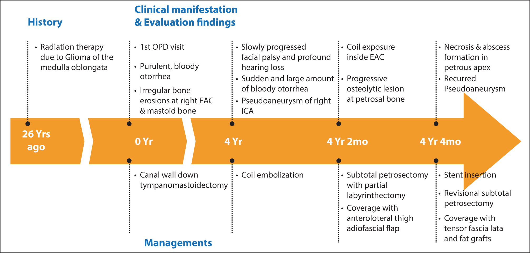
Fig. 7.
Clinical course of the patient. OPD: outpatient department, EAC: external auditory canal, ICA: internal carotid artery.
Download Original Figure
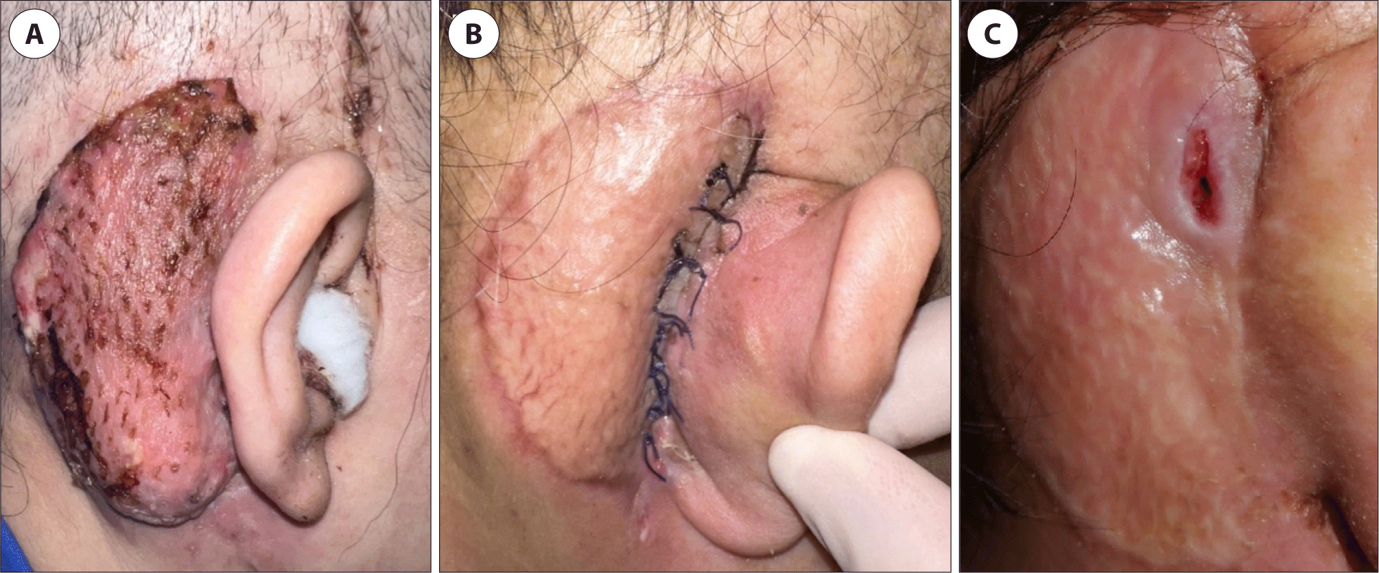
Fig. 8.
Clinical course of the external wound. A: External wound after first subtotal pestrosectomy. B: External wound after second subtotal petrosectomy. C: External wound of recent follow-up.
Download Original Figure
고찰
두개저 방사선골괴사는 두경부 부위에 대해 고용량 방사선 치료 후 괴사되는 과정으로 치료가 가장 어려운 합병증 중 하나로 여겨진다.1) 두개저 방사선골괴사의 진단은 임상적, 영상학적 및 병리학적 소견에 근거하는데, 가장 흔한 임상 증상은 두통이며, 악취, 가피 형성, 비출혈, 복시 등의 뇌신경 증상이 동반되는 경우가 흔하다.2) 영상검사의 전형적인 특징으로는 두개저에 인접한 뼈에서 경화성 또는 미란성 변화와 함께 괴사성 연조직이 동반된다.3,4) 방사선골괴사의 조직학적 특징으로는 과도한 혈류, 동맥염, 혈전 형성, 세포 손실, 저혈관성 병변, 골수강 내 지방 증가, 그리고 섬유화의 과정이 순서대로 나타난다.5) 두개저 방사선골괴사의 치료는 보전적 치료 방법과 수술적 방법이 있으며, 보전적 치료 방법으로는 전신 항생제, 항염증성 약물의 투여 및 고압산소치료가 있다.2)
두개저 방사선골괴사에서 약 50%는 내경동맥에 인접한 부위에서 괴사가 발생하는데, 경동맥 파열로 인한 과다 출혈이 사망 원인 중 가장 큰 비율을 차지한다고 알려져 있다.6) 내경동맥의 침범이 발생한 경우, 부골절제술과 같은 적극적인 치료에도 불구하고 경동맥 파열이 발생할 수 있다. 따라서 모든 환자에서 내경동맥에 대한 평가가 필요하며, 일부의 경우에는 혈관조영술을 시행하여 혈관의 주행을 확인하고 가성동맥류과 같은 병변이 없는지 확인하여야 한다. 만약 검사상 가성동맥류가 확인되거나, 내경동맥을 둘러싸는 뼈의 외벽이 파괴되어 내경동맥이 노출되거나, 괴사 조직으로 생각되는 병변과 내경동맥이 인접한 경우, 내경동맥의 파열을 방지해야 한다. 이를 위해서는 염증 부위를 제거하는 적극적인 수술적 개입이 필요하며, 수술 전에 내경동맥 평가를 위해 혈관 조영술을 고려해야 한다. 뇌에 저관류가 없는 경우 내경동맥에 혈관 내 코일 색전술을 시행 후 괴사 조직 제거 수술을 시행해 볼 수 있다.2) 또한 어떤 연구에서는 내경동맥에 혈관 스텐트를 삽입하고 2–3주 후에 수술을 진행하면 스텐트 주변에 신생혈관외막이 형성되어 병변으로부터 경동맥의 박리가 가능하다고 보고되고 있는데, 내경동맥이 심하게 비틀려 있는 경우 스텐트 삽입으로 인해 내경동맥이 파열될 수 있음에 주의하여야 한다.7)
본 증례 및 문헌 고찰을 통해 살펴본 바와 같이, 방사선골괴사가 의심되는 경우에는 내경동맥에 대한 평가가 반드시 필요하며, 내경동맥의 침범이 의심되는 경우 적극적인 검사 및 치료를 조기에 고려하여야 한다. 또한 초기 치료 후에도 지속적인 평가를 시행하여 재발 여부를 확인하고 염증의 진행을 막는 것이 내경동맥의 파열을 예방하는 데 중요한 것으로 사료된다.