Introduction
Osteomas are the most common paranasal sinus benign tumors and often occur in the frontal and ethmoidal sinus.1) They rarely exhibit symptoms because of their gradual increase in size and are mostly discovered accidentally during a radiographic examination. In advanced cases, osteomas may cause ocular protrusion, oculomotor dysfunction, and visual impairments, as well as symptoms caused by dural invasion such as meningitis and cerebrospinal fluid leakage.2) In particular, pneumocephalus caused by the frontal sinus or ethmoid sinus osteoma is extremely rare, but it can cause serious side effects, indicating the need for surgical resection using the safest method possible. In general, endoscopic removal is appropriate if the osteoma is located somewhere other than the frontal sinus. In ethmoid osteoma, the endoscopic approach is relatively simple. Additionally, the endoscopic approach allowed the removal of all ethmoid osteomas involving the anterior skull base or lamina papyracea.3) Herein, we report a case of a patient who visited the clinic with Broca’s aphasia, was diagnosed with pneumocephalus caused by an ethmoid sinus osteoma accompanied by mucocele, and was treated safely by only endoscopic sinus surgery.
Case Report
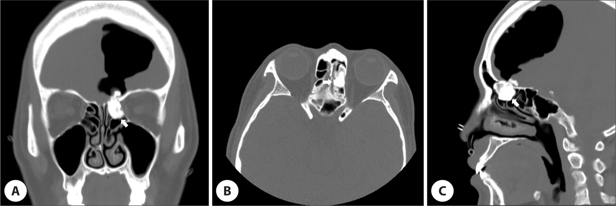
A 49-year-old Korean woman visited the emergency room of the hospital with aphasia without any accompanying symptoms. She had no specific underlying diseases, and it was the first event of aphasia. A brain computed tomography (CT) performed at the time of admission revealed pneumocephalus along with a mass on the left ethmoid sinus roof. At admission, the patient had Broca’s aphasia and general weakness but no other neurological symptoms, and she did not present any nasal symptoms. A paranasal sinus CT scan was performed, which revealed an ethmoid sinus osteoma measuring approximately 2.4×1.8 cm and invading the skull base from the left ethmoid sinus roof (Fig. 1). Soft tissue density was also observed on the upper side of the intracranial osteoma, and pneumocephalus findings were detected on the left side of the brain. A skull base defect caused by the osteoma was suspected as the cause of the pneumocephalus, and an endoscopic sinus surgery was planned to remove the osteoma and repair the defect.

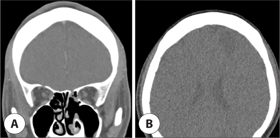
During operation, the osteoma in the ethmoid roof was exposed after ethmoidectomy (Fig. 2A). After confirming the osteoma on the ethmoid sinus roof, most of it was removed using a drill. Then, the attachment site of the osteoma to the skull base was exposed, and its remnant was removed using a curette to minimize damage to the dura. The removed osteoma had a smooth surface on the side of the ethmoid sinus and a cystic polyp on the intracranial component, which appeared as a soft tissue density on the CT scan (Fig. 2B). After the removal of the osteoma, a 2×1 cm defect occurred in the dura mater at the skull base attachment of the osteoma, and shrinkage of left anterior brain was observed (Fig. 2C). The surgery was completed after multilayer reconstruction of the defect using tachosil, duracell, and nasoseptal flap. The aphasia improved 3 days after surgery, and normal conversation was possible at postoperative 5 days. After surgery, she was discharged without any complications, including cerebrospinal fluid leakage, and the pneumocephalus showed improvement on a CT scan taken after 3 months (Fig. 3). There were no special problems or recurrence during the 3-year follow-up.


Discussion
Osteomas are benign, slow-growing lesions consisting of mature bone, which often occur in the paranasal sinuses.3,4) Paranasal sinus osteomas are most commonly found in the frontal sinus, followed by the ethmoid sinus, maxillary sinus, sphenoid sinus. There are three accepted theories for the etiology of paranasal sinus osteomas, viz., developmental, traumatic, and infectious.5) According to the developmental theory, the apposition of membranous and endochondral tissues traps some of the embryonic cells, eventually resulting in unchecked osseous proliferation.6) The traumatic theory and the infectious theory are highly similar, and both rely on an inflammatory process as the triggering force for bony tumor formation.5)
Most osteomas are asymptomatic, but as they grow and compress the surrounding structures, the inflammatory change caused by secondary infection can induce symptoms. According to the involved location of osteoma extension, patients can complain of various symptoms, including headache, facial pain, ocular pain, and proptosis. Pneumocephalus is an additional rare complication of frontoethmoid osteoma for which two possible theories have been proposed.7-10) The first theory is appropriately termed as “the inverted soda pop bottle phenomenon,” where the leakage of cerebrospinal fluid through a dural or arachnoid defect exerts a siphon effect to draw air up into the relatively low-pressure intracranial space. The second theory – the “ball valve” mechanism – postulates that the breached dura acts as a simple flap valve when the extracranial pressure exceeds the intracranial pressure.
Osteoma treatment can be controversial, but its principle involves periodic observation if asymptomatic, and surgical excision is considered in case symptoms develop. These symptoms include unexplained headaches, recurrent sinusitis, ocular symptoms, and central nervous system manifestations, and if the sinuses expand beyond their normal range, when the opening of the frontal sinus is located or increased by >50%, or when the nasofrontal duct is obstructed, surgical treatment is indicated for all cases of osteoma, irrespective of size. Surgical methods can vary depending on the location, size, and relationship with the surrounding tissues.3,4,9,10) The osteoplastic flap approach is the most commonly used technique for frontal sinus osteomas. This approach has several pros and cons. The advantages of this approach are that it provides excellent access to all aspects of the frontal sinus and makes reconstruction possible if needed. However, it has some disadvantages. This way may cause frontal pain, numbness, paresthesia, embossments and impose frontal sinus obliteration which entails increased risk for late mucocele formation. Small osteomas in the nasal cavity or ethmoid sinus, with minimal invasion to the surrounding areas, can be treated by appropriate removal using an endoscope and a drill. With the recent development of endoscopic procedures, there has been an increase in attempts using endonasal approaches.10) The advantages of endoscopic approach are as follows: It provides more detailed visualization, reduces intraoperative traumatic impact and bleeding, and decreases postoperative morbidity and duration of hospital day. However, intraoperative complications in some restricted areas may be difficult to manage by endoscopic approach, as it requires long procedure time, and sophisticated surgical training.
In the present case, initially, the neurosurgeon planned a transcranial approach, although endonasal approaches have several benefits such as shortened hospital stays, faster recovery, and less cosmetic issues. To date, not many cases of removal solely by endoscopic surgery have been reported, and in Korea, our case is the only one where the osteoma in the ethmoid sinus, which caused intracranial symptoms, was removed by only endoscopic surgery. Although rare, osteomas from the skull base can cause pneumocephalus; hence, we expect that this case report will be helpful when explaining treatment options.


