Case Report

비강 내 이소성 치아의 내시경적 제거 2예

조은현1https://orcid.org/0000-0002-0444-0981, 박두식1https://orcid.org/0000-0003-2112-3387, 위지혜1https://orcid.org/0000-0002-6513-7011, 이중섭1,*https://orcid.org/0000-0003-4763-2091
Eun Hyun Cho1https://orcid.org/0000-0002-0444-0981, Doo Sik Park1https://orcid.org/0000-0003-2112-3387, Jee Hye Wee1https://orcid.org/0000-0002-6513-7011, Joong Seob Lee1,*https://orcid.org/0000-0003-4763-2091
Author Information & Copyright
1한림대학교 의과대학 한림대학교성심병원 이비인후-두경부외과학교실
1Department of Otorhinolaryngology-Head and Neck Surgery, Hallym University Sacred Heart Hospital, Hallym University College of Medicine, Anyang, Korea
*Corresponding author: Joong Seob Lee, Department of Otorhinolaryngology-Head and Neck Surgery, Hallym University Sacred Heart Hospital, Hallym University College of Medicine, Anyang 14068, Korea, Tel: +82-31-380-3840, Fax: +82-31-386-3860, E-mail: apniosio@hallym.or.kr

© Copyright 2023 The Busan, Ulsan, Gyeoungnam Branch of Korean Society of Otolaryngology-Head and Neck Surgery. This is an Open-Access article distributed under the terms of the Creative Commons Attribution Non-Commercial License (http://creativecommons.org/licenses/by-nc/4.0/) which permits unrestricted non-commercial use, distribution, and reproduction in any medium, provided the original work is properly cited.

Received: Feb 20, 2023; Revised: Mar 29, 2023; Accepted: Mar 31, 2023

Published Online: Jun 30, 2023

ABSTRACT

An ectopic tooth is an eruption of a tooth into an abnormal location, such as the palate, sinonasal cavities, mandibular condyle, coronoid process, orbit, or skin. It can be found incidentally or be presented with a variety of symptoms, such as unilateral nasal obstruction, epistaxis, rhinorrhea, chronic nasal congestion, headache, or facial pain. Ectopic teeth are most commonly supernumerary, but may also be deciduous or permanent teeth. Possible causes of ectopic teeth in the sinonasal cavity could be genetic factors, cleft palate, odontogenic infections, trauma, crowding or displacement due to cysts or tumors, but the etiology is not yet fully understood. Early surgical removal is recommended to avoid possible complications. We report two cases of a child with an ectopic supernumerary tooth found in the nasal cavity, both of which were successfully removed via endoscopic surgery.

Keywords: Tooth eruption, ectopic; Tooth, supernumerary; Nasal cavity; Endoscopy

서론

이소성 치아란 정상적인 위치가 아닌 구개, 부비동, 하악골 관절돌기, 하악골 근육돌기, 안와, 비강, 피부 등의 위치에서 맹출된 치아를 말한다.13) 이소성 치아의 95%가 구개와 상악에 발생하며 비강 내 발생은 매우 드물다.24) 과잉치, 영구치, 유치 모두 이소성 치아로 발견될 수 있으나 과잉치가 가장 흔하다.2) 과잉치의 유병률은 0.1%–1%로 알려졌고 그중 극히 적은 수에서 비강 내 치아로 발현된다.2,5) 여성보다 남성에서 더 많이 발생하며 절반의 경우에서 성인이 되기 전에 발견된다.2,57) 가능한 원인으로는 유전적 요인, 발달장애, 외상으로 인한 치아 변위, 낭종, 감염, 치아 총생이나 잔존 유치로 인한 정상적 맹출의 방해 등이 있다.13,6,8) 소아에서는 구순구개열, 치조열 등의 구조적 발달이상을 가진 환아에서 더 흔한 것으로 보고되었다.13,6) 두 명의 소아 환자에서 발견된 비강 바닥에 위치한 이소성 치아 증례를 경험하여 문헌고찰과 함께 보고하고자 한다.

증례

증례 1

특이 과거력이 없는 8세 남자 환자가 점액성 비루 및 비폐색으로 개인 의원에서 진료 중에 발견된 비강 내 이물로 내원하였다. 비내시경 소견상 좌측 하비도와 비중격 사이에 유동성 있는 백색의 단단한 덩이가 관찰되었다(Fig. 1D). 전산화단층촬영을 시행하였고, 좌측 비강저에 치아로 추정되는 고밀도의 종물이 관찰되었다(Fig. 1B, C). 치관은 비강의 외측을 향하고 하비갑개를 압박하며 하비도 일부를 막고 있었다. 파노라마 X-선상 수평위(transverse)의 방사선 비투과성 과잉치 추정 물질이 관찰되었으며, 그 외 치아 발육 및 배열의 이상소견은 관찰되지 않았다(Fig. 1A). 악안면부 외상의 과거력이나 수술력, 구개열 등의 선천 기형 또한 없었다.

jcohns-34-2-47-g1
Fig. 1. Clinical and radiological findings of Case 1. A: Panoramic radiograph shows a horizontally directed tooth-like radiopacity in the left nasal cavity. B,C: Computed tomography (CT) scan revealed a calcified tooth-like mass in the left inferior nasal meatus, originating from the nasal floor near the septum. D: Hard whitish mass between inferior meatus and nasal septum of the left nasal cavity was observed. E: Two-month-postoperative endoscopic finding showing uneventful course of recovery. F: Gross specimen of the removed tooth, measuring 1.4×0.5×0.5 cm with incomplete root formation and no tubercles or developmental grooves.
Download Original Figure

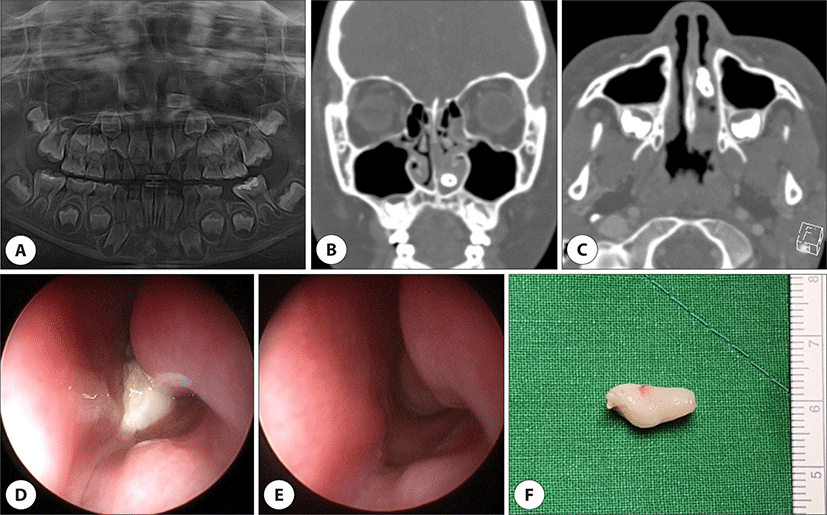
전신마취하 내시경적 제거술을 시행하였다. 종물은 비중격과 접하는 비강저에 기시부가 확인되었다. 기시부의 일부를 덮고 있는 종창된 점막을 함께 절제하였다. 제거된 종물은 1.4×0.5×0.5 cm 크기의 원추형에 가까운 형태였고 불완전한 치근 발달을 보였다(Fig. 1F). 수술 후 두 달간 추적 관찰하였으며, 비폐색 증상은 해소되었고 수술 부위는 합병증 없이 치유되었다(Fig. 1E).

증례 2

가와사키병의 과거력과 비알코올성 지방간염이 있는 10세 남자 환자가 개인 의원에서 우연히 좌측 비강 내 이물이 발견되어 의뢰되었다. 내시경 검사상 좌측 비강저 점막에 덮여 있는 단단한 원뿔모양의 종물이 관찰되었고, 하비갑개 전반부의 변형이 동반되었다(Fig. 2D). 전산화단층촬영에서 좌측 비강저와 비중격 사이의 절치공에서 기원하는 치아로 추정되는 골음영의 종물이 확인되었다(Fig. 2B, C). 파노라마촬영에서 이소성 치아 추정 물질 외에 상악과 하악의 나머지 치아 발육과 배열상태는 정상이었다(Fig. 2A). 역위된 정중과잉치(inverted mesioden)가 비강 내로 맹출된 것으로 진단되었다.

jcohns-34-2-47-g2
Fig. 2. Clinical and radiological findings of Case 2. A: Panoramic radiography shows a radiopaque mass superimposed upon the left permanent central incisor, which is erupting into the left nasal cavity. B,C: Computed tomography (CT) scan revealed a hyperdense, 1.2 cm-long mass in the left nasal cavity protruding from the incisive foramen. D: Endoscopic exam revealed a hard whitish mass partially covered with mucous membrane of the left nasal floor. E: Endoscopic exam showing well-healed mucosa 2 months after the surgery. F: Gross photo of a cone-shaped mesioden with complete root formation, measuring up to 2.0 cm.
Download Original Figure

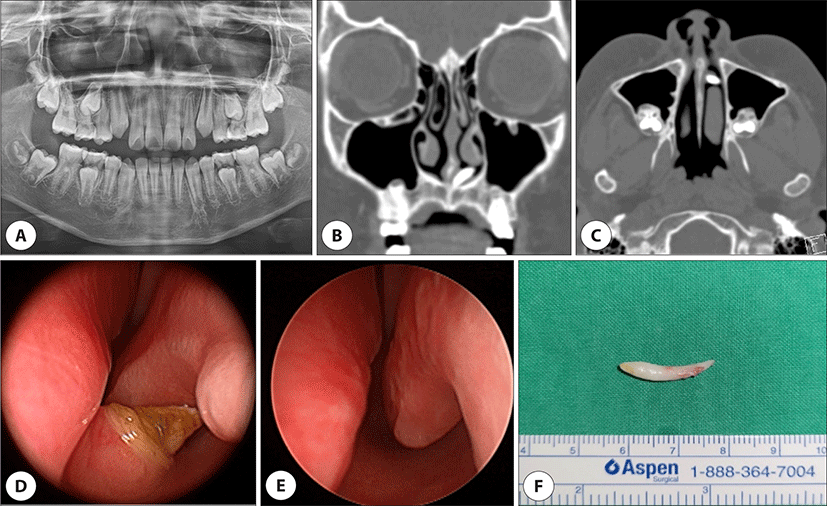
전신마취하 비내시경을 사용하여 이소성 치아를 제거하였고, 전비공 패킹으로 지혈하였다. 제거된 종물은 길이 2.0 cm에 달하는 백색의 원추형으로, 전형적인 정중과잉치 형태였다(Fig. 2F). 치아를 둘러싼 점막은 조직검사결과 만성 염증을 동반한 육아 조직소견을 보였다. 두 달 후 비강 내 점막은 특이소견 없이 치유되어 추적관찰을 종료하였다(Fig. 2E).

고찰

비강 내 이소성 치아는 대부분 과잉치로 보고되었다.1,3) 과잉치는 상악중절치 사이에 1개 또는 2개의 형태로 가장 흔히 발생하는데, 이를 정중치(mesiodens)라고 하며 주로 원추형태를 취한다.4,5) 이소성 치아는 주로 단일로 나타나며,2,3,6) 본 증례에서 제거한 비강 내 치아도 모두 단일의 과잉치였고 그중에서도 정중치였다.

비강 내 치아는 주로 무증상이지만 비폐색, 비충혈, 안면통, 두통, 재발성 코피, 악취나는 비루, 외비 변형, 코눈물관 폐색, 후각감소 등의 다양한 증상을 유발할 수 있다.2,3,6) 이소성 치아는 다른 증후군과 관련되어 발견되는 경우도 있는데, 대장 용종, 골종, 표피 낭종, 과잉치 등이 동반되는 가드너 증후군과 두개골, 쇄골, 치아의 비정상적인 성장을 특징으로 하는 쇄골두개이형성증이 대표적이다.5) 그 외에도 구순구개열, 다운 증후군, 스터지 웨버 증후군, 두개골간단 이형성증 등이 있다.4)

감별해야 할 진단 중에는 이물, 비석, 양성 혹은 악성종양, 석회화를 동반한 염증, 진균 감염, 골종, 치아종 등이 있다.1,2,6) 증상이 비특이적일 뿐만 아니라 비후성 점막에 일부 혹은 전체가 덮여 있는 경우가 많아 오진단되기 쉽다.1,6,8,9) 전산화단층촬영을 통해 확진이 이루어지며 맹출 부위 깊이, 주변 구조물과의 해부학적 관계를 평가하여 수술적 치료를 계획할 수 있다.2,6,9) 파노라마촬영은 전반적인 치아발달 상태를 확인하여 과잉치 여부 판별에 도움이 된다.2) 본 증례의 경우에도 치과적 검진 및 영상의학적 검사를 시행하여 과잉치로 판별할 수 있었다.

비강 내 이소성 치아는 개수, 방향, 맹출 여부 등에 따라 다양한 합병증이 발생할 수 있다.10) 비부비동염, 골수염, 비강 내 농양 및 천공, 구비강 누공, 눈물주머니염, 아스페르길루스증, 외비 변형, 정상치아 맹출의 방해 등이 보고되었다.2,6) 과잉치의 30%–60%에서 정상 맹출의 방해, 22%–63%에서 인접 영구치의 전위를 유발하며, 4%–9%에서 치성 낭종이 동반되는 것으로 알려졌다.11) Ryu 등은 정위보다 수평위의 정중치가, 시상면에서의 상하 위치로는 치근단부(near-apical), 치근단 상방(beyond-apical)보다 치경부(cervical)의 정중치가 인접 영구치의 맹출을 방해하여 상대적으로 합병증이 빈번하다고 보고하였다.12) 한편, 치근단 상방의 정중치는 비강 내로 맹출하거나 오래 매복되어 낭성 변화를 일으키는 문제를 유발할 수 있다.12)

수술 시기에 대해서 Park 등2)은 증상, 위치, 맹출 여부를 고려하여 치료를 정할 것을 권고하였다. 무증상의 미맹출 치아는 경과관찰을 할 수 있으나 영상의학적 검사를 포함한 추적관찰을 강조하였다.1,2) 하지만 합병증 예방을 위해서는 증상이 없더라도 조기 수술적 제거가 권고된다.1,4,6,7) Primosch11)는 즉시 제거를 권유하되 두 가지 예외를 두었다. 역위되지 않은 원추형 정중치는 예후가 좋으며 조기 맹출이 예상되므로 맹출을 기다려 볼 수 있고, 치근단 상방의 과잉치는 영구치의 맹출에 방해가 되지 않으므로 제거 시기가 중요하지 않다고 하였다. 단, 비강 내로 맹출하거나 다른 합병증을 유발하였을 경우에는 즉시 제거를 권유하였다.1,2,11) 저자들이 경험한 2예의 치아는 모두 정위가 아닌 역위 또는 수평위로 위치하였으며, 이미 비강 내로 맹출한 상태로 발견되었기에 즉시 제거를 요하였다. 첫 증례는 비폐색을 유발하였고 두 번째 증례는 맞닿은 하비갑개의 변형을 초래하여 합병증을 유발할 가능성이 있어 조기에 제거하였다.

내시경적 제거는 기존의 비외접근법에 비해 최소 침습적이며 수술 후 이환율, 합병증, 수술 시간 등에서 우수한 성적을 거두었다.1,2,6,7) 상악동 내 치아의 경우에도 개구비도단위와 가깝다면 기존의 Caldwell-Luc surgery보다 중비도 또는 하비도 상악동 개창술과 같은 내시경적 접근이 선호되는 추세이다.2,7,11) 이소성 치아가 비강 외측 벽에서 멀리 떨어져 있는 경우, 안와하벽 또는 비루관에 가까운 경우, 동반된 치성낭종의 크기가 클 경우에는 두 접근법을 병행하면 좋은 결과를 기대할 수 있다.13) 본 증례는 비강 바닥 전반부에 위치하였으며, 주변 조직과의 유착이나 비중격 또는 골부와의 강한 유합은 없었기에 비내시경하 쉽게 제거할 수 있었다.

비폐색 또는 비강 내 종물을 주소로 내원한 환자에서 이소성 치아 질환 및 관련 증후군을 염두에 두고 파노라마촬영, 전산화단층촬영 등의 포괄적인 검사를 시행해야 한다. 이소성 치아로 진단될 경우, 위치에 따라 본 증례와 같이 비내시경적 단순 제거를 통해 쉽게 치료가 가능하다.

Acknowledgements

Not applicable.

Funding Information

Not applicable.

Conflicts of Interest

No potential conflict of interest relevant to this article was reported.

Author Contribution

Conceptualization: Lee JS.

Data curation: Cho EH.

Formal analysis: Wee JH.

Methodology: Park DS.

Validation: Lee JS.

Investigation: Cho EH.

Writing - original draft: Cho EH, Park DS.

Writing - review & editing: Cho EH, Park DS, Wee JH, Lee JS.

Ethics Approval

Not applicable.

References

1.

Kim DH, Kim JM, Chae SW, Hwang SJ, Lee SH, Lee HM. Endoscopic removal of an intranasal ectopic tooth. Int J Pediatr Otorhinolaryngol 2003;67(1):79-81.

2.

Park HE, Park HJ, Bae JH, Lee SS. Clinical features of ectopic tooth occurring in sinonasal cavity. Korean J Otorhinolaryngol 2012;55(8):487-92.

3.

Smith RA, Gordon NC, De Luchi SF. Intranasal teeth: report of two cases and review of the literature. Oral Surg Oral Med Oral Pathol 1979;47(2):120-2.

4.

Kim YG, Kim SI, Kim DH, Kim SW. Incidental ectopic tooth in the nasal cavity and maxillary sinus. J Korean Skull Base Society 2017;12(1):42-5.

5.

Hong SC, Choo KB, Lee CH, Lee KI. A case of supernumerary tooth originated from the nasal cavity. J Clin Otolaryngol Head Neck Surg 2003;14(1):141-3.

6.

AlMulhim A, AlMomen A, AlKhatib A. Ectopic intranasal canine tooth in a child: a rare case report and literature review. Int J Surg Case Rep 2019;55:202-5.

7.

Levin M, Sommer DD. Endoscopic removal of ectopic sinonasal teeth: a systematic review. J Otolaryngol Head Neck Surg 2019;48(1):30.

8.

Jin KF, Lou ZC, Lou ZH. The diagnosis and management of supernumerary nasal teeth. J Laryngol Otol 2019;133(6):538-41.

9.

Min HJ, Oh SR, Kim KS. Endoscopic differences between intranasal ectopic teeth. Ear Nose Throat J 2020; 99(6):NP58-9.

10.

Omer RSM, Anthonappa RP, King NM. Determination of the optimum time for surgical removal of unerupted anterior supernumerary teeth. Pediatr Dent 2010;32(1): 14-20.

11.

Primosch RE. Anterior supernumerary teeth—assessment and surgical intervention in children. Pediatr Dent 1981;3(2):204-15.

12.

Ryu G, Song JS, Shin TJ, Hyun HK, Kim JW, Jang KT, et al. Retrospective study on three-dimensional characteristics of mesiodens using CBCT in pediatric dentistry. J Korean Acad Pediatr Dent 2021;48(1):77-94.

13.

Emanuelli E, Borsetto D, Brunello G, Sivolella S. Endoscopy-assisted removal through combined lower and middle meatotomies of an ectopic upper third molar in the sinus associated with a dentigerous cyst. Oral Maxillofac Surg Cases 2018;4(1):23-31.