Case Report

중이강 내 상피내암에 대한 광범위 절제술 후 재발한 상피암 1예

안현우1https://orcid.org/0009-0007-8037-5476, 김용완1https://orcid.org/0000-0002-6010-312X, 백무진1,*https://orcid.org/0000-0002-2492-8765
Hyeon Woo Ahn1https://orcid.org/0009-0007-8037-5476, Yong Wan Kim1https://orcid.org/0000-0002-6010-312X, Moo Jin Baek1,*https://orcid.org/0000-0002-2492-8765
Author Information & Copyright
1인제대학교 의과대학 해운대백병원 이비인후과학교실
1Department of Otorhinolaryngology, Inje University Haeundae Paik Hospital, Busan, Korea
*Corresponding author: Moo Jin Baek, Department of Otorhinolaryngology, Inje University Haeundae Paik Hospital, Busan 48108, Korea, Tel: +82-51-797-2290, Fax: +82-51-797-2304, E-mail: mjbaek@inje.ac.kr

© Copyright 2023 The Busan, Ulsan, Gyeoungnam Branch of Korean Society of Otolaryngology-Head and Neck Surgery. This is an Open-Access article distributed under the terms of the Creative Commons Attribution Non-Commercial License (http://creativecommons.org/licenses/by-nc/4.0/) which permits unrestricted non-commercial use, distribution, and reproduction in any medium, provided the original work is properly cited.

Received: Mar 20, 2023; Revised: Mar 27, 2023; Accepted: Apr 10, 2023

Published Online: Jun 30, 2023

ABSTRACT

Malignancy of the middle ear is rare and its incidence is 0.18 cases per million people, with its most common type being squamous cell carcinoma. The radiologic, surgical findings, and pathology reports has not been well characterized due to its low incidence. Squamous cell carcinoma in situ of the middle ear is even more so. Here, we report a case of a 69-year-old man who developed squamous cell carcinoma of middle ear 19 months after wide excision for squamous cell carcinoma in situ. The patient received concurrent chemoradiation therapy after revision operation and being evaluated as a stable disease for 30-months follow-up. This case represents the first reported occurrence of squamous cell carcinoma of the middle ear after wide excision for squamous cell carcinoma in situ. A diagnosis of squamous cell carcinoma should be considered in a patient with squamous cell carcinoma in situ of middle ear even after surgery.

Keywords: Carcinoma in situ; Carcinoma, squamous cell; Ear, middle; Recurrence

서론

중이강 내 악성 종물은 매우 드문 질환으로, 미국에서 100만 명 당 0.18명의 유병률을 보인다.1) 편평세포상피암(squamous cell carcinoma)이 가장 흔하며, 55.9%–62.8%의 비율을 차지한다.1,2) 편평상피세포암의 전구단계인 편평세포상피내암(squamous cell carcinoma in situ) 역시 중이강 내 발생하는 경우는 드물며, 일전 만성 중이염으로 고실성형술 중 비정상적인 고실 갑각부의 점막을 광범위하게 절제 후 편평세포상피내암으로 진단된 증례를 국내에서 처음으로 보고한 바가 있다.3) 편평세포상피내암의 치료로 알려진 광범위 절제술을 시행하였음에도 불구하고, 추적 관찰하던 도중 재발의심 소견으로 시행한 재수술에서 편평세포암이 진단되었기에, 저자들은 중이강 내 편평세포상피내암에 대한 광범위 절제술 후 재발한 편평세포상피암에 대한 1예를 보고하는 바이다.

증례

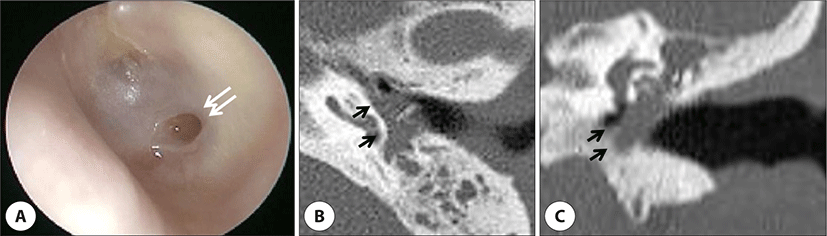
특이 병력 없는 67세 남자 환자로 30년 전부터 반복되는 좌측 이루로 본원에 내원하였다. 청력저하, 어지러움 등의 증상은 없었으며, 가족력상의 특이사항은 없었고, 전신상태는 양호하였다. 이내시경상 좌측 고막의 전하부에 이루를 동반한 천공소견(Fig. 1A)이 관찰되었다. 순음청력검사상 좌측 기도 역치는 36 dB, 골도 역치는 24 dB이었고, 측두골 전산화단층촬영에서 좌측 중이강과 유양동 내 연부조직 및 유돌봉소의 소실과 경화변성이 관찰되었다(Fig. 1B, C). 이에 만성 중이염 진단하에 좌측 고실 성형술 및 폐쇄형 유양동 삭개술을 시행하였다. 수술 시 갑각 주변에 단순 만성 중이염과는 다른 비정상적으로 두껍고 질긴 점막 소견이 있었으며, 안면신경와 및 고실동을 포함하여 다른 중이 공간으로의 침범은 확인되지 않았다. 이에 정상 점막을 포함하여 현미경하 관찰되는 모든 이상점막을 남김없이 광범위하게 절제하고 수술을 종결하였다. 조직검사상 불규칙한 배열을 가진 다핵각질세포가 관찰되었고 기저세포막은 침범되지 않은 소견을 보여 최종적으로 편평세포상피내암으로 진단되었다(Fig. 2).

jcohns-34-2-42-g1
Fig. 1. Preoperative otoscopic finding and temporal bone CT. A: Anteroinferior small perforation with discharge was observed at left tympanic membrane (arrows). B,C: Abnormal soft tissue density was seen in the left middle ear on CT (arrows). CT: computed tomography.
Download Original Figure
jcohns-34-2-42-g2
Fig. 2. Histopathologic examinations of first operation. Atypical tumor cells with intact basement membrane (arrows) which are positive for ki 67 gene expression (H&E, ×200; A), (ki-67, ×200; B).
Download Original Figure

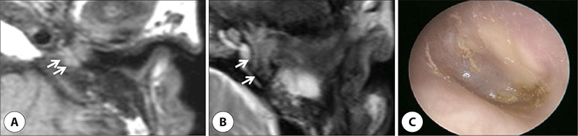
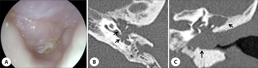
퇴원 후 첫 두 달은 일주일 간격으로 외래를 방문하여 드레싱을 시행하였으며, 수술 부위가 완전히 회복된 후 자기공명영상 촬영 시행 및 약 1년 뒤 추시 영상촬영을 계획하였다. 이후 1개월에서 4개월까지 기간을 늘려가며 경과를 추시하였다. 술 후 16개월째 추적관찰을 위해 촬영한 자기공명촬영상 중이강 내 잔존 연부조직의 크기 변화는 없으나, 술 후 변화 또는 잠복종양과의 감별은 불가능하다는 소견을 얻었다(Fig. 3). 술 후 19개월째 환자가 좌측 이통을 호소하여 시행한 이내시경상 좌측 고막 종창 소견이 관찰되었다(Fig. 4A). 측두골 전산화단층촬영에서 좌측 중이강과 외이도 부위에 연부조직 증가 및 하고실 하벽의 골미란 소견이 관찰되었으며(Fig. 4B, C), 이하선 및 경부 림프절 침범이 의심되는 소견은 없었다. 재발을 고려하여 좌측 고실 성형술 및 폐쇄형 유양동 삭개술 재수술을 계획하였다. 좌측 고막은 매우 두꺼우면서 심한 종창 소견이 관찰되었으며, 고막 근처에서 유래한 것으로 보이는 소엽상 종괴가 외이도 후상벽을 따라 외이도 입구까지 뻗어 있는 소견이었다. 중이 내는 육아종 및 종물이 후방부 고실동을 가득 메우고 추골병의 내측 및 하고실 근처까지 뻗어 있었으며, 동결편조직검사상 침윤성 편평상피세포암 소견을 얻어 종물을 주위 점막을 포함하여 남김없이 제거하였다. 중이의 후벽 하부 골성 고실륜을 제외하고 유양돌기동을 비롯한 기타 골 파괴 소견은 관찰되지 않았으며 이소골 상태는 양호하였다.

jcohns-34-2-42-g3
Fig. 3. Ear magnetic resonance imaging and otoscopic fining taken 16 months after first operation. No gross interval change of residual soft tissue lesion in left middle ear and mastoid (arrows) (T1WI; A), (T2WI; B). No newly appeared lesions or bulging at left tympanic membrane (C).
Download Original Figure
jcohns-34-2-42-g4
Fig. 4. Otoscopic finings (A) and temporal bone computed tomography (B,C) 19 months after first surgery. A: Newly appeared bulging was observed at left tympanic membrane. B,C: Increased amount of soft tissue density in entire left middle ear and lobulated soft tissue thickening in left external auditory canal which suggests possibility of recurred squamous cell carcinoma in situ.
Download Original Figure

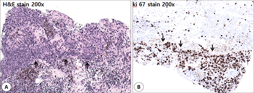
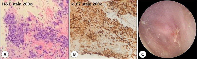
조직검사상 표피의 불규칙한 배열을 가진 비전형적인 다핵각질세포들이 기저막을 침범한 소견을 보였다. 면역조직화학염색에서는 p53(tumor suppressor protein) 음성반응을 보였으며, ki67(cell proliferation related protein)은 비전형 편평세포에서 양성반응을 보였다(Fig. 5A, B). 추가로 CK5/6과 p63 면역조직화학염색을 시행하였고, 양성 반응을 보이는 상피세포를 확인할 수 있어 최종적으로 편평세포상피암으로 진단되었다. 기타 외이도 및 유양돌기동 내 조직에서는 종양세포가 관찰되지 않았다.

jcohns-34-2-42-g5
Fig. 5. Pathology and otoscopic finding 30 months after revision surgery. Basement membranes are interrupted by tumor cells (H&E, ×200; A), (ki-67, ×200; B). There was no evidence of recurrence (C).
Download Original Figure

술 후 시행한 positron emission tomography 영상 및 복부, 흉부 전산화단층촬영상 원격전이 소견은 관찰되지 않았다. 이후 종양내과에서 Cisplatin 항암요법 1차례, FP(5-fluorouracil, cisplatin) 복합항암요법 1차례 및 방사선종양학과에서 30회에 걸쳐 총 6,000 cGY 방사선 치료를 포함하는 동시항암화학방사선요법을 시행하였다. 술 후 27개월 뒤 추적관찰을 위해 촬영한 자기공명촬영상 중이강 내 종양 재발 의심소견으로 시험적 고실개방술을 통한 조직검사를 시행하였다. 조직검사 결과 섬유 조직 외 특이소견은 관찰되지 않았으며, 현재까지 재발 없이 외래에서 경과관찰 중에 있다(Fig. 5C).

고찰

측두골 악성종양은 외이도에서 발생하는 경우가 많으며, 중이강 내 악성종양은 드문 것으로 알려져 있다.4) 그중 편평세포상피암이 55.9%–62.8%로 가장 흔하며,1) 5년 생존율 32%, 평균 생존기간 18.3개월로 예후가 가장 나쁜 편에 속한다.5) 재발률은 국소(local) 20.5%, 지역(regional) 5.5%, 원격(distant) 6.3%로 보고되고 있다.6)

편평세포상피내암은 암세포가 상피에는 존재하나 기저막까지는 침범이 되지 않은 편평세포상피암의 전구단계로서 3%–8%의 비율로 편평세포상피암으로 진행할 수 있으며,7) 일반적으로 광범위 절제술만으로 치료가 충분하다고 알려져 있다.8)

측두골 악성종양에 대한 여러 병기 분류법이 제시되었지만, 중이에서 발생한 악성종양에 대한 보편적으로 인정되는 병기 분류법은 현재로서는 없다.9) 보편적으로 쓰이는 Pittsburgh 분류법은 외이도에 중점을 두고 있기 때문에 중이 악성종양 질환에 대한 병기 분류 및 예후 예측, 치료 효과 판정에 적용하기에는 제한이 있다.10) 미국 국립암연구소에서 운영하는 SEER(The Surveilliance, Epidemiology, and End Results) 데이터를 기반으로 한 중이 악성종양의 범위에 따른 병기 분류법에 따르면, 본 증례의 환자는 원발부위에 국한되어 있었기 때문에, 국소병기(local disease)에 해당하였다.11)

중이 악성종양의 치료방침에 대한 보편적인 합의는 이루어지지 않았지만, 측두골을 침범했을 때 절제가능성이 있을 경우 일괴의 종양 절제와 필요 시 술 후 방사선치료를 병용하게 된다. 술식과 관련하여, Lichun 등은 유양동에 국한된 작은 크기의 초기 암에 대해 확장 고실유양동삭개술을 고려할 수 있으며, 종양이 외이도, 고실, 유양돌기, 측두하악관절, 측두골 인부 또는 추체부를 침범했을 경우 측두골 아전절제술 또는 전절제술을 시행할 것을 제안하였다.1) 술 후 방사선치료는 주로 진행된 병기(T3/T4), 절제연이 근접하거나 양성인 경우, 신경주위 침윤 또는 림프절 전이 소견이 있을 경우 시행한다.12)

본 환자의 경우 수술 중 고실동 침범 소견이 확인되었으나, 중이의 후벽 하부 골성 고실륜을 제외하고 기타 골 파괴 소견은 관찰되지 않았기 때문에 확장 고실유양동삭개술을 시행하였다. 기존 측두골 악성종양에 대한 보고들을 근거로 하였을 때, 술 후 방사선치료의 적응증에 해당하진 않았으나, 재발하였다는 점 및 수술 중 소견 등을 고려하여 종양내과 및 방사선종양학과 상의하에 술 후 동시항암화학방사선요법을 시행하였다.

편평세포상피내암은 일반적으로 광범위 절제술만으로 충분한 치료 효과를 보이지만 증례 환자에서 재발하였는데, 이는 다른 부위와는 다르게 편평하지 않으며 골봉소 및 중이열로 함입된 중이 점막 상피의 특징 때문일 것으로 추측된다. 따라서 술 중 중이강 내 이상 점막과 광범위 절제술 후 잔존 점막에 대한 적극적인 동결편조직검사가 종양의 악성 여부 및 추가적 점막 절제 범위를 판단하는 데 중요할 것으로 생각된다. 또한 원발 부위 및 점막상피가 함입되어 있는 부분에 대해 드릴을 이용하여 삭개하거나, 레이저를 이용하여 기화시키는 방법들을 고려해 볼 수 있겠으며, 이러한 방식이 재발률을 낮출 수 있을지에 대한 추가적 연구가 필요할 것으로 생각된다.

본 증례는 편평세포상피내암의 효과적 치료로 알려져 있는 광범위 절제술을 시행한 이후 발생한 편평세포상피암을 보고한 첫 증례이다. 따라서 중이강 내 발생한 편평세포상피내암의 경우 항상 재발을 염두에 두고, 중이강 내 구조적 특이성을 고려하여 보다 적극적인 조직검사와 광범위하며 공격적인 병변의 제거, 정기적인 고막소견 및 영상 검사 추적관찰이 필요할 것으로 생각된다.

Acknowledgements

Not applicable.

Funding Information

Not applicable.

Conflicts of Interest

No potential conflict of interest relevant to this article was reported.

Author Contribution

Conceptualization: Baek MJ.

Data curation: Kim YW.

Formal analysis: Baek MJ.

Methodology: Baek MJ, Kim YW.

Software: Baek MJ.

Validation: Kim YW.

Investigation: Ahn HW.

Writing - original draft: Ahn HY.

Writing - review & editing: Ahn HY, Kim YW, Baek MJ.

Ethics Approval

Not applicable.

References

1.

Feng L, Jin A, Dai B, Li Y, Guo Y, Wang D, et al. Outcomes of 18 cases with squamous cell carcinoma of middle ear who underwent both surgery and post-operative radiotherapy. Acta Otolaryngol 2016;136(2):141-3.

2.

Gurgel RK, Karnell LH, Hansen MR. Middle ear cancer: a population-based study. Laryngoscope 2009;119(10): 1913-7.

3.

Park BW, Baek MJ. A case of squamous cell carcinoma in situ of the middle ear. Korean J Otorhinolaryngol Head Neck Surg 2019;62(9):529-32.

4.

Ugumori T, Hyodo J, Hato N, Gyo K. Survival in primary carcinoma of the external and middle ear is strongly dependent on stage at diagnosis. Int J Otolaryngol Head Neck Surg 2013;2(6):221-7.

5.

Brant JA, Eliades SJ, Chen J, Newman JG, Ruckenstein MJ. Carcinoma of the middle ear: a review of the National Cancer Database. Otol Neurotol 2017;38(8):1153-7.

6.

Morris LGT, Mehra S, Shah JP, Bilsky MH, Selesnick SH, Kraus DH. Predictors of survival and recurrence after temporal bone resection for cancer. Head Neck 2012; 34(9):1231-9.

7.

Cox NH, Eedy DJ, Morton CA. Guidelines for management of Bowen’s disease: 2006 update. Br J Dermatol 2007;156(1):11-21.

8.

Shu MT, Lin HC, Yang CC, Chang KM. Squamous cell carcinoma in situ of the external auditory canal. J Laryngol Otol 2006;120(8):684-6.

9.

Clark LJ, Narula AA, Morgan DAL, Bradley PJ. Squamous carcinoma of the temporal bone: a revised staging. J Laryngol Otol 1991;105(5):346-8.

10.

Hirsch BE. Staging system revision. Arch Otolaryngol Head Neck Surg 2002;128(1):93-4.

11.

Alam U, Shafqat I, Ahsan S, Alonso J, Han AY, Mukdad L, et al. Survival in middle ear malignancy: a population-based analysis based on the SEER database. Otolaryngol Head Neck Surg 2021;165(1):122-8.

12.

Allanson BM, Low TH, Clark JR, Gupta R. Squamous cell carcinoma of the external auditory canal and temporal bone: an update. Head Neck Pathol 2018;12:407-18.