


Introduction
Management of penetrating neck injuries (PNI) is complicated and rarely experienced by medical personnel. Due to compact anatomical structure of neck, including major vascular, upper respiratory, digestive and neurological structure, usually injuries afflict multiple structures.1) PNI shows mortality rate of 3%–6%, caused by hemorrhagic shock, asphyxiation, spinal cord injury, air embolism or sepsis.2) Therefore, rapid systemic investigation, decision making and well-timed treatment is essential to prevent destructive result.
Once mandatory exploration was treatment of choice for injuries penetrated the platysma. Recent years, managing PNI changed its paradigm from mandatory exploration to selective management. In the selective management, physical examination and selective diagnostic studies, supported by clinicians, are used to manage patients. Initial management of PNI patients, airway, breathing, and circulation should be ensured by according to the Advanced Trauma Life Support (ATLS).3) After the initial management, selective management can be used on a patient with hemodynamically stable without any structure damages.
In discussion, a focused review on the initial airway management of the neck injury and unconventional surgical repair are described. This case showed a possible pitfall of airway management in an emergent situation. The first intubation trial through the lacerated trachea failed and possibly caused injuries to the patient. Comparison between intubation and surgical airway is also described. In severe laryngeal injury, a conventional treatment is the surgical reduction of larynx and laryngeal stent in place to ensure airway during recovery. In this case, no stent was used because of possibility of laryngeal stenosis, infection, and decrease in the vocal function was reported.4)
In Korea, most of PNIs are stab wounds. Open chainsaw injury to the neck is rarely reported. Here, we report a case of 59 years old carpenter who had suffered a penetrating neck trauma from kickback accident of chainsaw.
Case Report
A 59 years old construction worker presented at our emergency department with an open neck injury caused by a chainsaw. The patient was working with the chainsaw at construction site. The kickback accident happened to the patient which lead to the injury on his neck. The patient was brought to our department 30 minutes after the accident by paramedics. The injury was composed with superficial vertical laceration from right submental area to right of thyroid cartilage and deep & penetrating horizontal laceration on thyroid cartilage level were found on the patient. L-shaped laceration, total length of 25 cm injury was mainly on Zone II (Fig. 1A). Thyroid cartilage fracture was visualized below the vocal cord level. Diffuse hematoma was covering the wound. On admission, the patient was conscious and hemodynamically stable with a small bleeding from the wound. There was no active bleeding from his nose and mouth and O2 saturation was sustained at 98% with O2 mask. Initial blood pressure 100/70, respiratory rate 24, heart rate 85. Initial arterial blood gas analysis showed pH 7.48, pCO2/pO2 27/191 mmHg with 99% O2 saturation due to O2 mask. Little aspiration of blood was discovered, but the airway kept its patency.
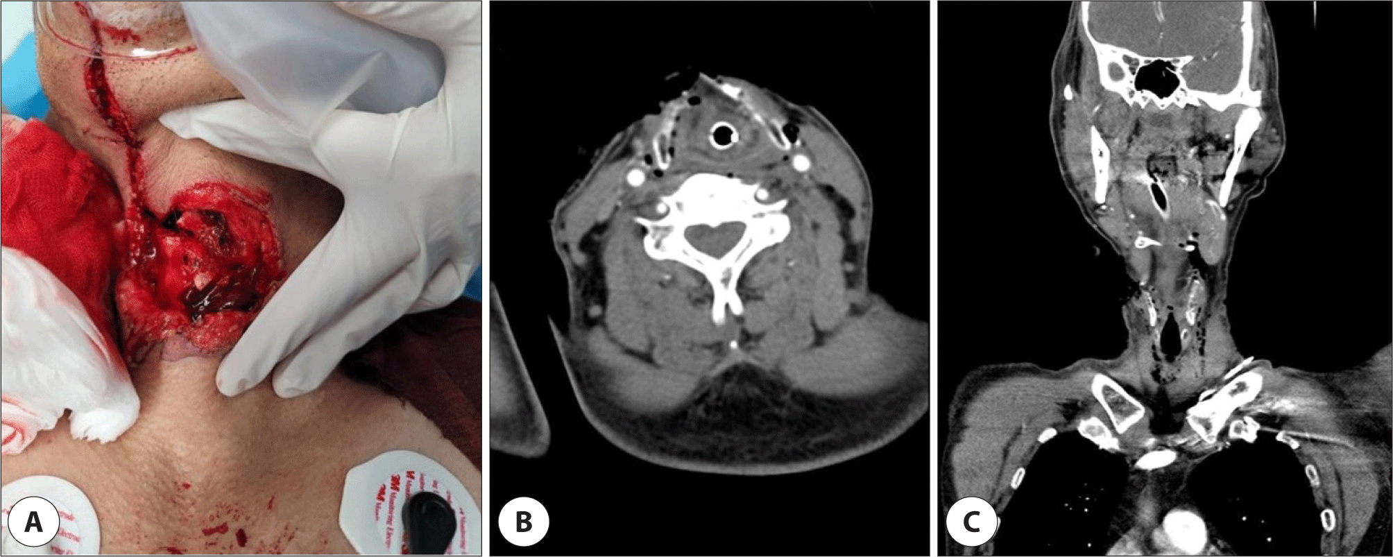
Neck computed tomography (CT) revealed diffuse emphysema and hematoma formation around the wound. Especially large soft tissue defect at anterior neck which exposed fractured thyroid cartilage (Fig. 1B, C). Also marked soft tissue swelling was visualized which lead to airway narrowing at oropharynx to larynx area. Otherwise, there was no major vessel injury.
According to ATLS, to ensure the airway stability, rapid sequenced intubation was done. At first, tube was inserted through lacerated lesion, which lead to vomiting food materials. Second, endotracheal intubation was successfully inserted. After intubation, the patient underwent mechanical ventilation with sedation. Bleeding was controlled with compression and circulation was secured proved by stable vital sign. No definite neurological deficit was found.
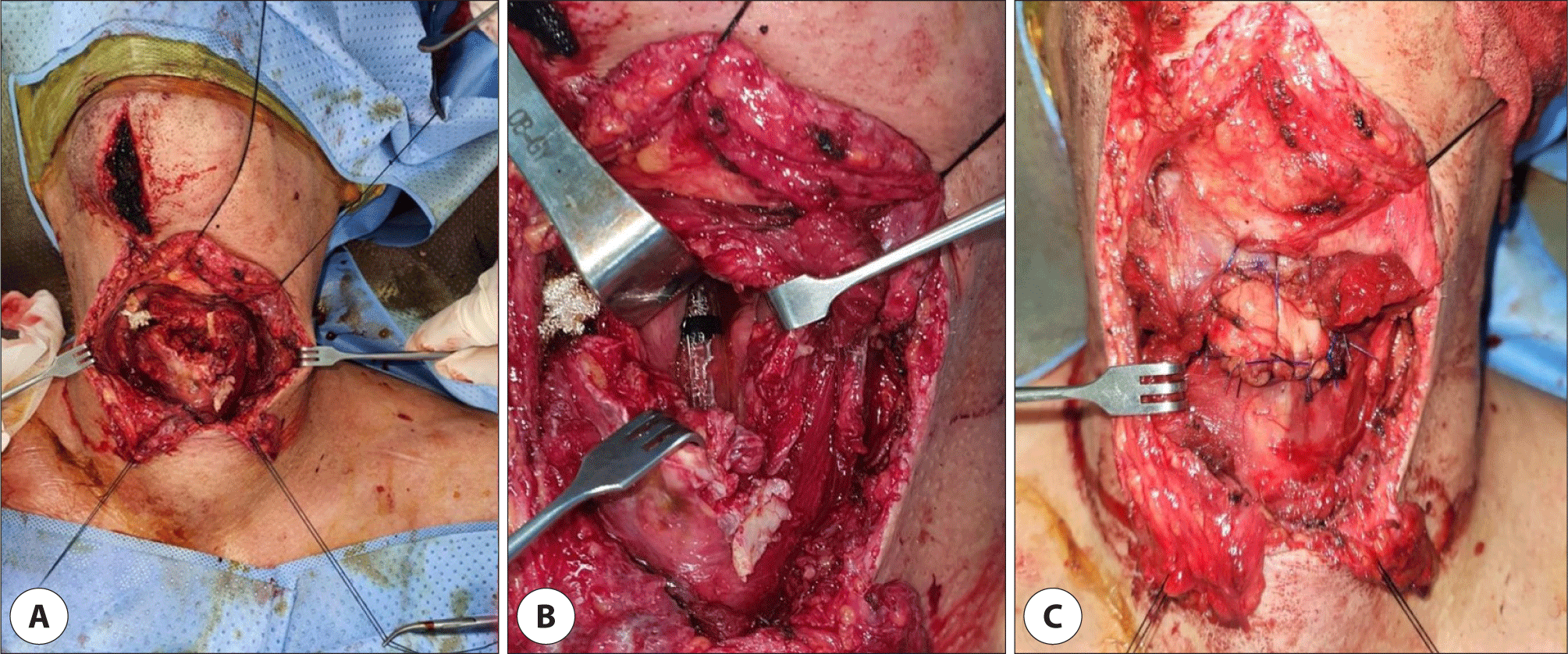
The patient underwent surgical exploration under general anesthesia in the operating room. Intraoperative findings: communicated fracture of the thyroid cartilage, no major vessel injury, and no esophagus injury. Upper epiglottis was lacerated and glottis was not injured including true vocal cord (Fig. 2A). In the operation, there were no injury to the both cervical vasculature and nerve. Fractured thyroid cartilage, especially upper portion, was removed and remain cartilage were suspended to hyoid bone. We sutured the remaining left portion of thyroid cartilage and right portion of cricoid cartilage with hyoid bone and remnant epiglottis according to cricohyoidoepigotopexy procedure (Fig. 2B, C). Nearby strap muscle, the sternohyoid muscle was used to reinforce cricohyoidoepigotopexy site. Laryngeal stent was not placed because there was no injury to the true vocal cord and integrity of larynx was well sustained by the pexy procedure. Drain was placed on the strap muscle and the wound was sutured layer by layer. Lastly, conventional tracheostomy was done to ensure long-term airway.
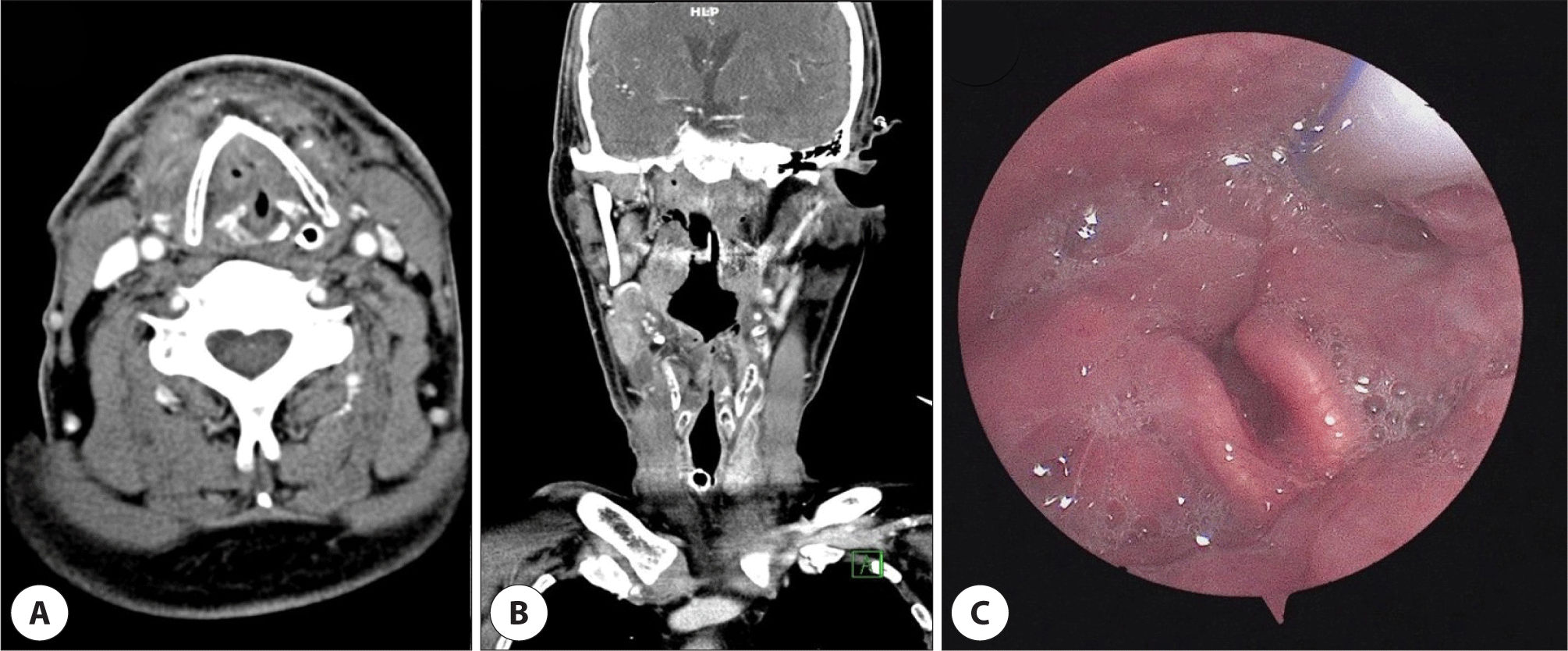
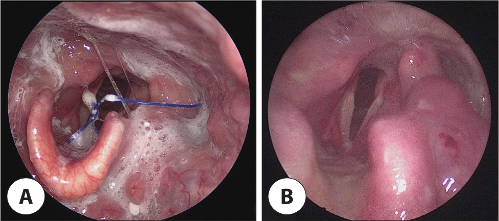
Admitted to intensive care unit after the surgery, the patient weaned mechanical ventilation at day 6. Wound swelling and pus was noted on the day 9. Neck CT was taken immediately and revealed fluid collection rather than abscess. The wound was irrigated and pus was drained and pus culture was done. The culture showed no growth. No more swelling was noted. Empirical antibiotics (ampicillin/sulbactam) was maintained. On day 14, follow up neck CT showed hyoid and cricoid/thyroid cartilage in good alignment and improved soft tissue swelling (Fig. 3A, B). On the same day, laryngoscopy showed saliva pooling and edematous larynx, and clear both vocal cord movement was noted (Fig. 3C). The patient was admitted to ENT (ear, nose, throat) ward from intensive care unit on day 16. Day 17, tracheostomy tube was removed, and no dyspnea was apparent. During postoperative care, there was a couple trials to swallowing which end up in aspiration. According to video-fluorescent swallowing study, the patient needed to keep tube feeding and longer time for swallowing rehabilitation. The patient was transferred to a special rehabilitation center at day 20. 3 months after the surgery, the patient visited our clinic for follow up and to remove remaining suture from the surgery. He had no difficulty in food swallowing, and speaking was fully recovered. Laryngoscopy showed well healed mucosa with remnant epiglottis. Laryngeal structures were well aligned (Fig. 4).
Discussion
In this report we present a case with a penetrating injury of neck cause by chainsaw. It was a quite rare event to manage chainsaw injury. Therefore, there were many debates about the first management at the emergency room. The first, intubation through the lacerated trachea might have caused additional injury to the patient. The patient vomited during the first intubation try and also, the injury caused by intubation try can cause more bleeding from the wound. Vomiting and bleeding both can cause serious aspiration problem to the patient. Most emergency physicians are capable of performing intubation, however bleeding, collapsed airway and possibility of spinal injury make intubation much harder.1) Therefore, use of fiberoptic or bronchoscopy guided intubation is more favorable to neck injury patients.5) Historically, there a debate on the management of compromised airway due to neck injuries. According to Gussack and Jurkovich,6) intubation is preferred because it is less invasive than surgical airway. On the other hand, some prefers surgical airway. The endotracheal tube insertion can cause various injuries including avulsion of the laryngeal mucous membrane, and creation of false passage.7) Many literatures review lead to the consensus on the intubation recommendation which includes intact trachea and larynx, visible airway by endoscopic inspection, and highly experienced physician.7) The second, the patient was managed in supine position, which is more vulnerable to aspiration than in head elevated position.
In severe laryngeal trauma cases, use of stent is the conventional method to ensure airway from the laryngeal stenosis after operations.8) Laryngeal stent is conventionally placed when multiple cartilage fracture which open reduction and internal fixation can’t ensure laryngeal bony structure stability and also when severe laryngeal laceration can cause stenosis.9) However, many studies have shown that use of stent may cause ulcer, or granulation at the site of placement. Also, surgical repair without stent showed similar or even better result of voice function and airway management compare to with stent.4) Some studies proposed causes of such outcome as the result of inflammation of nearby tissue caused by the stent.10) Or insertion of stent on severely injured larynx may have adverse effects on the recovery.11) Since the patient had only supraglottis injury without glottis, and subglottis injury, we assumed laryngeal reconstruction by supraglottic partial laryngectomy can be applied. As we expected, he showed prominent recovery after the laryngeal reconstruction without use of stent. In severe cases, it is almost mandatory to use the laryngeal stent to ensure the stability of larynx. This case showed the stability was ensured by partial laryngectomy with pexy procedure.
Physicians in Korea do not usually encounter a neck injury caused by chainsaw. Even if it is unusual, management should be done according to ATLS. Once airway and hemodynamic stability is acquired, following management including surgery can be done in a stable condition.