


Introduction
Pleomorphic adenoma is the most common benign tumor of the salivary glands. Pleomorphic adenoma mainly occurs in the major salivary glands and less in the minor salivary gland. If pleomorphic adenoma occurs in the minor salivary glands, the most commonly found site is the palate.1) In addition, it can be developed from lip, cheek, tongue, nasal cavity, larynx and nasopharynx. Pleomorphic adenoma rarely occurs in the nasopharynx. In particular, polymorphic adenomas from torus tubarius are extremely rare.2,3) We present a rare case of pleomorphic adenoma originating from torus tubarius in the nasopharyngeal cavity treated successfully via transnasal endoscopic resection.
Case Report
A 56-year-old female patient was referred to our otolaryngology department for chronic onset of nasal obstruction, postnasal drip, and progressive dysphagia that began several months ago.
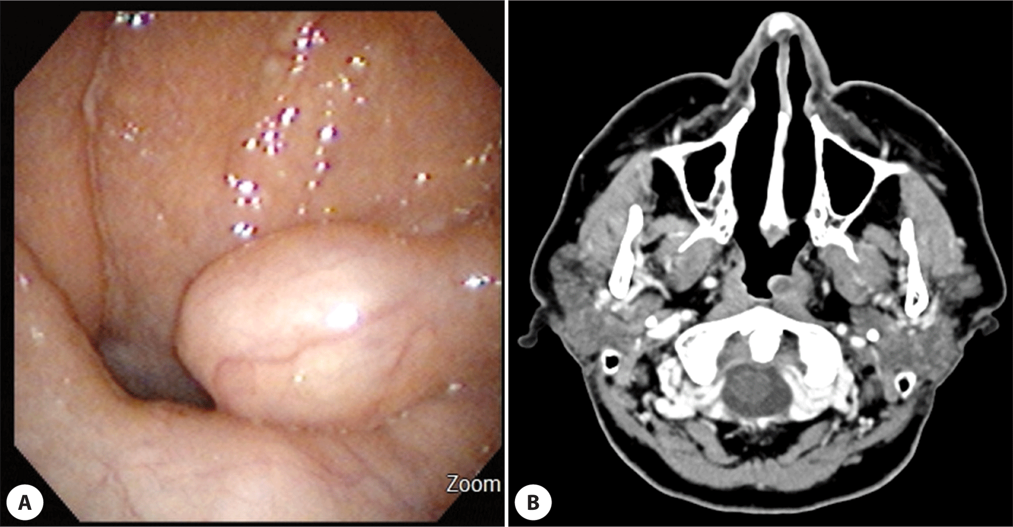
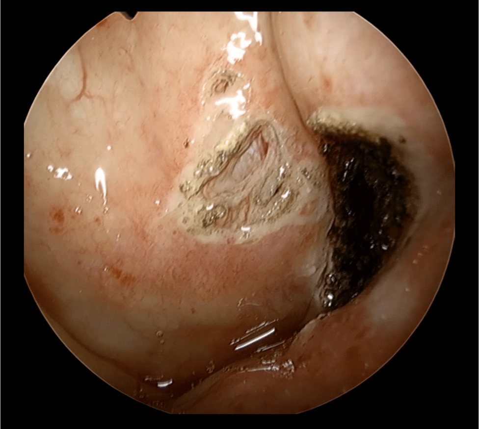
Diagnostic endoscopic examination of nasal cavities and nasopharynx revealed a smooth, ovoid, pink-white mass originating from the left tubarius (Fig. 1A). The tumor extended below the inferior part of torus tubarius without blocking the orifice of Eustachian tube. Computed tomography (CT) scan showed a 1.2×0.7×1.0 cm, well-defined non enhancing or barely enhancing lesion with rim enhancement at inferior aspect of left torus tubarius, which partially obstructed nasopharynx (Fig. 1B). Based on the CT scan, differential diagnosis of the nasopharyngeal mass can be cystic lesions such as mucosal retention cyst, Tornwaldt cyst, benign tumor of minor salivary gland and nasopharyngeal cancer. Preoperative biopsy was not performed on an outpatient basis because it was a small tumor with a smooth surface on endoscopy and there was little possibility of malignancy on CT. A transnasal endoscopic surgery was done under general anesthesia. Lateralization of right inferior turbinate was performed for wide surgical visualization. En block resection of the mass was done meticulously using bovie electrocauterization and straight cut forceps without damaging into the orifice of Eustachian tube (Fig. 2). For complete resection, it was excised that normal fat tissue around the E-tube orifice came out.
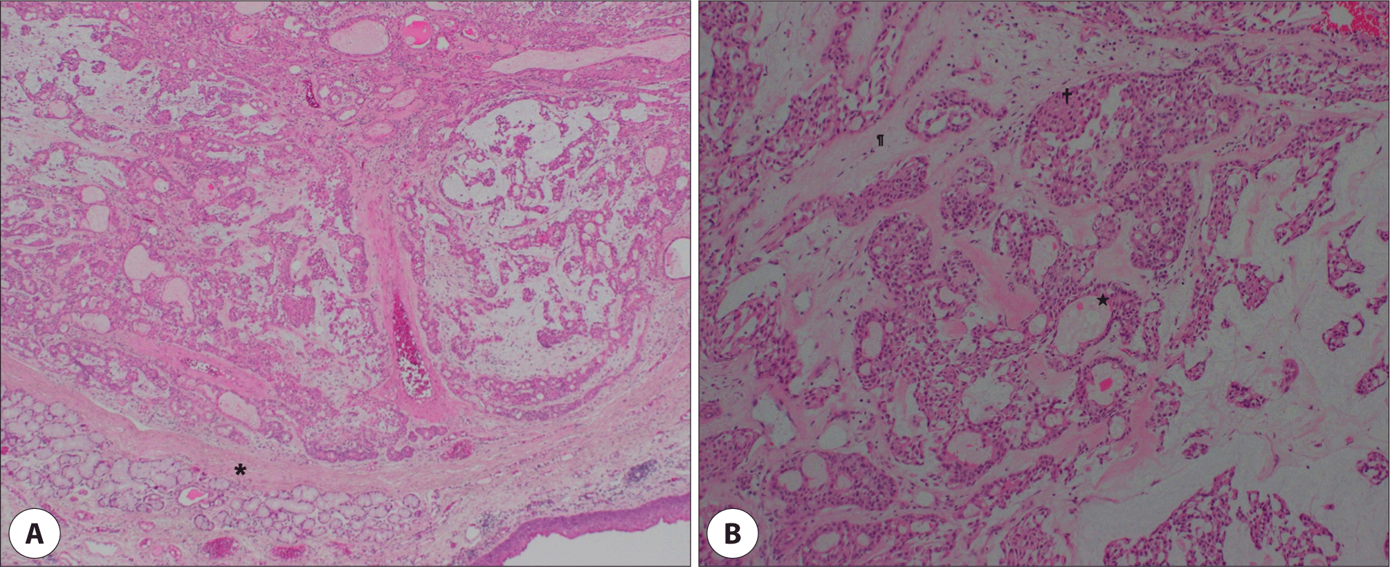
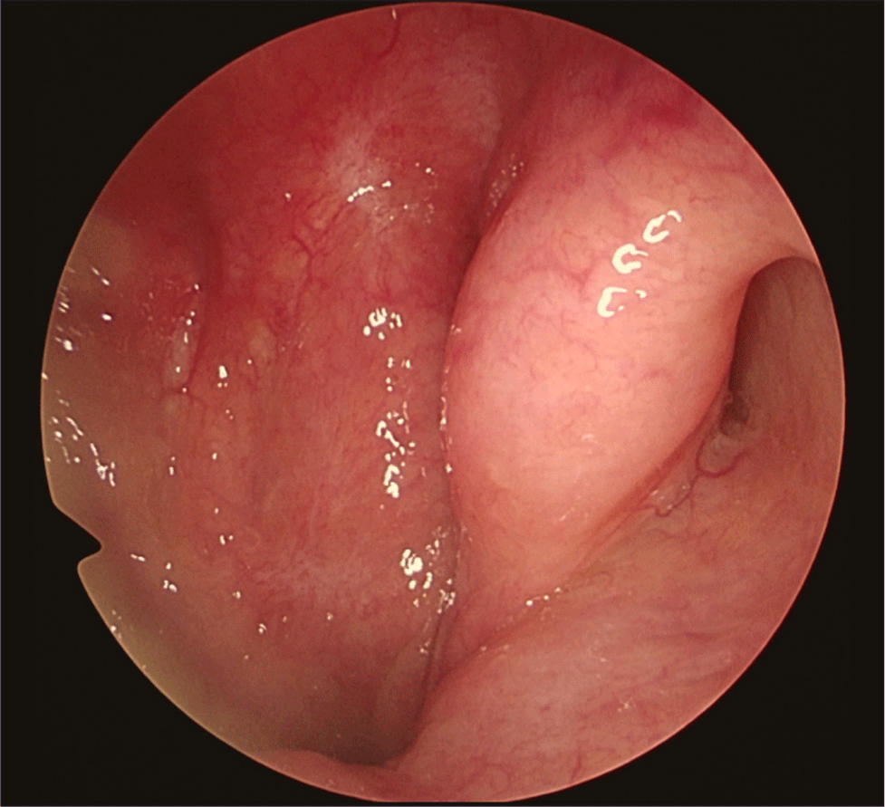
Histopathological examination revealed admixture of epithelial and mesenchymal components, characteristic of pleomorphic adenoma (Fig. 3). Immunohistochemical feature showed the positive in Cam5.2 and p63 marker and mucoid material without goblet cell appearing in alcian blue and mucicarmine stain was consistent of pleomorphic adenoma. A biopsy confirmed that the surgical margin was not involved. The patient’s symptoms disappeared immediately after the surgery and no complications developed. The patient has no evidence of recurrence at 12 months’ follow up with no dysfunction of the Eustachian tube (Fig. 4).
Discussion
Pleomorphic adenomas of the minor salivary glands can develop anywhere these glands are distributed. Li et al. reported the most common primary site for sinonasal/nasopharyngeal pleomorphic adenoma to be the nasal septum (61/115, 53.0%), followed by the lateral wall of the nasal cavity and the nasopharynx (each 16/115, 13.9%).4) The nasopharyngeal pleomorphic adenoma is uncommon and has rarely been described in the literature. Spiro et al. reported only one case of the nasopharyngeal pleomorphic adenoma among 492 cases that originated from the minor salivary gland.5) Especially, pleomorphic adenoma arising from torus tubarius is extremely rare with about 1 case being reported abroad and in Korea.2,3)
The symptoms of pleomorphic adenoma in the nasopharynx are nasal obstruction, nasal bleeding, dysphagia, and otitis media with effusion-induced hearing loss due to Eustachian tube obstruction.6) Although not all benign nasopharyngeal tumors block the Eustachian tube, a pleomorphic adenoma originating from torus tubarius requires ear examination because of its proximity to the orifice of the Eustachian tube. In this regard, if a patient with nasopharyngeal polymorphic adenoma has symptoms such as hearing loss/earfullness and signs of the retracted tympanic membrane or air-fluid level, it is recommended to perform audiometry and tympanometry. This patient didn’t have any ear symptoms.
Pleomorphic adenoma of the minor salivary gland shows a slight female predominance and affects the 4th to 5th decades.7) Diagnosis of nasal polymorphic adenoma can be difficult because the symptoms are nonspecific or asymptomatic. Nasal endoscopy and radiological assessment such as CT and magnetic resonance imaging (MRI) can be used to diagnose pleomorphic adenoma and evaluate tumor extension for deciding the appropriate surgical treatment. On CT images, these tumors show a well-defined, homogenous soft tissue mass with the remodeling of the surrounding bones.8) In MRI, variable enhancement patterns are shown due to the myxoid stroma and cellular component. MRI of pleomorphic adenoma may show either hypointense or isointense signal on T1 weighted images. Intensities can vary from isointense to hyperintense signals with heterogeneous enhancement on T2 weighted images.9)
The characteristic histologic finding of pleomorphic adenoma is an admixture of epithelial and mesenchymal components in a myxoid stroma within an incomplete fibrous capsule. Various cell patterns of the epithelial component can be observed, such as trabecular, ductal and cystic structures. Given that the rate of malignant transformation is 6% and up to 10% if the time period is over 15 years,10) an early and accurate diagnosis should be required through nasal endoscopy, image modalities and histologic examination.
The treatment of nasopharyngeal pleomorphic adenoma is complete surgical excision. If the tumor mass is connected to nasopharynx by a stalk, endoscopic resection could be performed.11) In addition, in the case of pleomorphic adenoma arising from the torus tubarius, a delicate resection is required to reduce complications such as postoperative adhesion and consecutive Eustachian tube dysfunction by preserving the cartilage around the torus tubarius. Recurrence is known to be rare, but a clear resection margin needs to be achieved to prevent recurrence. Furthermore, it is necessary to exclude the malignant transformation because carcinoma ex pleomorphic adenoma has relatively higher recurrence rate.4) There have been some reports that pleomorphic adenomas of the nasal cavity have a lower recurrence rate than those of the oral or parotid. It seems to be due to relatively few mucus and collagen substrates but higher cellularity.12) In our case, it is also expected to have a relatively low recurrence rate because it showed high cellularity histologically, and epithelium of the nasopharynx is an extension of the respiratory epithelium.
We successfully remove pleomorphic adenoma originated from torus tubarius using transnasal endoscopic resection. Transnasal endoscopic approach can be suitable for lowering postoperative complications and morbidity, by providing better visualization of surgical views.