Case Report

18년 뒤 재발한 비인두의 점액표피양암종 1예

허성재1,*https://orcid.org/0000-0002-1388-8075, 차은주1https://orcid.org/0000-0001-9632-0903, 김남균2https://orcid.org/0000-0001-8457-0403, 정지윤3https://orcid.org/0000-0001-6167-4998
Sung Jae Heo1,*https://orcid.org/0000-0002-1388-8075, Eun Joo Cha1https://orcid.org/0000-0001-9632-0903, Namkyun Kim2https://orcid.org/0000-0001-8457-0403, Ji Yun Jeong3https://orcid.org/0000-0001-6167-4998
Author Information & Copyright
1경북대학교 의과대학 이비인후-두경부외과학교실
2경북대학교 의과대학 내과학교실
3경북대학교 의과대학 병리학교실
1Department of Otorhinolaryngology-Head and Neck Surgery, Kyungpook National University School of Medicine, Daegu, Korea
2Department of Internal Medicine, Kyungpook National University School of Medicine, Daegu, Korea
3Department of Pathology, Kyungpook National University School of Medicine, Daegu, Korea
*Corresponding author: Sung Jae Heo, Department of Otorhinolaryngology-Head and Neck Surgery, Kyungpook National University School of Medicine, Daegu 41404, Korea, Tel: +82-53-200-3869, Fax: +82-53-200-3870, E-mail: blueskyhsj@hanmail.net

© Copyright 2022 xxxxxxxxxxxxxxx. This is an Open-Access article distributed under the terms of the Creative Commons Attribution Non-Commercial License (http://creativecommons.org/licenses/by-nc/4.0/) which permits unrestricted non-commercial use, distribution, and reproduction in any medium, provided the original work is properly cited.

Received: May 10, 2022; Revised: Jun 01, 2022; Accepted: Jul 12, 2022

Published Online: Sep 30, 2022

ABSTRACT

Mucoepidermoid carcinoma is a common cancer in the salivary glands, but very rarely in the nasopharynx. Since nasopharyngeal tumors often complain of only non-specific symptoms such as nasal obstruction or epistaxis, they are often discovered at an advanced stage, so proper diagnosis and treatment are important. We experienced a 68-year-old women who complained of nasal obstruction. She had a history of surgical removal of a low-grade mucoepidermoid carcinoma in the nasopharynx 18 years ago. On endoscopic examination, a mass was found in the same location of nasopharynx. It was confirmed that it had recurred as a high grade mucoepidermoid carcinoma, and was completely removed by an endoscopic approach. We report a case of recurred mucoepidermoid carcinoma of nasopharynx 18 years after surgery with a literature review.

Keywords: Carcinoma, mucoepidermoid; Nasopharynx; Transnasal endoscopic surgery

서론

비인두에 발생하는 원발 악성종양은 인구 10만 명당 1명 정도에서 발생하는 드문 질환으로 대부분은 상피세포 기원의 종양이다.1) 세계보건기구(WHO)는 비인두에 발생하는 악성종양을 암종, 유두 선암종, 침샘 종양, 육종/림프종 및 척수종으로 분류한다. 이중에서 원발 타액선 종양은 극히 드물 어 비인두암종의 약 0.48% 정도로 보고되고 있다.2) 현재까지 비인두에서 원발성으로 점액표피양암종(mucoepidermoid carcinoma)이 보고된 증례는 국내 문헌에서 2건으로 매우 드물기 때문에 조기에 의심하기 쉽지 않으며, 조직학적 진단 및 치료가 늦어질 경우 치명적인 결과를 초래할 수 있어 정확한 조기 진단과 치료가 필요하다.3,4)

저악성도 점액표피양암종의 경우 재발이 드물며, 특히 10년 이상 지난 후에 재발한 경우는 매우 드물다.5,6) 저자들은 비인두에 원발성 저악성도 점액표피양암종이 발생하여 수술적으로 제거한 지 18년이 지난 후 고악성도 점액표피양암종으로 재발하여, 비강접근법의 내시경적 수술을 통해 치료한 증례를 경험하였기에 문헌 고찰과 함께 보고하고자 한다.

증례

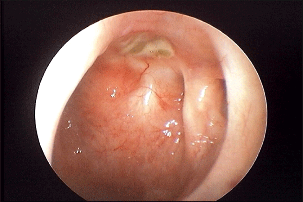
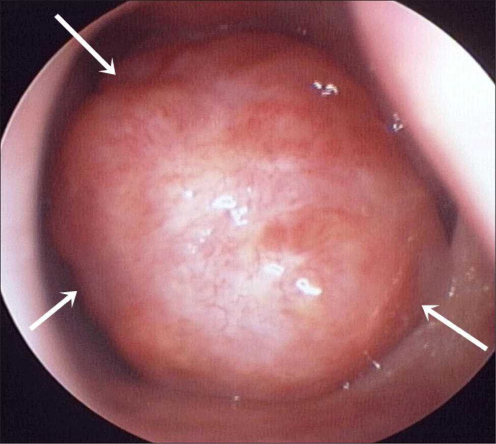
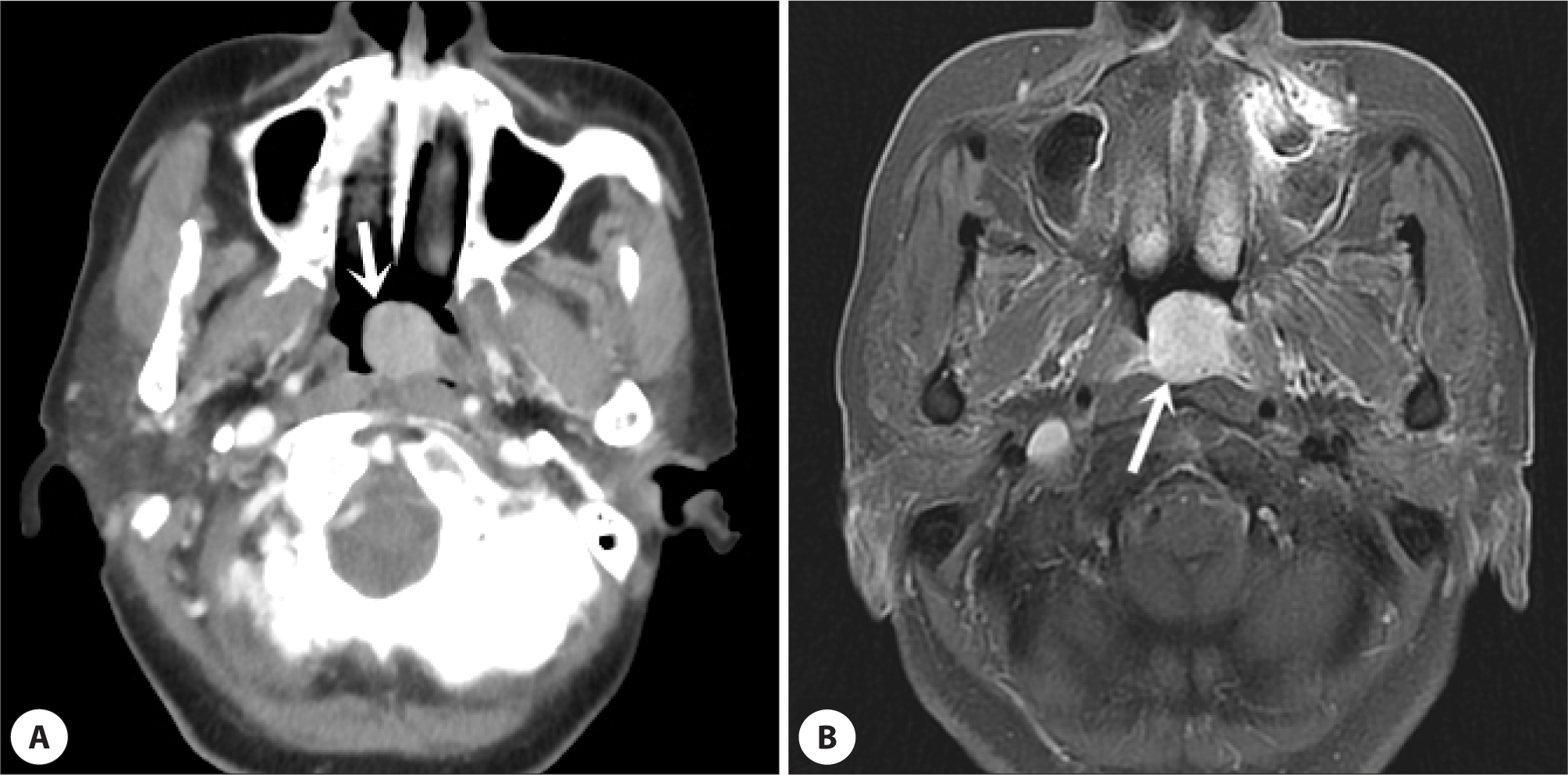
68세 여자 환자가 내원 6개월 전부터 시작된 좌측 코막힘을 주소로 내원하였다. 환자는 내원 18년 전 좌측 비인두의 종물에 시행한 조직검사에서 저악성도 점액표피양암종을 진단받고 내시경을 사용해서 비강 내 접근법으로 약 1 cm 이상의 안전역(safety margin)을 확보하여 절제술을 시행하였고, 조직검사 결과에서도 0.5 cm 이상의 안전역이 확보된 것으로 확인되었다. 그후 9년 동안 재발 소견이 없어 추적관찰을 종료하였다. 내원 시 시행한 비인두 내시경 검사에서 좌측 후비공 및 비인두에 경계가 매끈한 둥근 형태의 종물이 관찰되었다(Fig. 1). 조영증강 부비동 전산화단층촬영 및 자기공명영상에서 좌측 비인두에 2.0×1.6×1.6 cm 크기의 조영증강이 되는 종물이 관찰되었고(Fig. 2), 부인두공간 및 두개저 침범 소견은 확인되지 않았다(T1N0M0, Stage1). 비내시경하에 조직검사를 시행하였고 점액표피양암종 재발이 확인되었다. 양전자방출단층촬영에서 좌측 비인두의 악성 종양이 의심되었으나, 경부 림프절 및 원격 전이는 발견되지 않았다. 종물의 주변 조직 침범이 없고 경계가 비교적 명확하여 내시경을 통한 완전 절제가 가능하다고 판단되어, 전신마취하에 비내시경을 이용한 완전 절제를 시행하였다. 종물은 좌측 비인두 좌상방에서 기시하는 유경성의 형태로, 동결절편 검사를 통해 안전역을 확보하여 완전 절제를 시행하였다. 면역 조직화학염색결과 P63에 양성, P53 및 Ki-67에 일부 양성반응을 보였고, 점액 세포가 드물게 관찰되면서 낭성 병변은 거의 없이 주로 고형 성분으로 구성되었고, 비정형 세포가 관찰되면서 경계가 침습적인 양상을 보여서 고악성도 점액표피양암종으로 진단되었다(Fig. 3). 신경 주위 침범이나 림프맥관 침범은 없었고, 제거한 경계 부위에 종양은 관찰되지 않았다. 수술 후 66 Gy의 방사선 치료를 시행하였고, 2년의 추적관찰 기간 동안 재발 없는 상태로(Fig. 4) 경과관찰 중이다.
jcohns-33-3-144-g1
Fig. 1. Round, smooth surface mass (white arrows) observed in the nasopharynx.
Download Original Figure
jcohns-33-3-144-g2
Fig. 2. Contrast enhanced mass is located in the left nasophayngeal area (arrows). A: Axial CT image. B: Axial T2 MRI image. CT: computed tomography, MRI: magnetic resonance imaging.
Download Original Figure
jcohns-33-3-144-g3
Fig. 3. Macroscopic and microscopic findings of mass. Well-capsulated smooth surface 2 cm sized mass (A) is observed. In the microscopic examination, tumor cell nests that invade into the stroma in an irregular, infiltrative pattern are observed. Most of component is not a cystic component, but a solid nest or sheet form (H&E staining, ×40) (B). Tumor cells are mainly composed of epidermoid cells and intermediate cells, and cells (arrows) that appear to have intracytoplasmic mucin are often observed (H&E staining, ×200) (C).
Download Original Figure
jcohns-33-3-144-g4
Fig. 4. Postoperative endoscopic finding 2 years after surgery. Although some crusts are observed due to radiation therapy, no recurrence is observed.
Download Original Figure

고찰

점액표피양암종은 비인두선암종의 일부로 대부분 점막하 장액점액선에서 기원하고 주로 주타액선에서 발생하며 드물게 구강, 눈물샘, 비부비동 등의 소타액선에서도 발생한다.7,8) 비강 및 부비동에서 발생하는 부위는 상악동, 비강, 사골동 및 비인두 순으로 나타나며, 이러한 비인두 기원 점액표피양암종의 희소성으로 초기에 의심하기 어려워 진단이 늦어지는 경우가 많다.7)

비인두에 발생하는 악성종양은 무증상의 경부종괴, 비폐색, 코피, 이충만감의 증상이 주로 나타나며, 가장 흔한 편평세포암종은 조기에 경부 림프절 전이를 보이고 뇌신경마비, 두개 내 침범 등 진행된 상태로 발견되는 경우가 많다.9) 그러나, 비인두 타액선암은 유형에 따라 차이는 있지만 뇌신경마비는 드물며 경부 림프절 전이도 비교적 낮은 것으로 알려져 있다.10) 본 증례에서도 경부 림프절 전이 및 뇌신경마비는 관찰되지 않았고 증상은 비폐색만 있어서, 환자는 일반적인 비염으로 인한 코막힘으로 생각하고 증상 발현에서 진단까지 6개월이 소요되었다.

점액표피양암종은 조직학적 분화도에 따라 저악성도, 중등악성도, 고악성도로 구분한다. 저악성도 종양은 점액질이 많으며 낭성 변화가 관찰되지만, 고악성도 종양에서는 점액분비세포가 매우 적고 상피세포가 많다.11) 이와 같이 분화도에 따라서 종양세포의 성상에 차이가 있어서 같은 점액표피양암종이지만 자기공명영상의 T2 영상에서 저악성도 종양은 고음영으로 나타나지만, 고악성도 종양에서는 저음영으로 나타난다.12) 이와 같이 다양한 영상 소견을 보일 수 있기 때문에 진단을 위해서는 조직검사가 가장 중요하다. 치료 방법으로 저악성도의 경우 수술만으로 완치율이 높지만, 중등도에서 고악성도로 갈수록 종양의 크기가 크고 공격적인 특징을 가지기 때문에 완전한 수술적 제거와 수술 후 방사선 치료를 권장하고 있다.7) 현재 점액표피양암종의 방사선치료 효과에 대해서는 논란의 여지가 있으나 고악성도의 경우는 방사선에 치료 효과가 있는 것으로 알려져 있다.13) 본 증례는 초기 진단 시 저악성도 종양이었으나 재발하여 고악성도로 분화되었고 절제수술 후 방사선 치료를 추가로 시행하였다.

비인두암의 경우 해부학적 구조상 수술적 접근이 어렵고, 절개 범위가 크기 때문에 수술적 치료가 어려운 경우가 많지만, 본 증례의 경우 유경성의 종물이며 영상학적 검사에서 경계가 비교적 깨끗하였기 때문에 외부 절개 없이 내시경적 접근법으로 수술이 가능하였다. 종물의 크기가 크거나 시야 확보가 잘되지 않는 경우 후방 비중격절제술을 추가로 시행하여 내시경절제를 할 수도 있다.2) 최근 내시경접근법 두개저 수술의 발전으로 앞으로 비인두암의 수술에서 내시경 수술의 역할은 갈수록 커질 것으로 보인다.

비인두에 발생한 점액표피양암종의 증례가 매우 드물어서 비인두에 발생한 경우의 재발률을 알기는 어렵지만, 일반적인 점액표피양암종의 재발률은 약 10% 정도이며,5) Triantafillidou 등이 보고한, 구강에서 발생한 점액표피양암종의 16예 중 1예에서 10년이 지나서 재발한 것으로 보고하였다.6) 재발을 높이는 인자에는 흡연, 높은 TNM(tumor, node, metastasis) 병기, 신경 주위 침범, 림프맥관 침범, 고악성도의 조직분화도, 제거된 조직 변연 침범(margin positive) 등이 있다.5,6) 본 증례에서 처음 점액표피양암종이 진단되었을 때는 T1N0M0으로 Stage 1로 낮은 병기에 신경 및 림프맥관 침범이 없었으며 조직학적으로 저악성도였고, 제거된 조직 침범이 관찰되지 않아서 재발률이 높지 않을 것으로 예상되었지만, 18년이 지나서 재발하였다. 본 증례의 재발 원인으로는 18년의 시간이 지났기 때문에 불완전 절제의 가능성보다, 아직 잘 알려지지 않았지만 점액표피양암종을 유발할 수 있는 유전적 소인과 같은 요소가 영향을 주었을 것으로 생각된다. 비인두에서 점액표피양암종이 발견되면, 오랜 시간이 지나서도 재발할 수 있는 가능성을 염두해두고 장기간 추적관찰이 필요할 것으로 생각한다.

Acknowledgements

Not applicable.

Funding Information

Not applicable.

Conflicts of Interest

No potential conflict of interest relevant to this article was reported.

Author Contribution

Conceptualization: Heo SJ.

Data curation: Heo SJ, Cha EJ, Kim N, Jeong JY.

Formal analysis: Heo SJ, Cha EJ, Kim N, Jeong JY.

Methodology: Heo SJ, Cha EJ, Kim N, Jeong JY.

Writing - original draft: Heo SJ, Cha EJ

Writing - review & editing: Heo SJ, Cha EJ, Kim N, Jeong JY.

Ethics Approval

Not applicable.

References

1.

Chang ET, Adami HO. The enigmatic epidemiology of nasopharyngeal carcinoma. Cancer Epidemiol Biomarkers Prev 2006;15(10):1765-77.

2.

Booth JR, Unsal AA, Tadros S, Byrd JK, Kountakis SE. Salivary gland cancers of the nasopharynx: a population-based analysis of 383 cases. Otolaryngol Head Neck Surg 2019;161(3):442-9.

3.

Park K, Cho SH, Paik SS, Tae K. A case of mucoepidermoid carcinoma in nasopharynx. Korean J Otolaryngol Head Neck Surg 2009;52(4):382-5.

4.

Yoon S, Kim M, Kim M, Hong H, Song K. A case of nasopharyngeal mucoepidermoid carcinoma in a 5 year-old female patient. Korean J Head Neck Oncol 2018;34(2):89-92.

5.

Mimica X, Yuan A, Hay A, Katabi N, Zanoni DK, Valero C, et al. Mucoepidermoid carcinoma: evaluating the prognostic impact of primary tumor site. Oral Oncol 2021;123:105602.

6.

Triantafillidou K, Dimitrakopoulos J, Iordanidis F, Koufogiannis D. Mucoepidermoid carcinoma of minor salivary glands: a clinical study of 16 cases and review of the literature. Oral Dis 2006;12(4):364-70.

7.

Hemalatha AL, Sharath Kumar HK, Geetanjali S, Giripunja M, Shashikumar SD. Nasopharyngeal mucoepidermoid carcinoma: a common entity at an uncommon location. J Clin Diagn Res 2014;8(1):164-5.

8.

Kusafuka K, Takizawa Y, Iida Y, Ebihara M, Onitsuka T, Kameya T. Primary nasopharyngeal mucoepidermoid carcinoma in Japanese patients: two case reports with histochemical and immunohistochemical analysis and a review of the literature. Virchows Arch 2007;450(3):343-8.

9.

Lee AWM, Lin JC, Ng WT. Current management of nasopharyngeal cancer. Semin Radiat Oncol 2012; 22(3):233-44.

10.

Liu TR, Chen FJ, Zhang GP, Yang AK. Different therapeutic strategies in primary salivary gland-type nasopharyngeal carcinomas. Curr Opin Otolaryngol Head Neck Surg 2011;19(2):87-91.

11.

Eveson JW, Cawson RA. Salivary gland tumours. A review of 2410 cases with particular reference to histological types, site, age and sex distribution. J Pathol 1985;146(1):51-8.

12.

Kashiwagi N, Dote K, Kawano K, Tomita Y, Murakami T, Nakanishi K, et al. MRI findings of mucoepidermoid carcinoma of the parotid gland: correlation with pathological features. Br J Radiol 2012;85(1014):709-13.

13.

Ollero JM, Morón AH, Luis AM, Sánchez SM, Nazarewsky AA, López MJS, et al. Nasopharyngeal mucoepidermoid carcinoma: a case report and review of literature. Rep Pract Oncol Radiother 2012;18(2):117-20.