Case Report

외이도 연골부 및 골부의 변형을 동반한 이구선종 1예

이종규1https://orcid.org/0000-0002-6754-1114, 김정연2https://orcid.org/0000-0002-7539-9242, 이주엽1https://orcid.org/0000-0002-5330-5743, 장영수1,*https://orcid.org/0000-0003-4491-5088
Jong Gyu Lee1https://orcid.org/0000-0002-6754-1114, Jung Yeon Kim2https://orcid.org/0000-0002-7539-9242, Ju Yeob Lee1https://orcid.org/0000-0002-5330-5743, Young-Soo Chang1,*https://orcid.org/0000-0003-4491-5088
Author Information & Copyright
1인제대학교 상계백병원 이비인후과학교실
2인제대학교 상계백병원 병리학교실
1Department of Otorhinolaryngology-Head and Neck Surgery, Sanggye Paik Hospital, College of Medicine, Inje University, Seoul, Korea
2Department of Pathology, Sanggye Paik Hospital, College of Medicine, Inje University, Seoul, Korea
*Corresponding author: Young-Soo Chang, Department of Otorhinolaryngology-Head and Neck Surgery, Sanggye Paik Hospital, College of Medicine, Inje University, Seoul 01757, Korea, Tel: +82-2-950-1104, Fax: +82-2-935-6220, E-mail: s5636@paik.ac.kr

© Copyright 2022 xxxxxxxxxxxxxxx. This is an Open-Access article distributed under the terms of the Creative Commons Attribution Non-Commercial License (http://creativecommons.org/licenses/by-nc/4.0/) which permits unrestricted non-commercial use, distribution, and reproduction in any medium, provided the original work is properly cited.

Received: Jun 03, 2022; Revised: Jun 18, 2022; Accepted: Jul 14, 2022

Published Online: Sep 30, 2022

ABSTRACT

Ceruminous adenoma is a well-circumscribed tumor that originates from apocrine glands located in the external auditory canal (EAC). It is a rare disease that accounts for 5.7% of all EAC and auricular tumors. It usually presents as slow-growing tumor accompanying several ear symptoms; mass, fullness, hearing loss, pruritus and otorrhea. If the detection of the tumor is delayed, the tumor can obstruct the EAC completely and its further growth may lead to deformation of surrounding structures such as the cartilaginous or bony portion of EAC. The wide local excision is the treatment of choice for benign ceruminous gland tumors and thorough histopathologic review is essential in that there is possibility of recurrence and malignant transformation. Herein, we present a case of ceruminous adenoma with cartilage and bony deformation in the EAC with a literature review.

Keywords: Adenoma; Cerumen; Ear canal; Neoplasm recurrence, local

서론

이구선종(ceruminous adenoma)은 외이도 연골부의 이구선에서 기시하는 비교적 경계가 명확한 종괴로, 발생 연령은 평균 55세이며 성별에 따른 발생 빈도의 차이는 나타나지 않는다.1) 외이도 및 이개에서 발생하는 종괴의 5.7%를 차지하는 드문 질환으로,2) 전 세계적으로 150여 건, 국내에서는 단 6건만 보고되었다.35) 외이도 연골부에서 발생하지만 외이도 연골을 직접적으로 침범하지는 않는 형태로 나타난다.

외이도라는 제한된 공간 내에서 발생하기 때문에 비교적 작은 크기의 종물인 경우가 많으나, 발견이 늦어질 경우 외이도를 완전히 폐색하며 난청, 이루, 간헐적 통증과 같은 비특이적 증상을 나타낼 수 있고, 지속적으로 성장하여 외이도 연골부의 변형이나 중이강 침범 및 악성 종양이 동반될 수 있다. 현재까지 보고된 이구선종 증례 대부분은 외이도에 국한된 경우였으며, 국내에 보고된 5개의 증례 역시 종양이 외이도에 국한되어 있었으며 1예의 경우 중이강을 침범하였다. 최근 저자들은 외이도 연골부 및 골부의 변형을 동반한 이구선종 1예를 경험하였기에 문헌고찰과 함께 보고하는 바이다.

증례

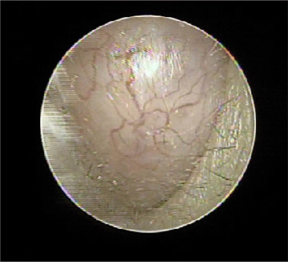
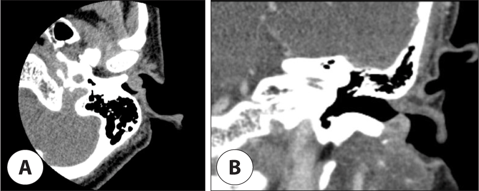
53세 여환이 내원 수개월 전부터 점차 커지는 좌측 귀 안의 종물을 주소로 내원하였다. 환자의 과거력상 특이 소견은 없었고, 내원 시 시행한 이학적 소견상 좌측 외이도 하방에서 기시하여 외이도 입구를 가득 메운 단단한 연분홍 종물이 보였으며, 고막은 관찰되지 않았다(Fig. 1). 측두골 전산화단층촬영에서 좌측 외이도를 막으며 연골부에 국한된 연부조직 음영이 관찰되었으나, 외이도 골부, 중이, 내이 및 유양동은 정상 소견이었다(Fig. 2). 청력저하나 어지럼증, 이루 등의 증상은 없었다.
jcohns-33-3-130-g1
Fig. 1. Oto-endoscopic image shows an oval shape mass arising from a postero-inferior potion of the left external auditory canal with smooth surface.
Download Original Figure
jcohns-33-3-130-g2
Fig. 2. Preoperative image finding. Non-enhanced temporal bone computed tomography (CT). A : Axial view shows that a soft tissue density is observed in the left external ear canal, B : Coronal view shows about 1 cm sized ovoid mass arising from anterior inferior wall of the left cartilaginous external auditory canal. Enhanced mass was observed.
Download Original Figure

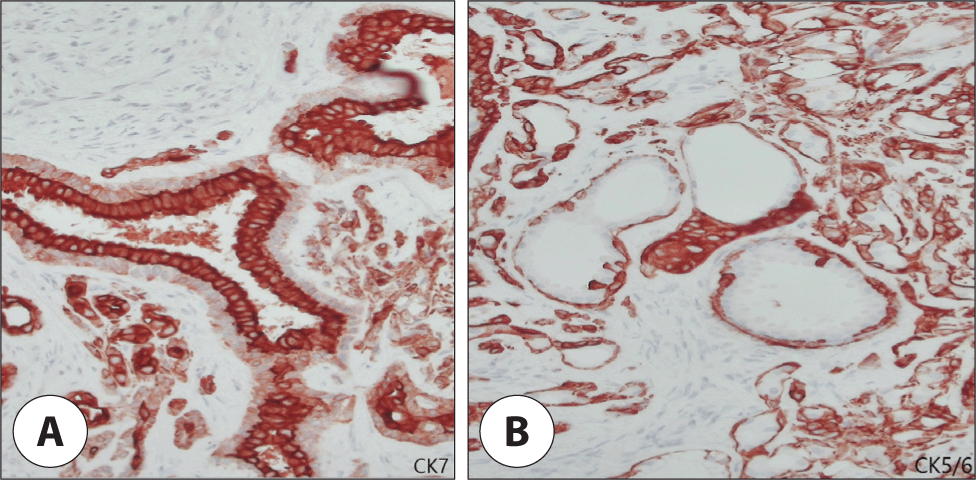
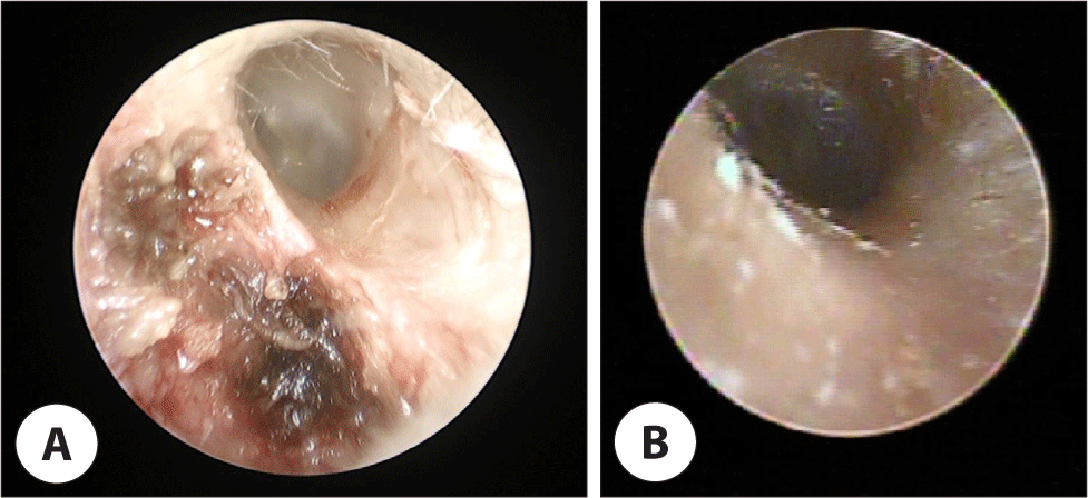
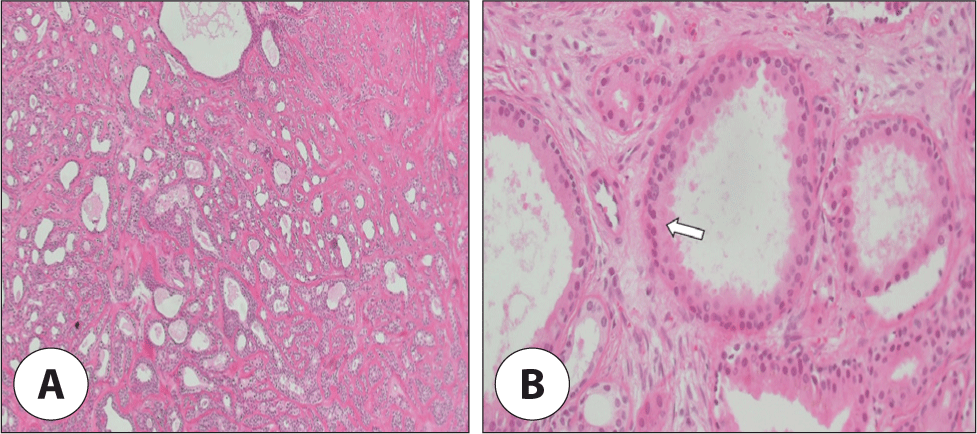
국소마취하에 내시경 귀수술 장비를 이용하여 종물의 기시부에 절개를 시행하였다. 종물은 외이도 하방에 폭이 다소 좁은 줄기부분을 가지고 있었으며, 이 부분을 절개 후 단단한 종물을 따라 외이도 하방 연조직을 박리하였다. 외이도 연골부는 외이도 종물에 의하여 5 mm 깊이로 파괴되어 있었고, 외이도 골부 외측면 하방 역시 일부 외이도 종물에 의하여 파괴된 모습을 보였다. 고막은 정상 소견이었으며, 이루나 이구색전은 관찰되지 않았다. 종물의 표면을 따라 종물을 완전히 절제하였으나, 광범위한 외이도 연골부 및 골부의 결손이 관찰되어(Fig. 3A) 젤폼 삽입 후 수술을 종료하였다. 병리조직학적 검사상 다양한 크기의 이구선 증식이 보이고(Fig. 4), 면역화학적 검사에서도 내측의 유선세포(luminal cell) 및 바깥쪽의 납작한 기저세포(basal cell)의 증식이 관찰되어(Fig. 5) 이구선종을 진단할 수 있었다. 병리학적 검사상 전체적으로 캡슐이 존재하지 않고 상피세포와 명확하게 구분이 되어 있고 경계가 잘 형성되어 있었으나, 파괴된 외이도 골부 외측면에 인접한 종괴는 경계가 잘 형성되어 있으나 다소 매끈하게 잘린 부분이 있었다. 이 부분을 절개 변연부(resection margin)로 보았을 때 일부 잔재 조직이 있을 가능성을 고려하여, 추가적인 변연절제술을 위하여 2차 수술을 계획하였다. 1차 수술 후 3주 경과 후 2차 수술이 진행되었고, 국소마취로 진행되었다. 이전 수술 부위의 회복된 피부 조직을 절개하고, 연골부와 골부에 대한 추가적인 제거 및 조직검사를 시행하였다. 외이도 골부와 주변 피부 조직의 추가 절제를 통하여 충분한 변연절제를 시행하였고, 젤폼 삽입 후 수술을 종료하였다. 2차 병리조직학적 검사상 이구선종 잔여 조직은 관찰되지 않아 완전한 제거 및 충분한 변연절제가 이뤄졌음을 확인하였다. 1주 경과 후 피부는 완전히 회복되었으며(Fig. 3B), 술 후 검사한 순음청력검사 결과에서 좌측 골도 청력 역치 10 dB, 기도 청력 역치 10 dbB로 이상소견을 보이지 않았고, 재발에 대한 외래 추적관찰을 시행 중이다.
jcohns-33-3-130-g3
Fig. 3. Pre- and postoperative findings of the left EAC. A : Intraoperative endoscopic finding of the left EAC after the complete resection of the tumor. Deformation of cartilage and bone of the EAC was observed. B : 2 months after endoscopic mass excision; endoscopic image shows left EAC with healed mucosa. EAC: external auditory canal.
Download Original Figure
jcohns-33-3-130-g4
Fig. 4. Pathologic findings. A : Various sizes of glandular proliferations and the myxoid nature of the stroma were observed (H&E staining, ×100). B : The well-differentiated glands consist of two layers; inner columnar eosinophilic epithelium and outer myoepithelial layer (H&E staining, ×400). White arrow indicates ceruminous decapitation secretion in the luminal cells.
Download Original Figure
jcohns-33-3-130-g5
Fig. 5. Immunohistochemical analysis. Differential immunohistochemical staining highlights (A) the luminal cells CK7 (×100) (B) while CK5/6 (×100) accentuated the basal cells.
Download Original Figure

고찰

Wetli 등은 1972년에 종양의 현미경적 소견, 행태 및 치료 반응에 따라 외이선종을 (1) 선종(adenoma), (2) 다형성선종(pleomorphic adenoma), (3) 선양낭포암종(adenoid-cystic carcinoma), (4) 선암종(adenocarcinoma)의 4가지 형태로 구분하였다.2) 이후 2그룹이 추가되어 현재 세계보건기구에서는 6가지로 구분하고 있으며 이구선종(ceruminous adenoma), 이구선 다형성종(pleomorphic adenoma), 이구선 유두상 한선낭포선종(syringocystadenoma papilliferum), 이구선 암종(adenocarcinoma), 이구선 선양낭포암종종(adenoid cystic carcinoma), 이구선 점액상피양암종(mucoepidermoid carcinoma) 등이 있다.6) 이 중 선양낭포암종의 빈도가 가장 높고 선암종과 더불어 악성의 양상을 가지며, 양성인 선종, 다형성 선종과의 비는 2.5:1로 보고하였다. 일반적으로 양성 이구선종의 임상 증상은 외이도의 무통성 종물이며, 외이도 폐쇄에 따른 청력 감소가 동반되거나 이차적인 외이도염, 이구색전이 발생할 수 있다. 그러나 이구선 기원의 종양은 다른 부위의 아포크린 종양보다 악성의 비율이 높기 때문에 심한 통증, 혈성 이루, 뇌신경 마비의 증상이 동반될 경우 악성의 가능성을 의심해야 한다.7,8)

드물게 중이강 내에서 발생하는 증례가 보고된 바 있으나,4,9) 대부분의 이구선종은 외이도에서 발견되며, 서서히 자라나는 형태이기 때문에 외이도 내에 국한되는 형태로 발견되어, 기시부와 그 변연 절제를 통하여 완전 절제가 가능하다. 그러나 본 증례에서와 같이 외이도의 완전 폐색 이후에도 지속적으로 성장을 할 경우 기시부의 연골 및 골 변형이 동반될 수 있다. 이러한 연골 및 골 변형이 동반되는 소견이 발견될 경우, 우선 종양 절제술 후 병리조직학적 검사를 통하여 잔여 조직의 가능성과 악성 여부를 평가하는 것이 필요하다. 이구선종은 비교적 경계가 명확하지만 캡슐이 존재하지 않아(non-encapsulated, well-circumscribed tumor) 표면의 침범이 동반될 수 있음과 일부 이구선종에서는 악성과 양성 조직이 혼재되어 나타남을 명심하고, 전체 조직에 대한 평가를 통하여 악성 동반 여부와 해부학적 위치에 따른 잔여 조직 의심 부위를 확인해야 한다.10) 따라서 외이도 연골부 및 골부의 변형을 동반한 이구선종은 병리조직학적 검사 결과에 따라 추가적인 변연절제술 및 재건술을 통하여 이구선종의 재발 가능성을 낮추고 외이도 변형을 교정하는 것이 필요하다.

외이도에 발생한 종괴에 대하여 감별진단해야 할 양성 종양으로 외골증, 연골종, 점액종, 연골피부염, 혈관종, 근종, 지방종, 섬유종이 있으며, 악성 종양으로는 육종, 기저세포암과 구별하여야 한다. 최근 Das 등은 세침흡입검사를 통하여 수술 전 이구선종을 진단하여 종괴의 크기에 의한 불필요한 광범위한 변연절제술이나 후이개 접근을 최소화할 수 있다고 보고하였다.11) 이구선종은 굉장히 드문 질환이지만, 흩어진 형질세포양 세포(plasmacytoid cell)와 섬유근육체 기질 파편(fibromyxoid stromal fragment)에 강하게 결합된 상피세포(epithelial cell)와 근상피세포(myoepithelial cell), 유두 형성(papillae formation)과 같은 병리조직학적 소견이 이구선종에 특징적임을 고려하여 세침흡입검사 시 진단할 수 있으나, 전술한 바와 같이 캡슐이 형성되는 형태의 종괴가 아니기 때문에 완전한 조직 절제술 후 조직 변연주로의 조직 침범 여부를 명확히 확인하는 것이 필요하다. 외이도 양성 종양의 경우, 광범위한 국소 절제가 재발을 방지하는 데 필요하다. 특히 이구선 선종의 경우 잔류 조직이 남는 경우 국소 재발의 원인이 될 수 있다. 본 증례에서 술자는 내시경적 접근으로 종괴 절제를 진행하였으며, 과도한 외이도 상피의 손상을 막기 위하여 수술 시야에 다소 제한이 있었지만, 종괴의 내측면 제거 시 내측에서 외이도 골부와 연골부를 따라 종괴의 하방 및 외측으로 종괴 표면을 절제하였기 때문에, 병리조직검사상의 종괴의 하방 변연부에서 매끈하게 잘린 부분이 파괴된 외이도 골부 및 연골부로의 침범 조직의 잔류 가능성을 배제할 수 없다고 판단하여 2차 수술을 계획하였다. 이구선 선종의 예후는 좋은 것으로 알려져 있으나 치료 없이 둘 경우 약 70%에서 악성으로 변화함을 명심해야 한다.11)

현재까지 보고된 이구선종 증례 대부분은 외이도에 국한되고 기시부 및 변연 절제술로 치료할 수 있었으나, 저자들은 외이도의 완전 폐색과 외이도 연골부 및 골부의 변형을 동반한 증례를 경험하였기에 문헌고찰과 함께 보고하는 바이다.

Acknowledgements

Not applicable.

Funding Information

Not applicable.

Conflicts of Interest

No potential conflict of interest relevant to this article was reported.

Author Contribution

Conceptualization: Chang YS.

Data curation: Lee JG, Kim JY, Lee JY, Chang YS.

Formal analysis: Kim JY, Chang YS.

Writing - original draft: Lee JG, Chang YS.

Writing - review & editing: Lee JG, Kim JY, Lee JY, Chang YS.

Ethics Approval

Not applicable.

References

1.

Thompson LDR. Ear ceruminous adenoma. Ear Nose Throat J 2011;90(7):304-5.

2.

Thompson LDR, Nelson BL, Barnes EL. Ceruminous adenomas: a clinicopathologic study of 41 cases with a review of the literature. Am J Surg Pathol 2004; 28(3):308-18.

3.

Kim SW, Choi JH. Ceruminous adenoma mimicking otofuruncle in the external auditory canal. Korean J Otorhinolaryngol Head Neck Surg 2011;54(7):501-3.

4.

Seo YJ, Park JH, Han JS, Park KH. A case of confusing ceruminous adenoma of external auditory canal. J Korean Skull Base Soc 2020;15(2):122-5.

5.

Yeo SC, Lee HJ, Kim JY, Kim DW, Seo JH, Ahn SK. A case of ceruminous adenoma arising from the external auditory canal. Kosin Med J 2018;33(2):228-34.

6.

Sandison A, Thompson LDR. Tumours of the external auditory canal: ceruminous adenoma. In: El-Naggar AK, Chan JKC, Grandis JR, Takata T, Slootweg PJ, editors. WHO classification of head and neck tumours. 4th ed. Lyon: International Agency for Research on Cancer; 2017. p. 265.

7.

Mansour P, George MK, Pahor AL. Ceruminous gland tumours: a reappraisal. J Laryngol Otol 1992;106(8): 727-32.

8.

Mills RG, Douglas-Jones T, Williams RG. ‘Ceruminoma’ – a defunct diagnosis. J Laryngol Otol 1995; 109(3):180-8.

9.

Jeong H, Noh H, Kim CH. Middle ear ceruminous gland adenoma obstructing the eustachian tube orifice. Case Rep Otolaryngol 2021;5987353.

10.

Nagarajan P. Ceruminous neoplasms of the ear. Head Neck Pathol 2018;12(3):350-61.

11.

Das R, Nath G, Bohara S, Raghuvanshi S. Ceruminous adenoma: a rare tumor diagnosed on cytology with histological correlation. J Cytol 2017;34(3):168-70.