Original Article

Treatment of Paranasal Sinus Indolent Mucormycosis

Yong-Wan Kim1https://orcid.org/0000-0002-6010-312X, Myoung Joo Kang2https://orcid.org/0000-0002-4334-4028, Yeo Myeong Lee3https://orcid.org/0000-0002-6431-1452, Seong Kook Park3,*https://orcid.org/0000-0003-3903-8412
Author Information & Copyright
1Department of Otolaryngology Head and Neck Surgery, Inje University Haeundae Paik Hospital, Busan, Korea
2Department of Hemato-Oncology, Inje University Haeundae Paik Hospital, Busan, Korea
3Department of Otolaryngology Head and Neck Surgery, Inje University Busan Paik Hospital, Busan, Korea
*Corresponding author: Seong Kook Park, Department of Otolaryngology–Head and Neck Surgery, Busan Paik Hospital, College of Medicine, Inje University, Busan 47392, Korea. Tel: +82-51-890-6379, Fax: +82-51-892-3831, E-mail: sinus4@paik.ac.kr

© Copyright 2022 The Busan, Ulsan, Gyeoungnam Branch of Korean Society of Otolaryngology-Head and Neck Surgery. This is an Open-Access article distributed under the terms of the Creative Commons Attribution Non-Commercial License (http://creativecommons.org/licenses/by-nc/4.0/) which permits unrestricted non-commercial use, distribution, and reproduction in any medium, provided the original work is properly cited.

Received: Nov 26, 2021; Revised: Jan 03, 2022; Accepted: Feb 03, 2022

Published Online: Mar 31, 2022

ABSTRACT

Background and Objectives: Mucormycosis of the nasal cavity and paranasal sinuses is a rare but highly aggressive especially in patients with diabetes or immunosuppressed patients. However, chronic non invastive type of mucormycosis can be observed in immunocompetent patients. In this study, we investigated indolent mucormycosis cases and assessed the clinical and radiological outcomes of indolent mucormycosis of paranasal sinus in healthy patients treated by endoscopic sinus surgery (ESS) alone. Materials and Methods: A retrospective chart analysis of 9 patients with the diagnosis of indolent mucormycosis of paranasal sinus and treated by ESS between 2007 and 2017 was performed. The data were collected from the medical records: age, sex, clinical presentations, pre- and postoperative endoscopic findings, underyling diseases, pathology, pre- and postoperative radiological findings. Radiologic images were reviewd to assess the involved side and sinus, bony sinus wall changes. Results: The histopathologic findings revealed mucormycosis with broad, non-septated, right-angled hyphae. Although the diagnosis of mucormycosis, the patients were not any antifungal agents after surgery. There was no disease progression and recurrence. Conclusion: In the case of paranasal sinus mucormycosis, ESS alone is thought to be sufficient to the treatment for indolent cases in immunocompetent patients without evidence of preoperative computed tomography and endoscopic findings of invasion and antifungal treatment may not be necessary.

Keywords: Endoscopic surgery; Fungi; Indolent; Mucormycosis; Paranasal sinuses

Introduction

Mucormycosis of the nasal cavity and paranasal sinuses is a rare but opportunistic infection of the class Phycomycetes, order Mucorales. It is highly aggressive and causes a rapidly progressive, life-threatening disease, especially in patients with diabetes or immunosuppressed patients. The most effective treatment consists of immediate surgical debridement, administration of systemic antifungal drugs, recovery of compromised immunity.1,2) However, asymptomatic or chronic indolent type of mucormycosis can be observed in immunocompetent patients.13) There is no consensus of treatment for indolent sinonasal mucormycosis patients.

In this study, we collected 9 cases of mucor fungal balls and assessed the clinical and radiologic results of indolent mucormycosis of paranasal sinus in healthy patients treated by endoscopic sinus surgery (ESS) alone.

Materials and Methods

A retrospective review of nine patients with the diagnosis of chronic indolent mucormycosis of paranasal sinus and treated by ESS alone between 2007 and 2017 was performed. Institutional Review Board approval was obtained from the (IRB number 2018-0153). Informed patient consents were waived because it was a retrospective chart review. The following data were collected from the medical records: age, gender, clinical presentations, pre- and post ESS endoscopic findings, underyling diseases, histopathology, pre- and postoperative radiologic findings.

Radiologic images were reviewed to assess the involved side and sinus, changes of bony sinus wall.

Results

Patient clinical characteristics

Nine patients were identified as having a diagnosis of indolent paranasal sinus mucormycosis. They consists of 2 men and 7 women. The mean age was 65.7 (range, 48–77 years). Our patients have nasal symptoms included facial pain, nasal stuffiness, postnasal drip and foul odour. All patiens had previously healthy and reported no underlying disease, medication, facial trauma with the exception of four patients with well controlled two hypertension patients and two diabetes patients (Table 1).

Table 1. Patient clinical characteristics
Pt no Age (y)/ gender Clinical symptoms With NP Underlying disease Involved sinus F/U (mon) Recurrence
1 77/F Facial pain N Hypertension Lt. MS 5 None
2 70/F Nasal stuffiness, foul odour Y Diabetes Lt. MS 5 None
3 59/F Facial pain N None Lt. MS 11 None
4 75/F Facial pain Y Hypertension Lt. MS 13 None
5 48/M Nasal stuffiness, postnasal drip Y None Rt. MS 8 None
6 68/F Nasal stuffiness, postnasal drip Y None Rt. MS 12 None
7 67/F Foul odour, postnasal drip N None Lt. MS 17 None
8 67/F Foul odour, postnasal drip N Diabetes Lt. MS 10 None
9 60/M Nasal stuffiness, postnasal drip Y None Lt. MS 24 None

All patients were treated with endoscopic sinus surgery alone and all survived.

Pt no: patient number, y: year, mon: month, F/U: follow up, F: female, Lt: center, MS: maxillary sinus, M: male, Rt: right, NP: nasal polyp.

Download Excel Table

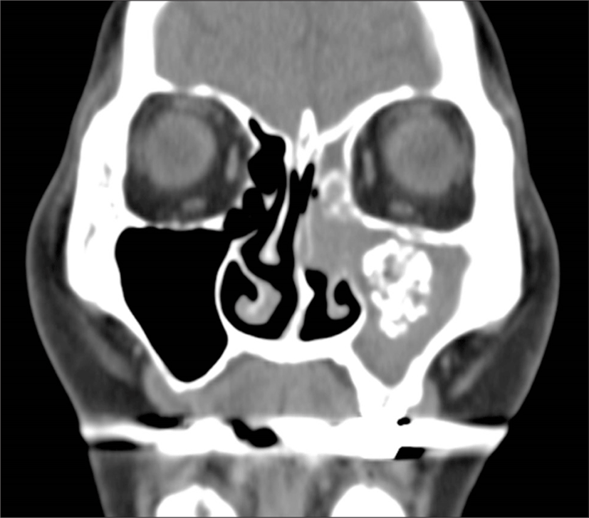
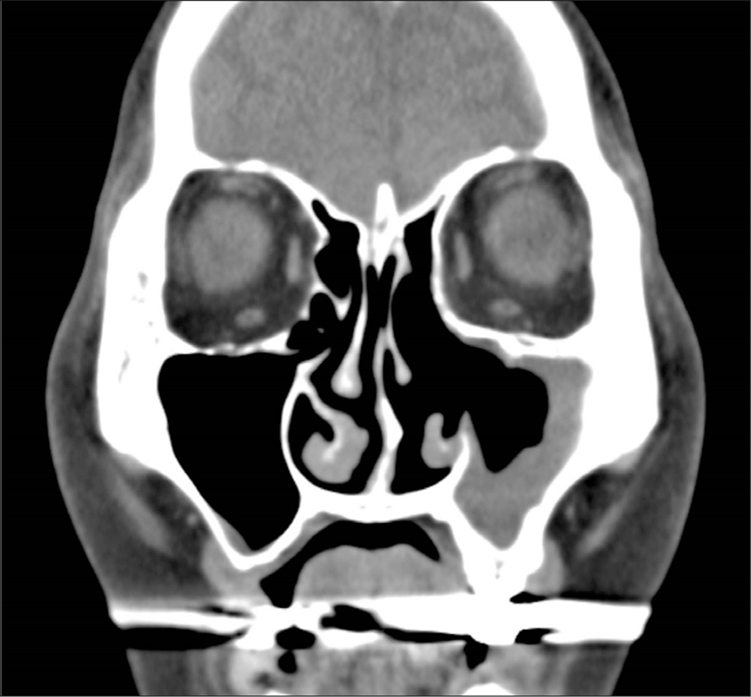
Preoperative paransal sinus computed tomography (CT) showed total or partial opacification of the unilateral maxillary sinus with focal calcifications without bony destruction of the involved sinus or invasion of the orbit or brain in all patients (Fig. 1). Five patients had inflammatory nasal polyps in the involved paranasal sinus (Fig. 2).

jcohns-33-1-3-g1
Fig. 1. Pre-operative, computed tomography image of the paranasal sinus in coronal view showing total opacification of the center maxillary sinus with focal calcifications, without bony destruction or invasion of the orbit or brain.
Download Original Figure
jcohns-33-1-3-g2
Fig. 2. Pre-operative endoscopic image showing nasal polyp in the center middle meatus. There were no fungal materials observed at this image. IT: inferior turbinate, MT: middle turbinate, NP: nasal polyp, S: septum.
Download Original Figure
Outcomes

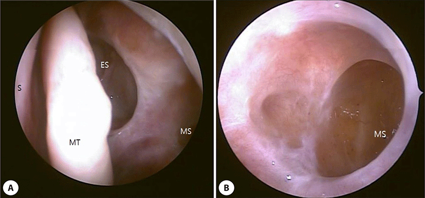
All patients underwent ESS based on clinical symptoms, paranasal CT with suspicision of fungal sinusitis. Surgical procedures included uncinectomy, ethmoidectomy and middle meatal antrostomy with removal of clay like, thick brownish- green material. After surgery, the patients were given antibiotics for 3 days and then discharged for general protocol for fungal ball.

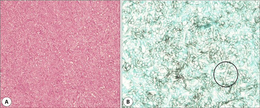
The histopathologic findings showed mucormycosis with broad, non-septated, right-angled hyphae (Fig. 3). In spite of diagnosis of mucormycosis, the patients were not any antifungal agents after surgery.

jcohns-33-1-3-g3
Fig. 3. Histopathology showing irregular, nonseptated and broad, hyphaes with right angle branching, consistent with mucormycosis (H&E, ×200) (GMS_400).
Download Original Figure

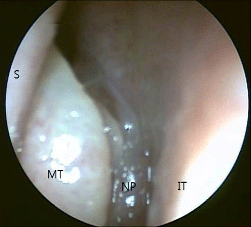
All patients had monthly angled endoscopic follow up (Fig. 4) and postoperative 3 months paranasal CT follow up (Fig. 5). The average follow up duration was 11.7 months. There was no recurrence.

jcohns-33-1-3-g4
Fig. 4. Post-operative endoscopic image taken four months after endoscopic sinus surgery showing no recurrence in the center nasal cavity and maxillary sinus. Below image was taken under 70 degree angled endoscope. ES: ethmoid sinus, MS: maxillary sinus, MT: middle turbinate, S: septum.
Download Original Figure
jcohns-33-1-3-g5
Fig. 5. Post-operative, paranasal sinus computed tomography image taken two months after endoscopic sinus surgery, showing no recurrence or residual calcified lesion in the center maxillary sinus, except remained post-operative change and mucosal thickening.
Download Original Figure

Discussion

Mucormycosis is a clinical disease caused by fungi from 4 families of the Mucorales, which is a member of the class Zygomycets. The zygomycetes are hyaline fungi commonly found in breads and fruits. They are ubiquitous in soil, in vegetation, and in the air, making them frequent inhabitants of the upper airway mucosa but do not become an infectious source in healthy people.1,4) They become pathogenic when the patient’s resistance has been changed.4)

Some studies suggest that mucormycosis is an opportunistic infection in approximately 5% to 12% of all fungal infections in the high-isk group.5)

Rhinocerebral or craniofacial zygomycosis is a common form and usually fatal or fatal. Typically, the patient is at high risk for leukopenia or metabolic acidosis. Diabetes mellitus is the single most common disease associated with the development of invasive mucormycosis.5,6) In this study, two patients were presented with diabetes mellitus, but all were well-controlled, and did not show any signs of acidosis and appeared to be non- invasive fungal ball.

Rhinocerebral mucormycosis is the most common form, accounting for one-third to one-half of all mucormycosis.5) The first symptom of rhinocerebral murcomycosis is sinusitis or periorbital cellulitis,7,8) followed by suffusion of conjunctiva, blurry vision, and soft tissue swelling after ocular pain, facial pain, facial numbness.9,10) If not treated, the infection spreads from the ethmoid sinus to the orbit, resulting in extraocular muscle dysfunction and proptosis. Severe chemosis may also be seen. Involvement of the controlateral eye is an ominous finding suggesting cavernous sinus thrombosis when bilateral proptosis, chemosis, vision loss, and ophthalmoplegia occur.5) Progressive vision loss may be due to optic nerve involvement or infarction1013) or cavernous sinus thrombosis. Upon naso-endoscopic examination, mucosal findings may appear normal at the very early stages of fungal spread. If the infected tissue then progress through an erythematous phase, ultimately forms black, necrotic eschar as the thrombosed blood vessels and tissue infarction occurs.14,15)

However, indolent rhinocerebral mucormycosis in immunocompetent patients is often characterized by subtle symptoms and signs.16,17) The symptoms in this study were facial pain, nasal stuffiness, foul odor, postnasal drip and non - specific features and similar pattern to chronic sinusitis and fungal ball. No necrotic eschar was observed in the endoscopic findings.

The treatment of invasive mucormycosis requires reversal of the predisposing factors, aggressive surgical removal of infected tissue and systemic antifungal medical therapy.5,16)

There is no consensus on the extent of surgery in the treatment of mucormycosis. Endoscopic surgery is best suitable for earlier and limited disease. In particular, there is uncertainty about the appropriate treatment of limited sinonasal mucormycosis in obvious healthy subjects. These patients are more benign and tend to be localized. Some reports showed that limited mucormycosis can be cured with surgical debridement alone2,18) or with combination of surgery following a course of amphotericin B.16) In previous study, authors reported that indolent mucormycosis of paranasal sinus in four case series immunocompetent patients could be treated by endoscopid sinus surgery alone, and antifungal drugs may not be needed.2) In this study, we showed that, in nine case series, paranasal indolent mucormycosis of healthy patient could be treated by ESS alone.

Conclusion

Paranasal sinus mucormycosis is very rare in healthy patients. This study still includes a small number of patients. However, ESS alone is thought to be sufficient for the treatment of chronic indolent cases in immunocompetent patients without evidence of invasion and antifungal treatment may not be necessary.

Acknowledgements

Not applicable.

Funding Information

Not applicable.

Conflicts of Interest

No potential conflict of interest relevant to this article was reported.

Author Contribution

Conceptualization: Park SK.

Data curation: Lee YM.

Formal analysis: Lee YM.

Methodology: Kim YW.

Investigation: Lee YM.

Writing - original draft: Kim YW, Kang MJ.

Writing - review & editing: Kim YW, Kang MJ, Lee YM, Park SK.

Ethics Approval

Institutional Review Board approval was obtained from the Inje University Busan Paik Hospital (IRB number 2018-0153). Informed patient consents were waived because it was a retrospective chart review.

References

1.

Kim ST, Kim WS, Lee HH, Kim JY. Successful treatment of invasive rhinopulmonary mucormycosis with an indolent presentation by combined medical and surgical therapy. J Craniofac Surg 2013;24(2):e182-4.

2.

Jung H, Park SK. Indolent mucormycosis of the paranasal sinus in immunocompetent patients: are antifungal drugs needed? J Laryngol Otol 2013;127(9):872-5.

3.

Bobey AB, O’Brien EK, Richardson BE, Baker JJ, Poage DP, Leopold DA. The changing face of paranasal sinus fungus balls. Ann Otol Rhinol Laryngol 2009; 118(7):500-5.

4.

Szalai G, Fellegi V, Szabó Z, Vitéz LC. Mucormycosis mimicks sinusitis in a diabetic adult. Ann N Y Acad Sci 2006;1084(1):520-30.

5.

Spellberg B, Edwards J Jr, Ibrahim A. Novel perspectives on mucormycosis: pathophysiology, presentation, and management. Clin Microbiol Rev 2005;18(3):556-69.

6.

O’Neill BM, Alessi AS, George EB, Piro J. Disseminated rhinocerebral mucormycosis: a case report and review of the literature. J Oral Maxillofac Surg 2006; 64(2):326-33.

7.

Dhiwakar M, Thakar A, Bahadur S. Improving outcomes in rhinocerebral mucormycosis - early diagnostic pointers and prognostic factors. J Laryngol Otol 2003; 117(11):861-5.

8.

Talmi YP, Goldschmied-Reouven A, Bakon M, Barshack I, Wolf M, Horowitz Z, et al. Rhino-orbital and rhino-orbito-cerebral mucormycosis. Otolaryngol Head Neck Surg 2002;127(1):22-31.

9.

Peterson KL, Wang M, Canalis RF, Abemayor E. Rhinocerebral mucormycosis: evolution of the disease and treatment options. Laryngoscope 1997;107(7):855-62.

10.

Thajeb P, Thajeb T, Dai D. Fatal strokes in patients with rhino-orbito-cerebral mucormycosis and associated vasculopathy. Scand J Infect Dis 2004;36(9):643-8.

11.

Hussain S, Salahuddin N, Ahmad I, Salahuddin I, Jooma R. Rhinocerebral invasive mycosis: occurrence in immunocompetent individuals. Eur J Radiol 1995; 20(2):P151-5.

12.

O’Brien TJ, McKelvie P. Rhinocerebral mucormycosis presenting as periorbital cellulitis with blindness: report of 2 cases. Clin Exp Neurol 1994;31:68-78.

13.

Sponsler TA, Sassani JW, Johnson LN, Towfighi J. Ocular invasion in mucormycosis. Surv Opthalmol 1992; 36(5):P345-50.

14.

Husain S, Alexander BD, Munoz P, Avery RK, Houston S, Pruett T, et al. Opportunistic mycelial fungal infections in organ transplant recipients: emerging importance of non-Aspergillus mycelial fungi. Clin Infect Dis 2003;37(2):221-9.

15.

Petrikkos G, Skiada A, Sambatakou H, Toskas A, Vaiopoulos G, Giannopoulou M, et al. Mucormycosis: ten-year experience at a tertiary-care center in Greece. Eur J Clin Microbiol Infect Dis 2003;22(12):753-6.

16.

Ketenci I, Ünlü Y, Şentürk M, Tuncer E. Indolent mucormycosis of the sphenoid sinus. Otolaryngol Head Neck Surg 2005;132(2):341-2.

17.

Ferguson BJ. Mucormycosis of the nose and paranasal sinuses. Otolaryngol Clin North Am 2000;33(2):349-65.

18.

deShazo RD, O’Brien M, Chapin K, Soto-Aguilar M, Gardner L, Swain R. A new classification and diagnostic criteria for invasive fungal sinusitis. Arch Otolaryngol Head Neck Surg 1997;123(11):1181-8.