


Introduction
Melanotic oncocytic metaplasia (MOM) of the nasopharynx is an extremely rare benign lesion. Since the first report by Shek et al. in 1995, only 36 cases have been reported in seventeen English literatures to date.1-17) Macroscopically, it is usually observed as small sized brown to black colored mucosal lesions with or without slight elevation. In this study, we report an additional new case represented as a rare form and review of the literature.
Case Reports
A 76-year-old man was referred to our hospital complained with globus pharyngeus for 3 months which was not improved even with medication of local clinic. The patient had no other specific underlying disease, except hypertension and gout which was regularly managed. He was a smoker with consumption of 3 cigarettes per day for 40 years (6 pack-year).
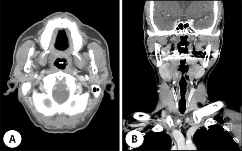
A flexible fiberscope revealed three cystic mass around the epiglottis. And also a cystic mass was found near the left Eustachian tube opening in the nasopharynx, incidentally (Fig. 1A). Neck Computed Tomography (CT) with contrast showed focal protrusion at left nasopharyngeal wall with no evidence of definite enhancement (Fig. 2). Laryngeal Microscopic Surgery (LMS) was planned to remove epiglottic cysts as treatment. At the same time, endoscopic approach excisional biopsy about nasopharyngeal cystic mass was performed. The lesion was completely removed using upward cup forceps and microdebrider. The cystic mass was soft and contained viscous discharge like pus.
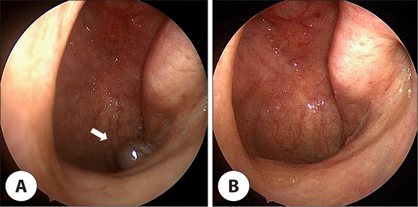
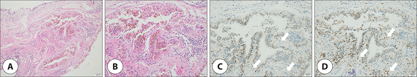
Microscopically, oncocytic cells appeared as a distinct population of well-formed glandular structures demarcated from normal respiratory epithelium. Oncocytic cells had uniformly abundant eosinophilic granular cytoplasm on hematoxylin and eosin (H&E) staining. Scattered brown pigments were also noted in the cytoplasm of oncocytic cells (Fig. 3A, B). Immunohistochemistry analysis revealed dendritic melanocytes in the basal layer of oncocytic gland, which were negative for HMB-45 stain, but positive for S-100 stain (Fig. 3C, D). Although oncocytic metaplasia occurring in melanoma has been reported, this lesion can be easily distinguished from melanoma because there is no malignant component. Finally, the lesion was diagnosed as MOM of the nasopharynx. On the other hand, the pathological results of the lesions around epiglottis were diagnosed as epidermal inclusion cysts. The lesion of nasopharynx was completely removed and the follow-up endoscopy showed no evidence of recurrence at 18 months after surgery (Fig. 1B).
Discussion
MOM is characterized by the presence of both oncocytic change and melanin pigmentation of epithelium simultaneously.7) Oncocytic cells have undergone cytoplasmic changes and are considered to be related to an aging process, because the cells are discovered predominantly in elderly persons.18) Oncocytic metaplasia is most frequently encountered in certain epithelial organs, such as the salivary gland, the parathyroid gland, the thyroid gland, and the kidney,6) but also is not uncommon in the upper respiratory tract.4) However, it is extremely rare to observe the melanotic variant concurrently in the nasopharynx. Our study is the 37th case reported in the English literature.
The origin of the melanin pigment in this lesion is still unknown. But we identified that oncocytes in MOM of the nasopharynx lack premature melanosomes, in which melanin is produced. Therefore, the melanin pigment in oncocytes is believed to be passed on from the adjacent melanocytes via their dendrites. And this transfer of melanin seems to occur after the onset of oncocytic metaplasia.4,7) Microscopically, the melanocytes with their dendritic process could be positive by immunohistochemical staining for S-100 and Melan-A, but negative for HMB-45.12,15) In addition, histological appearance, which is positive in Fontana-Masson stain whereas negative in Berlin blue stain (Prussian blue stain), confirms that these pigments are melanin.7,9,15)
We reviewed the literature of MOM of the nasopharynx including our cases.1-17) They are summarized in Table 1, which lists age, sex, site, symptom, endoscopic finding, number of lesions, clinical impression and smoking history.
| Case | Sex/ age | Site | Symptom | Endoscopic finding | Number | Clinical impression | Smoking history |
|---|---|---|---|---|---|---|---|
| 11 | M/67 | ETO | Serous otitis media | 2 mm brown nodule | Single | Nasopharyngeal carcinoma | Unknown |
| 21 | M/63 | ETO | Tinnitus | 2 mm brown nodule | Single | Nasopharyngeal carcinoma | Unknown |
| 32 | M/69 | NPx | None | Small black lesions with slightly elevation | Multiple | Not documented | Unknown |
| 43 | M/70 | ETO | Tinnitus | 1-2 mm brown nodules | Multiple | Nevus | Unknown |
| 54 | M/64 | ETO / NC / Suprapharynx | Discomfort of the throat | Small, flat elevations with brown discoloration up to a few millimeters | Multiple | Malignant tumor | Unknown |
| 65 | M/79 | ETO | Hearing loss / Otorrhea / Otalgia | A pigmented lesion | Single | Not documented | Unknown |
| 76 | M/62 | ETO | Discomfort of the ear | 5 mm black nodule & small spots with black discoloration | Multiple | Melanoma | Unknown |
| 87 | M/80 | NC / Pharynx | Hoarseness | Lesions with a few millimeters in size, brown to black in color | Multiple | Melanoma | Smoker (25PY) |
| 97 | M/69 | ETO | Rhinorrhea | Lesions with a few millimeters in size, brown to black in color | Single | Melanoma | Smoker (120PY) |
| 107 | M/74 | Suprapharynx | Epistaxis | Lesions with a few millimeters in size, brown to black in color | Single | Tumor | Unknown |
| 117 | F/74 | ETO | Discomfort of the throat | Lesions with a few millimeters in size, brown to black in color | Multiple | Melanoma | Unknown |
| 127 | M/68 | NPx | None | Lesions with a few millimeters in size, brown to black in color | Single | Tumor | Unknown |
| 137 | M/56 | ETO | Hemoptysis | Lesions with a few millimeters in size, brown to black in color | Single | Not documented | Unknown |
| 147 | M/63 | ETO | Epistaxis | Lesions with a few millimeters in size, brown to black in color | Single | Melanoma | Unknown |
| 158 | M/ND | NPx | None | Pigmented lesions | Multiple | Not documented | Unknown |
| 169 | M/58 | NPx | Epistaxis | Small, irregular, black-pigmented lesions | Multiple | Not documented | Unknown |
| 1710 | M/73 | TT | Nasal obstruction / Chill | Black nodules measuring several millimeters | Multiple | Melanoma / Hemangioma | Non-smoker |
| 1811 | M/72 | TT | Headache / Hearing loss | Dark blue colored mucosal lesions | Multiple | Melanotic dysplasia | Smoker (100PY) |
| 1911 | M/71 | TT / Soft palate | Hoarseness | Black mucosal lesions | Multiple | Nasopharyngeal pigmented lesion | Smoker (40PY) |
| 2011 | M/51 | TT | Tongue pain | Dark colored spots | Multiple | Melanotic oncocytic metalplasia of nasopharynx | Unknown |
| 2112 | M/63 | NPx | Epistaxis | Black nodules up to several millimeters | Multiple | Melanosis / Melanoma | Smoker (80PY) |
| 2213 | M/57 | TT | None | 2-3 mm flat, elevated lesions with brown to black discoloration | Multiple | Melanoma | Smoker (60PY) |
| 2314 | M/60s | TT | None | Black nevus-like lesion | Single | Not documented | Unknown |
| 2415 | F/70 | ETO | None | 2 mm black nodules | Multiple | Melanosis | Smoker (12.5PY) |
| 2515 | F/61 | ETO | Hoarseness | 2 mm black nodule | Single | Melanoma | Smoker (46PY) |
| 2615 | M/74 | NPx | Hoarseness | 2 mm black nodules | Multiple | Melanoma | Smoker (50PY) |
| 2716 | M/57 | NPx | Nasal obstruction | Flat lesion with pigmentation | Single | Not documented | Smoker (ND) |
| 2816 | M/61 | NPx | Neck mass | Mass without pigmentation | Single | Not documented | Smoker (ND) |
| 2916 | M/69 | NPx | Rhinorrhea | Flat lesion with pigmentation | Multiple | Not documented | Smoker (ND) |
| 3016 | M/56 | ETO | Epistaxis | Flat lesion with pigmentation | Single | Not documented | Smoker (ND) |
| 3116 | M/58 | NPx | Cranial nerve VII palsy | Flat lesion with pigmentation | Single | Not documented | Smoker (ND) |
| 3216 | M/52 | ETO | Tinnitus | Flat lesion with pigmentation | Multiple | Not documented | Smoker (ND) |
| 3316 | F/77 | NPx | Hoarseness | Nodule with pigmentation | Single | Not documented | Smoker (ND) |
| 3416 | M/59 | NPx | Nasal obstruction | Flat spot with pigmentation | Single | Not documented | Smoker (ND) |
| 3516 | M/59 | ETO | Tinnitus | Nodule without pigmentation | Single | Not documented | Smoker (ND) |
| 3617 | M/75 | ETO | Epistaxis / Rhinorrhea / Cough / Sputum | Brown nodules | Multiple | Nevus / Melanoma | Smoker (75PY) |
| 38 (present) | M/76 | ETO | Globus pharyngeus | Cystic mass | Single | Cystic mass | Smoker (6PY) |
The age of patients ranges from 51 to 80 years with a mean of 65.9 years, except for two patients who do not be documented the exact age. This disease predominantly occurred in males (34/37), with a male to female ratio of almost 8:1. Interestingly, all the patients reviewed are from East and Southeast Asia, including Korea, Japan, China, Taiwan, and Hongkong. Limited geographical distribution like that implies the possibility that the occurrence of MOM of the nasopharynx is associated with genetic predisposition. In general, oral melanosis as physiological pigmentation is known to occur more frequently in darker skinned individuals due to increased melanocytic activity,19) but further studies would be needed to find out why this disease has only been observed in Asians so far.
A single or multiple lesions (18 cases and 19 cases, respectively) was mainly distributed around the lateral wall of the nasopharynx (17 cases in Eustachian tube opening, 4 cases in torus tubarius, and 13 cases in nasopharynx, respectively). In addition, it also could be found in nasal cavity, suprapharynx, and soft palate. The lesions appeared as distinctive nodules, flat spots, or mucosal abnormalities including irregularity or slight elevation, which usually have brown to black colored pigmentation and small size (a few milimeters) on endoscopic examination. However, in three cases including ours, the lesion was observed without gross pigmentation. Another unique feature of our case is that its shape was observed as a cystic pattern. Histologically, oncocytic lesions shows cystic growths that contain papillary projections lined by the characteristic cell, the oncocyte.18) One case was reported as a cystic pattern in the non-MOM of the nasopharynx.16) But such form has not been reported in the case of MOM of the nasopharynx.
In fact, patients visited the hospital with complain of various symptoms. Mass forming lesions can cause obstructive symptoms, while flat lesions are more likely to have no symptoms. In particular, if the mucous membrane around the Eustachian tube opening is affected, ear or nose symptoms may occur.10,16) Symptoms related to the ear were tinnitus (4 cases), otitis media including otorrhea and otalgia (3 cases), and hearing impairment thought to be caused by exudation (2 cases). Characteristically, when the lesions with elevation such as nodule were located in the Eustachian tube opening, more than half of cases (5/9) showed ear related symptoms. And nose related symptoms were epistaxis (6 cases), rhinorrhea (3 cases), and nasal obstruction (3 cases). Other symptoms such as hemoptysis (2 cases), hoarseness (5 cases), and discomfort of the throat (3 cases) were also expressed, including our case with globus pharyngeus. However, nine patients did not complain of any discomfort, and some of the symptoms described above are considered to have little direct relationship with the lesion because of size, location and shape. It would be reasonable to judge that it was found by accident during the examination.
MOM of the nasopharynx can resemble melanoma on endoscopy, with the presence of pigmentation and multifocality which simulates satellite lesions in melanoma.10) In fact, 63.6% (14/22) of the cases documented suspected that the lesion had the potential for malignancy. It is essential to make differential diagnosis between these two diseases because the treatment and the prognosis of each disease are very different. All the reported cases of MOM of the nasopharynx followed a benign clinical course. To date, concurrence or subsequent development of malignant neoplasm has not been reported in patients with this disease. Simple excision and periodic follow-up is a suitable treatment of choice for MOM of the nasopharynx.13)
The exact pathogenesis of MOM is unknown. Sakaki et al. hypothesized that smoking may be a predisposing factor for MOM.7) Of the 20 patients with information about smoking history, all but one patient were smokers. In addition, it is notable that the smoking period was over 40 years. The incidence and intensity of pigmentation in smoker’s melanoma is closely related to duration and amount of tobacco smoking.19) Smoking-associated melanosis is due to increased melanin production by melanocytes and its deposition within the basal cell layer and lamina propria. This process is thought to be a result of either the noxious chemicals in cigarette smoke or heat stimulating melanocytes to protectively produce melanin.20) Nicotine, which exhibits affinity for intracellular melanin, disrupts the melanin-containing cells, causing the migration of melanosomes and subsequent melanization.21)
Oncocytic metaplasia of the nasopharynx and Warthin tumor of the parotid gland share several common features. Both diseases have circumscribed lesions and lymphocytic stroma, occur mainly in the geriatric population, have a benign course, and are associated with smoking.17) And also these are the same pathological entity in different anatomical situation. Therefore, it is possible that the oncocytic metaplasia of the nasopharynx is an early stage form of Warthin tumor.22) In review of literature, one patient who visited asymptomatically except neck mass was associated with concomitant Warthin tumor.
Conclusion
Although oncocytic change with melanotic variant in the nasopharynx is an extremely rare condition, it is a disease that must be differentiated from malignant melanoma. However, the clinical impression of it may be confused due to the morphological diversity seen by endoscopic examination. In particular, as in the case of this report, it may be more difficult to differentiate if it has atypical form such as cyst without pigmentation. Therefore, MOM of the nasopharynx must always keep in mind the diversity of its forms in order to determine accurately the direction of diagnosis and treatment in the clinical setting.