


Introduction
A paratracheal cyst is an air-filled cyst of the trachea line by respiratory ciliated epithelium. Most paratracheal cysts are found incidentally on the neck or thoracic computed tomography (CT) scans. They are found more frequently on the right side of the trachea, in the region of the thoracic outlet and usually asymptomatic.1,2) They may, however, function as a reservoir for secretion, thereby chronic infection could occur.3,4) Although respiratory symptoms may be caused by the infected cysts, there are little knowledge and interest in this clinical entity. Also, the anatomic location of the cysts could make it difficult to decide on surgical intervention. Therefore, there are only few cases reports about infected paratracheal cyst with surgical treatment, and especially in the case of fungal infection. Here, we report a case of fungal infection of the paratracheal air cyst.
Case Report
A 75-year-old woman complaining of progressive dyspnea that had occurred the previous day visited the emergency room. It was difficult to breathe even when lying still, and the dyspnea became worse when exhaling. She had hypertension and diabetes as underlying disease.
The arterial blood gas analysis (ABGA) showed low pO2 (35.8 mmHg) with normal pH (7.425), pCO2 (32.2 mmHg) and HCO3- (20.7 mmol/L). The body temperature was 37.8℃, and other vital signs were stable. Diffuse lower neck swelling with overlying skin redness was observed on physical examination. Dyspnea was aggravated in supine position and a weak crackle lung sound was auscultated. There were no abnormal findings in the oral cavity or pharynx. On the fiberoptic endoscopic examination, larynx including vocal cord movement was intact.
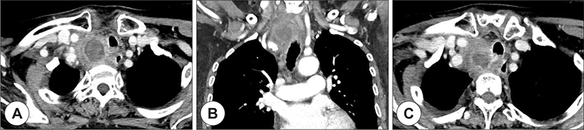
On the neck CT scan, 6.2×3.1×3.5 cm sized enhancing mass with central low density was found at the right superior mediastinal area (Fig. 1). The trachea has deviated to the left and anterior sides, and was narrowed at the thyroid gland level. The mass was suspected to be associated with the tracheal membrane. There was a small amount of pleural effusion and atelectasis at the right lung.
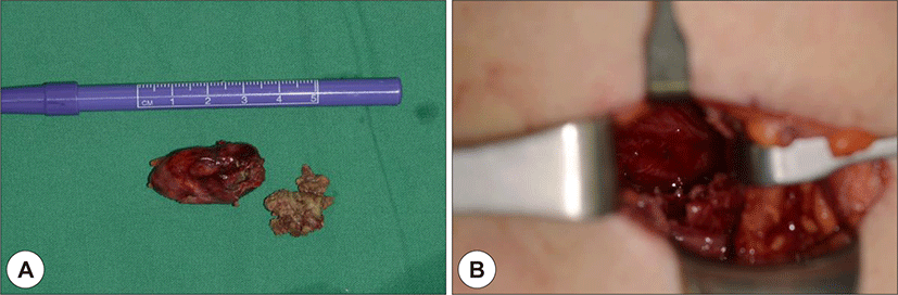
Antibiotics (ampicillin/sulbactam) was administrated intravenously, and it was planned to perform surgical excision or incision/drainage as soon as possible. After a 6 cm incision at the right lower neck, the right paratracheal area was dissected under general anesthesia. The mass wall could be identified, but it adhered to surrounding soft tissues. Careful dissection was done along the capsule of the mass. The mass was continued to the posterior tracheal wall. After separation of the mass from the trachea, the mass was ruptured and yellowish cheese-like dirty material was expelled out from the mass (Fig. 2). The right recurrent laryngeal nerve could not be identified due to surrounding soft tissue inflammation and adhesion. After removal of mass, a penrose drain was inserted and vacuum-assisted closure (V.A.C.) system was applied to the operation site because air leakage could occur, although no defect was found at the trachea wall on surgical field.
The patient was taken to the intensive care unit, closely monitored for breathing for a day, and then transferred to a general ward on 2 days after surgery after removal of vacuum drainage because no air leak was observed and acute inflammation was rapidly improved. Penrose drain was removed on 4 days after surgery. The patient complained of mild discomfort of breathing but it was resolved after seroma aspiration on 2 days after surgery. However, right vocal cord palsy was observed with the laryngoscopic examination.
Because inflammatory signs and respiratory discomfort were nearly disappeared, the patient was discharged 7 days after surgery, although right vocal cord was still paralyzed.
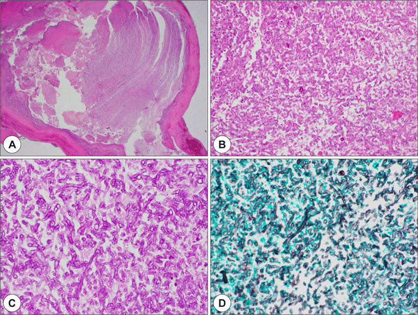
Pathologic examination revealed the material from the cystic mass was a fungal organism (Aspergillus species). It was strongly stained with PAS and Gomori staining (Fig. 3).
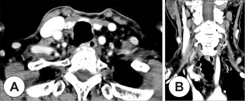
On a CT scan follow-up 3 months after surgery, there was no inflammation sign, but air pocket was observed at the right paratracheal area (Fig. 4). It was decided to observe the patient without further surgical treatment because the patient did not complain of any symptoms although right vocal cord movement was slightly decreased.
Discussion
Paratracheal air cysts (PAC) are aggregates of air close to the trachea which was covered with ciliated columnar epithelium.2) Most PACs are detected incidentally on thoracic CT scans and most of them are asymptomatic. Previous studies have reported that the prevalence of PACs was estimated from 0.3% to 3.9%.5,6) On the other hand, some reports reported prevalence as high as 8.1% by advanced CT scan resolution with thinner section.2,7)
As it is thought that PAC may act as a reservoir for respiratory secretions, it could be infected and cause respiratory symptoms such as cough, sputum, hemoptysis, and fever.8,9) Although there are some case reports of infected PACs with these symptoms, there were few reports of the infected cyst causing compression symptoms like dysphagia or dyspnea. Moreover, there were no reports of fungal infection of the PAC. As far as we know, this is the first report of a paratracheal cyst with fungal infection.
Generally, surgical treatment of PACs is uncommon, both because most PACs are asymptomatic and the majority of the patient is elderly with poor respiratory function. Furthermore, some reports asserted that infected PACs would be primarily managed with conservative treatment including antibiotics and O2 supply if needed.8,10) In addition, endobronchial ultrasound-guided transbronchial fine needle aspiration was used effectively along with pharmacological treatment in some cases.11,12) These conservative methods can be effective for conventional infection of PACs. In this case, however, the patient complained of progressive dyspnea and the tracheal deviation was found due to the large infected PAC. Moreover, abscess formation was strongly suspected in the CT scan. Considering these situations, it was thought that surgical treatment would be inevitable. After successful surgical excision of PAC, symptoms including dyspnea were improved.
Another notable characteristic in this case was the size of the cyst. Most PACs are less than 10 mm in size. The size of PAC was reported from 1.5 mm to 20 mm (mean, 5.6 mm) in axial and from 2.3 mm to 25 mm (mean, 8.0 mm) in coronal.5,13) Interestingly, the size of the cyst in this case was 40×39 mm in CT measurement.
Furthermore, we applied V.A.C system postoperatively. It was developed in the early 1990s, and has been used for infected wound in various surgical field.14,15) Recently, several reports showed good results in controlling infection and wound healing of intractable deep neck infection, necrotizing fasciitis, esophageal fistula, pharyngocutenous fistula or flap necrosis.16-19) Also in this case, infection was well controlled after surgery and V.A.C system. Additionally, although tracheal wall defect was not found during the surgery, there may be a risk of undetected fistula. We judged that V.A.C system would be better than high-pressure drain tube like hemo-vac for the fistula and nerve function preservation.20) Fortunately, air leakage was not observed after the surgery and the patient’s condition was improved, so we could remove V.A.C system on 2 days after surgery.
According to our experience, in a patient who complains of symptoms such as cough, fever and dyspnea with normal lung and laryngopharyngeal examination, it should be suspected of an infected PAC. If an infected cyst is suspected, appropriate drainage or cyst excision should be considered.