증례

이하선에 재발한 호산성 과립세포종과 결절성 호산성 과립세포증식증 1예

정남윤1https://orcid.org/0000-0003-3375-5365, 노미숙2https://orcid.org/0000-0002-5676-5569, 이동근1,*https://orcid.org/0000-0002-7296-1420
Nam Yoon Jung1https://orcid.org/0000-0003-3375-5365, Mee Sook Roh2https://orcid.org/0000-0002-5676-5569, Dong Kun Lee1,*https://orcid.org/0000-0002-7296-1420
Author Information & Copyright
1동아대학교 의과대학 이비인후-두경부외과학교실
2동아대학교 의과대학 병리학교실
1Department of Otorlaryngology-Head and Neck Surgery, Dong-A University College of Medicine, Busan, Korea
2Department of Pathology, Dong-A University College of Medicine, Busan, Korea
*Corresponding author: Dong Kun Lee, Department of Otorhinolaryngology-Head and Neck Surgery, College of Medicine Dong-A University, 26, Daesingongwon-ro, Seo-Gu, Busan 49201, Korea, Tel: +82-51-240-5428·Fax: +82-51-253-0712·E-mail: chaos001@hanmail.net

© Copyright 2021 The Busan, Ulsan, Gyeoungnam Branch of Korean Society of Otolaryngology-Head and Neck Surgery. This is an Open-Access article distributed under the terms of the Creative Commons Attribution Non-Commercial License (http://creativecommons.org/licenses/by-nc/4.0/) which permits unrestricted non-commercial use, distribution, and reproduction in any medium, provided the original work is properly cited.

Received: Aug 13, 2021; Revised: Sep 16, 2021; Accepted: Nov 04, 2021

Published Online: Dec 31, 2021

ABSTRACT

Oncycytoma is a tumor which is abundunt with granular and eosinophilic cytoplasm and mitochondria. It can be found in various organs such as the kidney, thyroid gland, pancreas, and breast. Salivary gland is reported to be involved with oncocytoma in less than 1% among the salivary gland tumors. A 65-year-old female visited presenting with recurrent lump of the left parotid gland. She had been diagnosed with oncocytoma after left superficial parotidectomy 8 years ago. Physical examination revealed multiple painless tumors in the left parotid gland. Ultrasound and computed tomography showed multiple mass in the left parotid gland. The patient had a revision parotidectomy. Pathology confirmed an oncocytoma and nodular oncocytic hyperplasia. We report a rare case of recurrent oncocytoma and nodular oncocytic hyperplasia from the parotid gland and report the case with related literature. (J Clinical Otolaryngol 2021;32:302-307)

Keywords: Adenoma, Oncocytoma; Nodular oncocytic hyperplasia; Parotid gland

서 론

호산성 과립세포종(oncocytoma)은 조직학적으로 과립성, 호산성의 세포질과 풍부한 미토콘드리아를 보이는 상피세포로 이루어진 종양이다.1-3) 신장, 침샘, 갑상선, 췌장, 유방 등 여러 장기에서 발견되며,3) 침샘에서는 침샘 기원 종물 중 1% 미만을 차지하는 드문 질환이다.1,2) 결절성 호산성 과립세포증식증(nodular oncocytic hyperplasia)은 침샘에서는 0.1% 정도의 빈도를 보이는 매우 드문 질환으로, 미만성, 다발성의 호산성 과립세포 증식 소견을 보이는 비증식성 병변이다.4)

65세 여자 환자가 8년 전 호산성 과립세포종으로 좌측 이하선 부분 절제술을 시행한 후 재발한 좌측 이하선 종물로 내원하였다. 신체 진찰 상 무통성 이하선 종괴를 보였으며, 초음파 및 전산화단층촬영 상 양측 이하선에서 여러 개의 종물이 확인되었다. 우선 종물의 크기가 크고, 재발소견을 보인 좌측 이하선의 종물 제거술을 시행하였으며, 조직검사 상 결절성 호산성 과립세포증식증 및 과립세포종으로 진단되었다.

이하선의 결절성 호산성 과립세포증식증은 해외에서는 13례 보고된 바 있으나,5-17) 국내에서는 처음 보고되는 증례로, 이하선의 호산성 과립세포종 재발과 동반된 결절성 호산성 과립세포증식증 환자를 경험하였기에 문헌 고찰과 함께 보고하고자 한다.

증 례

65세 여자가 좌측 이하선 부위 종물과 불편감을 주소로 본원 외래로 내원하였다. 환자는 약 8년 전 다른 술자에게 좌측 이하선 종물로 진료 후, 좌측 이하선 천엽 부분 절제술(superficial partial parotidectomy)을 받은 후, 조직검사 상 호산성 과립세포종(oncocytoma)으로 진단되었던 과거력이 있었다.

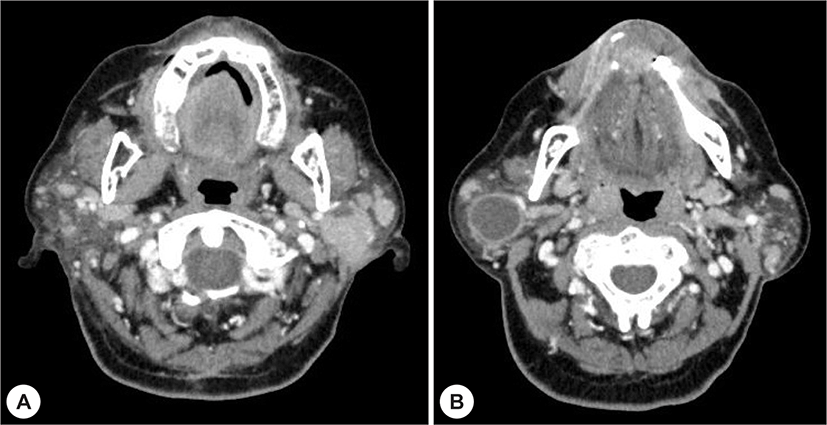
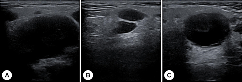
신체 검사에서 좌측 이하선 부위에는 3개의 종물이 만져졌으며, 우측 이하선 부위에는 1개의 종물이 만져졌다. 통증이나 압통은 없었으며, 주변 조직과 유착된 소견은 보이지 않았다. 목 전산화 단층촬영에서는 양측 이하선에서 일부에서 낭성 변화를 보이는 불균질한 조영 증강을 보이는 종물들이 확인되었다(Fig. 1). 초음파 검사 상 양측 이하선에 다발성의 저에코성 종물이 확인되었고(Fig. 2), 이들에 대한 세침흡인 세포검사를 시행하였으며, 악성종양의 소견은 없고 와르틴 종양에 부합하는 조직 소견이라는 판독을 받았다. 좌측 이하선에 다발성 종물이 있었고, 환자가 좌측 이하선 부위의 불편감을 호소하였고, 이전 좌측 이하선 수술 이후 재발되었기 때문에 재수술로 인한 안면신경 손상의 위험성이 증가하는 가능성 등을 종합적으로 고려하여 좌측 이하선을 우선 수술한 후 우측 이하선을 차례로 수술하기로 계획하였다.

jcohns-32-3-302-g1
Fig. 1. Computed tomography of oncocytoma and nodular onococytic hyperplasia. Multiple tumors of various sizes (radius ranging from 8 mm to 26 mm) are in the left parotid gland (A). A cystic mass, about 20×25×26 mm is in the right parotid gland (B).
Download Original Figure
jcohns-32-3-302-g2
Fig. 2. Ultrasonography of oncocytoma and nodular onococytic hyperplasia. Multiple cystic masses are shown in the left parotid gland (A, B). A cystic mass is in the right parotid gland (C).
Download Original Figure

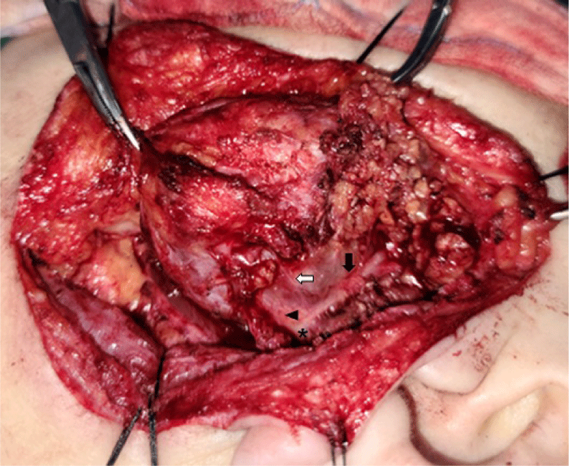
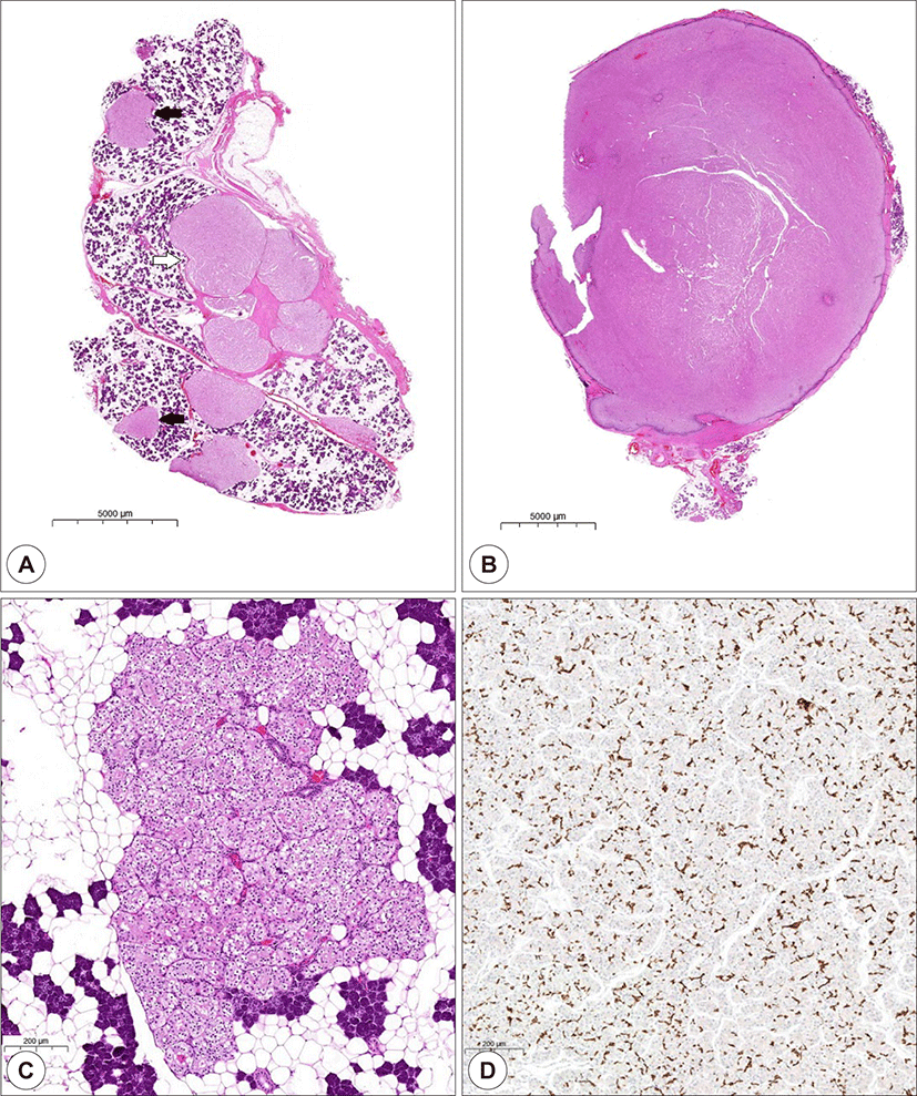
수술은 변형 블레어 절개를 통한 좌측 이하선 재절제술을 시행하였고, 안면신경 보존을 위해 수술 중 신경 모니터링을 위해 NIM-Neuro 3.0 system(Medtronic, Jacksonville, FL, USA)의 근전도 감시 장치를 이용하였다. 이하선을 노출시키자 이하선 천엽(superficial lobe)이 일부 남아 있었다. 잔존 이하선 천엽에서 종물이 여러 개 확인되었으며, 가장 큰 종물은 심엽(deep lobe)에 안면신경으로 둘러싸여 있었다. 안면신경의 측두분지, 관골분지, 협부분지, 하악분지, 경부분지를 확인 및 보존하면서 이하선 천엽의 잔존부위와 심엽의 종물을 안전역을 확보하여 제거하였고, 안면신경 각 분지의 전도가 정상임을 확인하였다(Fig. 3). 수술 부위의 함몰을 최소화하기 위해 무세포 동종 진피(acellular dermal matrix)인 MegaDerm®(L&C Bio, Seongnam, Korea)을 적용하였다. 조직검사 결과, 좌측 이하선 천엽의 여러 개의 종물들은 결절성 호산성 과립세포증식증(nodular oncocytotic hyperplasia)로 확인되었으며, 심엽에서 제거한 종물은 호산성 과립세포종으로 확인되었다(Fig. 4).

jcohns-32-3-302-g3
Fig. 3. Intraoperative finding. The facial nerve trunk (astrix) is shown after dissecting the remnant superficial lobe of the parotid gland. It divides into a branch of temporal and zygomatic branch (black arrow), a thin branch of buccal branch (white arrow), and a branch of marginal mandibular and cervical branch (arrowhead).
Download Original Figure
jcohns-32-3-302-g4
Fig. 4. Pathologic finding of oncocytoma and nodular oncocytic hyperplasia. The specimen shows a mixture of the oncocytoma (white arrow) and nodular onococytic hyperplasia (black arrow) (H&E stain, bar=5,000 μm) (A). An oncocytoma is an encapsulated tumor with oncocytes which is characterized by abundant eosinophilic cytoplasm (H&E stain, bar=5,000 μm) (B). A nodular oncocytic hyperplasia shows a cluster of oncocytes without encapsulation (H&E stain, bar=200 μm) (C). The oncocytes of oncocytoma and nodular oncocytic hyperplasia are positive for CK5/6 (CK5/6, bar=200 μm) (D).
Download Original Figure

수술 후 환자는 House-Brackman grade 3 정도의 좌측 아랫입술의 마비 소견을 보여 문헌상으로는 안면신경 마비의 회복에 차이가 없지만, 경험적으로 고용량 스테로이드 치료를 시행하였으며, 수술 후 2주째 정상으로 회복되었다. 현재 수술 6개월째 재발 등의 소견 없이 외래 경과관찰 중이며, 우측 이하선 종물에 대한 수술은 추후 계획 예정이다.

고 찰

침샘 종양은 조직학적으로 다형선종, 와르틴 종양, 기저세포종, 호산성 과립세포종 등의 양성 종양과 점액표피양암종, 선양낭성암종 등의 악성 종양이 있다.1) 이 중 호산성 과립세포종은 조직학적으로 세포질이 호산성이며, 미토콘드리아와 과립이 풍부한 상피세포가 특징적인 종물로 침샘 종양 중 1% 미만으로 발견된다.1-3) 결절성 호산성 과립세포증식증은 0.1% 정도로 매우 드물게 보고되는 병변으로, 다발적으로 침샘 조직을 대체하는 호산성 과립세포를 보이며, 잔존하는 장액선을 침범하는 소견을 나타낸다.4)

호산성 과립세포는 노화 등의 원인으로 세포가 손상된 이후 그 기능을 잃으면서 화생(metaplasia)을 통해 생기는 것으로 생각된다.4,18) 호산성 과립세포 내의 미토콘드리아에서 후천성 기능장애가 발생하고, 잘못된 대사과정의 결과로 호산성 과립세포종과 결절성 호산성 과립세포증식증이 발생한다는 가설이 제기되고 있으나, 정확히 밝혀진 바는 없다.4,18)

세계보건기구의 분류에 따르면 호산성 과립세포종은 결절성 호산성 과립세포증식증(nodular oncocytic hyperplasia), 호산성 과립세포종(oncocytoma), 호산성 과립세포암종(oncocytic carcinoma)로 분류된다.4,19) 호산성 과립세포종은 조직학적으로 미토콘드리아가 풍부한 호산성 세포질과 하나의 두드러진 인(nucleolus)을 보이는 세포핵을 가진 세포가 규칙적으로 배열된 특징을 보이며, 섬유질의 피막으로 둘러싸여 주변 조직과 구별된다.19,20) 결절성 호산성 과립세포증식증은 침샘 내에 호산성 세포가 증식하면서 결절성 병변을 보이지만, 피막이 없어 호산성 과립세포종과 달리 진단된다.4,19) 이 때문에 주변의 침샘조직을 침범하는 등의 소견을 보인다.4) 호산성 과립세포암종은 호산성 과립세포가 피막이 없는 상태로 비정상적인 세포 증식, 국소적 침범, 신경 주위 침범, 림프관 또는 혈관 침범 등의 주변 조직으로 침윤성 성장을 보일 때 진단할 수 있으며, 이하선에서 가장 흔하게 발견된다.19,20)

임상증상은 다른 양성 침샘 종양과 비슷하다.5,-7,12) 신체 진찰에서 무통성, 유동성 종물이 흔하게 관찰되며, 7% 정도에서 양측성으로 나타난다.5-7,12) 그 외에도 종물의 크기가 커짐에 따라 통증, 안면마비 등이 나타날 수도 있다.5-7,12) 본 증례에서도 환자는 무통성 이하선 종물을 주증상으로 호소하였다.

진단을 위한 검사로는 경부 전산화단층촬영 및 경부 자기공명영상을 고려할 수 있다.2-4) 호산성 과립세포종은 전산화단층촬영 상 불균질한 조영증강을 보이는 단발성 또는 다발성 종괴나 소엽상으로 나타나기도 하며, 일부에서는 낭성 변화를 보이기도 한다.2,4) 자기공명영상에서는 경계가 분명하고, 균질한 조영증강을 보이는 종물을 확인할 수 있으며, 와르틴 종양(Warthin’s tumor)이나 기저세포선종(basal cell adenoma) 등과 감별이 필요하다.2,3) 본 증례에서도 전산화단층촬영 상 불균질한 조영증강을 보이고, 일부에서 낭성 변화를 보이며, 양측성으로 발생한다는 점에서 호산성 과립세포종 외에도 와르틴 종양을 의심할 수 있었다.

그 외에도 진단을 위해 초음파와 초음파 유도 하 세침흡인 세포검사를 시행할 수 있다.2,3,6) 초음파에서는 이하선의 호산성 과립세포종은 경계가 분명한 저에코성 종물로 보여 다른 이하선 종물과 감별이 어려울 수 있다.2,3,6) 초음파 유도 하 세침흡인 세포검사 상 호산성 과립세포를 확인할 수 있으나, 이는 호산성 과립세포종 이외에도 와르틴 종양, 세엽세포암종(acinic cell carcinoma), 점액표피양암종(mucoepidermoid carcinoma) 등 여러가지 상황에서 발견될 수 있는 소견으로, 호산성 과립세포종으로 진단을 내리기에는 불충분한 경우가 많다.2,3,6) 이 중 가장 흔하게 발견되는 와르틴 종양과 구별할 수 있는 특징은 조직 내 림프구의 유무이며, 호산성 과립세포종은 조직내에 림프구가 없는 반면, 와르틴 종양에서는 림프구가 풍부하게 발견된다.4) 이 때문에 대부분의 환자에서 수술 후 조직검사를 통해 정확한 진단이 내려진다.2-4,6) 본 증례에서도 수술 전 시행한 세침흡인 세포검사에서 와르틴 종양에 부합하는 소견을 보여 수술 후 진단과는 다른 소견을 보였다.

호산성 과립세포종의 가장 좋은 치료방법은 완전한 수술적 절제이다. 20% 이하에서 재발이 보고되고 있으나, 불완전한 절제가 원인으로 생각되며, 악성화 가능성은 낮은 것으로 보고되고 있다.1,2,4,6) 결절성 호산성 과립세포증식증의 수술 후 재발은 추후 연구가 필요한 실정이다.6,7) 이하선의 호산성 과립세포종은 위치에 따라 이하선 천엽절제술 또는 전절제술을 시행할 수 있으며, 수술 전 임상 및 영상 검사를 통해 수술적 절제 범위가 결정된다.1,2,6)

호산성 과립세포종은 수술 후 방사선 치료나 항암 치료 등의 추가적인 치료는 필요하지 않으나, 추후에 호산성 과립세포암종이 발생한 증례들이 드물게 보고되어 장기적인 추적관찰이 필요할 것으로 생각된다.5,6,9,11)

호산성 과립세포암종은 드물게 보고되는 질환으로 병인이나 치료 등에 관한 연구가 필요한 실정이나, 예후가 불량하여 조기에 외과적 치료를 시행하며, 추가로 방사선 치료와 항암제 치료 등을 고려하는 적극적인 치료를 권고하고 있다.6,20)

본 증례에서는 8년 전 좌측 이하선의 호산성 과립세포종으로 수술한 이후 재발한 종물을 주소로 내원한 환자에서 호산성 과립세포종의 재발과 더불어 결절성 호산성 과립세포증식증이 추가로 진단되었다. 수술적 완전 절제 이후 재발이나 합병증이 없는 상태로, 정기적인 추적관찰 및 남은 우측 이하선 종물에 대한 치료 계획을 수립할 예정이다. 이하선에 호산성 과립세포종이 다발성으로 발생한 경우, 결절성 호산성 과립세포증식증을 고려하여야 하겠다.

Acknowledgements

Not applicable.

Funding Information

This work was supported by the Dong-A University research fund.

Conflict of Interest

No potential conflict of interest relevant to this article was reported.

Author Contribution

Conceptualization: Lee DK.

Data curation: Lee DK.

Validation: Jung NY, Lee DK.

Writing - original draft: Jung NY, Roh MS.

Writing - review & editing: Jung NY, Roh MS, Lee DK.

Ethics Approval

Not applicable.

References

1).

Vlachaki E, Tsapas A, Dimitrakopoulos K, Kontzoglou G, Klonizakis I. Parotid gland oncocytoma: a case report. Cases J 2009;2(1):6423.

2).

Sharma V, Kumar S, Sethi A. Oncocytoma parotid gland. Ann Maxillofac Surg 2018;8(2):330-2.

3).

Ak 1 IB, Altay C, Güler E, Çamlıdağ İ, Harman M, Danacı M, et al. Discrimination of oncocytoma and chromophobe renal cell carcinoma using MRI. Diagn Interv Radiol 2019;25(1):5-13.

4).

Shellenberger TD, Williams MD, Clayman GL, Kumar AJ. Parotid gland oncocytosis: CT findings with histopathologic correlation. AJNR Am J Neuroradiol 2008;29(4):734-6.

5).

Hung CJ, Liu YH, Liao JB. Nodular oncocytic hyperplasia of bilateral parotid glands with parapharyngeal space extension. Ear Nose Throat J 2020.

6).

Akbulut A, Kaptan ZK, Yasaran M, Karadas H, Koca G, Korkmaz M. Metachronous bilateral multifocal nodular oncocytic hyperplasia of the parotid gland. J Stomatol Oral Maxillofac Surg 2019;120(6):598-600.

7).

Rooper LM, Onenerk M, Siddiqui MT, Faquin WC, Bishop JA, Ali SZ. Nodular oncocytic hyperplasia: can cytomorphology allow for the preoperative diagnosis of a nonneoplastic salivary disease? Cancer Cytopathol 2017;125(8):627-34.

8).

Palmer TJ, Gleeson MJ, Eveson JW, Cawson RA. Oncocytic adenomas and oncocytic hyperplasia of salivary glands: a clinicopathological study of 26 cases. Histopathology 1990;16(5):487-93.

9).

Goyal R, Ahuja A, Gupta N, Singh G, Vaiphei K. Multifocal nodular oncocytic hyperplasia in parotid gland: a case report. Acta Cytol 2007;51(4):621-3.

10).

Sato S, Kishino M, Ogawa Y, Nakatsuka S, Hoshida Y, Ogawa I, et al. Multifocal nodular oncocytic hyperplasia of bilateral parotid glands: a case report with a histological variant of clear cells. Pathol Res Pract 2011;207(7):452-5.

11).

Trabelsi A, Trak-Smayra V, Bosq J, Janot F, Vuong PN, Casiraghi O. Multifocal nodular oncocytic hyperplasia of the parotid: case report. Ann Pathol 2002;22(3):215-8.

12).

Slater L. Bilateral multifocal parotid tumors: acinic cell carcinomas versus nodular oncocytic hyperplasia. J Oral Maxillofac Surg 2013;71(4):655.

13).

Bonfils P, de Preobrajenski N, Miranda L, Halimi P, Bigorgne C, NGuyen B. Multifocal nodular oncocytosis hyperplasia of the parotid gland. Ann Otolaryngol Chir Cervicofac 2007;124(2):76-9.

14).

Hyde J, Takashima M, Dodson B, Said S. Bilateral multinodular oncocytomas of the parotid arising in a background of bilateral oncocytic nodular hyperplasia. Ear Nose Throat J 2008;87(1):51-4.

15).

Zhu Y, Cresswell M, Charnock JM, Reece P. Familial bilateral nodular oncocytic hyperplasia of the parotid gland in mother and son. BMJ Case Rep 2021;14(4):e240980.

16).

Hammami B, Thabet W, Kallel R, Boudawara T, Mnejja M, Charfeddine I. Bilateral multifocal nodular oncocytic hyperplasia of the parotid gland: a rare entity. Pathologica 2021;113(2):131-5.

17).

Takeda Y, Suzuki A, Ishikawa G. Nodular hyperplasia of oncocytes in mouse submandibular glands. J Oral Pathol Med 1985;14(2):182-9.

18).

Capone RB, Ha PK, Westra WH, Pilkington TM, Sciubba JJ, Koch WM, et al. Oncocytic neoplasms of the parotid gland: a 16-year institutional review. Otolaryngol Head Neck Surg 2002;126(6):657-62.

19).

Seethala RR, Stenman G. Update from the 4th edition of the World Health Organization classification of head and neck tumours: tumors of the salivary gland. Head Neck Pathol 2017;11(1):55-67.

20).

Ardekian L, Manor R, Peled M, Laufer D. Malignant oncocytoma of the parotid gland: case report and analysis of the literature. J Oral Maxillofac Surg 1999;57(3):325-8.