증례

비전정에서 발생한 신경섬유종 1예

김보수1https://orcid.org/0000-0003-4159-7336, 이근익1https://orcid.org/0000-0002-7924-5646, 김보미2https://orcid.org/0000-0003-3776-8622, 김용완1,*https://orcid.org/0000-0002-6010-312X
Bo-Soo Kim1https://orcid.org/0000-0003-4159-7336, Keun-Ik Yi1https://orcid.org/0000-0002-7924-5646, Bomi Kim2https://orcid.org/0000-0003-3776-8622, Yong Wan Kim1,*https://orcid.org/0000-0002-6010-312X
Author Information & Copyright
1인제대학교 해운대백병원 이비인후과학교실
2인제대학교 해운대백병원 병리학교실
1Department of Otolaryngology-Head and Neck Surgery, Inje University College of Medicine, Busan, Korea
2Department of Pathology, Haeundae Paik Hospital, Inje University College of Medicine, Busan, Korea
*Corresponding author: Yong Wan Kim, Department of Otolaryngology-Head and Neck Surgery, Haeundae Paik Hospital, Inje University College of Medicine, 875, Haeun-daero, Haeundae-gu, Busan 48108, Korea, Tel: +82-10-6765-1770·Fax: +82-51-797-0298·E-mail: kimyw@paik.ac.kr

© Copyright 2021 The Busan, Ulsan, Gyeoungnam Branch of Korean Society of Otolaryngology-Head and Neck Surgery. This is an Open-Access article distributed under the terms of the Creative Commons Attribution Non-Commercial License (http://creativecommons.org/licenses/by-nc/4.0/) which permits unrestricted non-commercial use, distribution, and reproduction in any medium, provided the original work is properly cited.

Received: Aug 10, 2021; Revised: Sep 01, 2021; Accepted: Nov 09, 2021

Published Online: Dec 31, 2021

ABSTRACT

We report on a case of neurofibroma arising from the nasal vestibule. A 73-year-old male had complained of progressive swelling of nasal vestibule over 10 years. Sinus endoscopy and computed tomography presented the mass in the nasal vestibule. We resected the mass and the pathologic diagnosis was the neurofibroma. Neurofibroma arising in the area of the nose is extremely rare. We discuss the clinical and pathological characters of neurofibroma arising in the nasal cavity. (J Clinical Otolaryngol 2021;32:261-264)

Keywords: Neurofibroma; Nose; Endoscopy

서 론

말초신경초에서 기원하는 종양은 크게 양성 종양과 악성 종양으로 나눌 수 있으며, 양성 종양에는 신경섬유종과 신경초종 그리고 악성 종양에는 신경원성 육종이 있다.1) 특히, 신경섬유종은 드문 종양으로 비강 및 부비동에서 드물게 발생하며,2) 신경초세포 및 신경외배엽에서 유래된 신경주위세포들에서 기원한다. 이는 단독 또는 다발성으로 발생하며, 비강에서는 대부분 단일 병변으로 극히 드물게 발생한다.3) 최근 저자들은 73세 남자 환자의 비전정에 발생한 신경섬유종 1예를 비내시경 수술로 치료하였기에 보고하고자 한다.

증 례

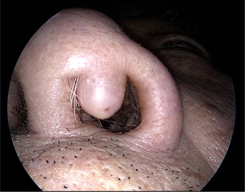
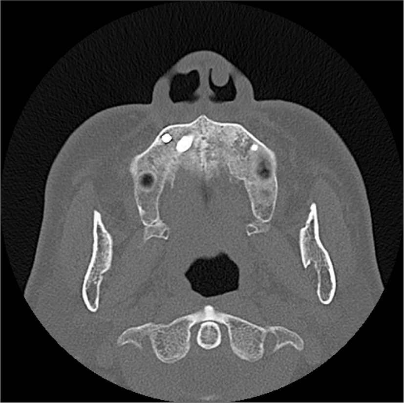
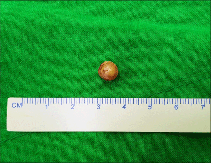
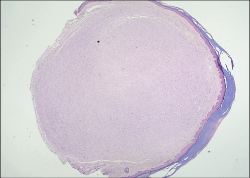
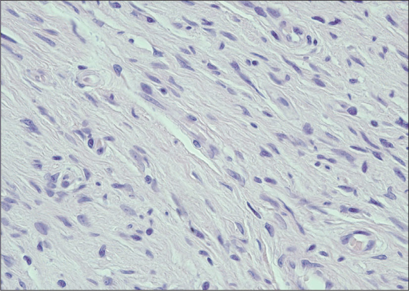
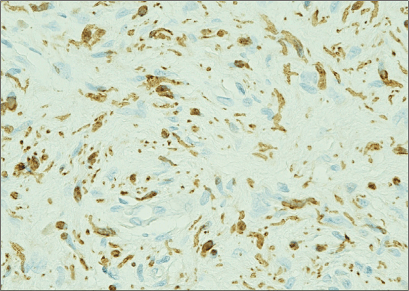
73세 남자가 10년 전 발생한 좌측 비전정 종물을 경과관찰 중 개인의원에서 이에 대한 수술을 권유받고 본과로 의뢰되었다. 기저질환으로 고혈압과 당뇨가 있었고, 가족력상 특이소견은 없었다. 과거력상 2015년 좌측 엄지발가락 타박상으로 입원치료한 것 외에 특이소견은 없었으며, 신체검사상 피부에 밀크커피색 반점이나 국소 임파절 종대소견은 관찰되지 않았다. 비내시경상 좌측 전방 비전정 직경 1 cm의 폴립양 살색 종물이 관찰되었다(Fig. 1). 얼굴뼈 전산화 단층촬영에서 약 8×8×7 mm 크기의 둥근 경계를 가진 연조직 종물이 좌측 비공에서 비강으로 나와있었다(Fig. 2). 우측으로의 비중격 만곡이 관찰되었으며, 비중격이나 비외측벽의 골미란, 골파괴 소견 및 안와나 두개내 침범은 보이지 않았다. 전신 소견은 양호하였으며, 혈액검사상 platelet function test(C/Epi)>300 이외 이상소견은 없었다. 전신마취 하 좌측 전방 비전정 종물에 대해 타원형의 절개 후 절제생검을 시행하였다. 종물의 악성화를 의심할만한 소견은 없었으며, 출혈 부위는 전기적 소작술을 시행하였으며, 제거된 종물을 조직 검사 의뢰하였다(Fig. 3). 단면은 경계가 좋고 연한 노란색으로 점액양이며, 만졌을 때 고무와 같이 딱딱한 소견이었다(Fig. 4). 헤마톡실린에오진 염색 표본에서, 저배율에서는 경계가 좋지만 피막이 없는, 고형성 종괴가 중층편평상피 아래에서 관찰되었다. 고배율에서 병변은 주로 타원 모양이거나 길쭉한 모양의 세포로 구성되어 있었으며, 다량의 아교질과 섞여 있었고, 세포이형성이나 세포분열은 관찰되지 않았다(Fig. 5). 신경능 표지 항원인 S-100단백질은 면역조직화학염색에 양성이었다(Fig. 6). 최종적인 진단은 신경섬유종이었으며, 외래 추적관찰 중 술 후 9개월 동안 비전정 협착 등의 술후 합병증 또는 비내시경 검사상 재발 소견을 보이지 않았다.

jcohns-32-3-261-g1
Fig. 1. It is skin color polypoid mass about 1 cm in diameter in left anterior nasal vestibule.
Download Original Figure
jcohns-32-3-261-g2
Fig. 2. Facial bone CT - Axial view.
Download Original Figure
jcohns-32-3-261-g3
Fig. 3. The mass in nasal vestibule after excision.
Download Original Figure
jcohns-32-3-261-g4
Fig. 4. Hematoxylin and eosin stain reveals an well demarcated unencapsulated polypoid mass overlied by an intact squamous mucosa. Hematoxylin and eosin ×12.5.
Download Original Figure
jcohns-32-3-261-g5
Fig. 5. Schwann cells and fibroblasts are intermixed with collagen. Hematoxylin and eosin ×400.
Download Original Figure
jcohns-32-3-261-g6
Fig. 6. S-100 antibody reacts to lesional Schwann cells. S100×400.
Download Original Figure

고 찰

비부비동관의 양성 말초신경초종양에는 신경섬유종과 신경초종이 있으며, 드물게 발생하지만, 신경섬유종보다는 신경초종이 흔하다. 말초신경에서 유래된 신경섬유종은 비부비동관 내의 다른 곳보다 비전정 주위에서 흔하다.4,5) 남녀사이의 차이는 없으며, 발생 평균 나이는 46세이며, 환자들은 대개 비폐색, 비출혈, 통증을 호소한다. 전반적인 평균 크기는 3.1 cm이다.6,7) 신경섬유종의 발생은 크게 단발성과 다발성으로 나뉜다. 다발성으로 발생하는 경우는 제 1형 신경섬유종증과 제 2형 신경섬유종증이 있다. 신경섬유종의 10%는 신경섬유종증과 관련하여 발생한다고 알려져 있으며, 비강이나 부비동에 발생하는 신경섬유종은 대부분 단발성이며, 비강과 사골동의 동시침범이 가장 흔하다. 다발성의 경우에는 다수의 밀크커피색반점과 피부나 피하에 생기는 신경섬유종이 특징적이며, 거의 대부분 가족력이 있다.8) 신경섬유종은 코막힘, 비출혈 등의 증상을 나타낼 수 있으며, 부비동을 침범한 경우 협부의 종창, 압통 등의 비특이적 증상을 나타낸다. 특히 부분적 악성화가 되었을 경우에도 안면 종창이나 통증을 유발할 수 있으므로 진단시 주의해야 한다.4) 본 증례 환자는 커피색 반점이나 피부나 피하에 신경섬유종이 관찰되지 않았고, 가족력도 없었으며, 별다른 증상 호소는 없었다.

악성화에 대한 명확한 배제를 위해서는 면역조직화학검사를 포함한 조직학적 검사 및 완전 절제 후의 소견을 포함한 총체적인 조직검사가 필요하다.9) 비부비동 원발 양성 종양에 대해 신경초종과 신경섬유종을 감별하는 것은 중요하다.1) 신경초종이 단발성이고 피막에 잘 싸여 있으며, 개개의 신경섬유가 실질을 통과하지 않고, 종물을 싸고 있는 형태로 악성변화가 거의 없고, 출혈이나 낭포성 변화를 보이는 경우가 많다. 반면 신경섬유종은 다발성이 흔하고 피막형성이 불확실하며, 개개의 신경섬유가 종물내를 통과하므로 박리가 더욱 어렵다. 또한 12%의 악성화 변화가 보고되며, 퇴행성 변화가 드물어 무증상이 많다.10,11) 신경섬유종은 신경초종과 달리 피막이 없어 침습성이 강하기 때문에 외과적 완전 절제가 반드시 필요하며, 국소 절제시 재발률이 높다.12) 동통을 동반하거나 방사선 검사에서 악성화가 의심될 경우에는 더욱 광범위한 수술이 필요하다.1) 수술로 인한 반흔에 의해 비전정 협착(nasal vestibular stenosis)이 유발된다13)고 알려져 있어, 특히 연조직 삼각(soft tissue triangle) 부분에 유의해야 한다. 비중격과 측하방 비익연골에 비해, 측상방 비익연골의 지지력이 부족하기 때문에, 비전정 협착은 비전정의 측상방에서 대개 시작한다.14) 비전정의 내벽이 손상되면 2차적으로 육아조직 및 섬유조직이 증식하면서 비전정 협착이 유발14,15)하며, 부분적 또는 윤상의 반흔이 추가적인 수축력을 만들고, 비익연골의 약한 부분을 비틀리게 한다.14) 조직 손실과 반흔으로 인한 수축력 이외에도 흡기 때 발생하는 일정한 음압이 손상된 비전정을 더욱 수축하게 한다16)고 알려져 비전정 수술시에는 주의를 기울여야 하고, 수술 후에도 합병증 발생 여부를 확인해야 한다. 조직학적으로는 신경초종에서 Verocay body가 관찰되고, Antoni A type, Antoni B type 조직 소견을 보이고, 신경섬유종에서 신경초 세포, 신경섬유, 섬유아세포가 관찰된다.17-20) 본 증례의 경우 육안적으로 모두 절제하였지만, 신경섬유종 특성상 피막이 없어 재발가능성을 완전히 배제할 수 없고, 재발과 비전정 협착 등의 합병증 발생 여부에 대한 추가적인 추적관찰이 필요할 것으로 생각된다.

Acknowledgements

Not applicable.

Funding Information

Not applicable.

Conflicts of Interest

No potential conflict of interest relevant to this article was reported.

Author Contribution

Conceptualization: Kim YW.

Data curation: Kim BS, Yi KI.

Methodology: Kim BS, Kim YW.

Investigation: Kim B, Kim BS.

Writing - original draft: Kim BS, Yi KI, Kim YW.

Writing - review & editing: Kim BS, Yi KI, Kim YW, Kim B.

Ethics Approval

Not applicable.

References

1).

Noh H, Park YJ. A neurofibroma arising from the nasal septum: a case report. J Clin Otolaryngol Head Neck Surg 2006; 17(2):261-5.

2).

Donnelly MJ, Al-Sader MH, Blayney AW. Benign nasal schwannoma. J Laryngol Otol 1992;106(11):1011-5.

3).

Jeong JH, Keum HS, Kim KR, Park YW. Solitary neurofibroma of the nasal cavity: transnasal endoscopic excision. Korean J Otolaryngol Head Neck Surg 2005;48(9):1169-72.

4).

Perzin KH, Panyu H, Wechter S. Nonepithelial tumors of the nasal cavity, paranasal sinuses and nasopharynx. A clinicopathologic study. XII: Schwann cell tumors (neurilemoma, neurofibroma, malignant schwannoma). Cancer 1982;50(10): 2193-202.

5).

Azani AB, Bishop JA, Thompson LDR. Sinonasal tract neurofibroma: a clinicopathologic series of 12 cases with a review of the literature. Head Neck Pathol 2015;9(3):323-33.

6).

Kuroda N, Kazakov DV, Hes O, Michal M, Goda M, Miyazaki K, et al. Hybrid peripheral nerve sheath tumor of the nasal cavity showing schwannomatous, neurofibromatous, and perineuriomatous areas. Med Mol Morphol 2010;43(2): 82-5.

7).

Biswas D, Mal R. Bilateral solitary maxillary sinus neurofibroma. Ear Nose Throat J 2010;89(1):E1-2.

8).

Frosch MP, Anthony DC, DeGirolami UL. The central nervous system. In: Kumar V, Abbas AK, Fausto N, editors. Robbins and cotran pathologic basis of disease. 7th ed. Philadelphia, PA: Elsevier Saunders; 2005. p. 1347-419.

9).

Perzin KH, Pushparaj N. Nonepithelial tumors of the nasal cavity, paranasal sinuses, and nasopharynx: a clinicopathologic study. XIV: chordomas. Cancer 1986;57(4):784-96.

10).

Seo YI, Nam SY, An KH, Kim SY, Lee KS. Extracranial nerve sheath tumors of the head and neck. Korean J Otolaryngol Head Neck Surg 1997;40(6):908-13.

11).

Gooder P, Farrington T. Extracranial neurilemmomata of the head and neck. J Laryngol Otol 1980;94(2):243-9.

12).

Lee KC, Lee SC, Jin SM, Lee YB. A neurofibroma arising from the nasal septum. Korean J Otolaryngol Head Neck Surg 1998;41(6):795-8.

13).

Yoon BW, Kim DW, Choi SJ, Cho KS. Latrogenic nasal vestibular stenosis after maxillofacial reconstructive surgery. Braz J Otorhinolaryngol 2018;84(1):126-30.

14).

Daines SM, Hamilton GS III, Mobley SR. A graded approach to repairing the stenotic nasal vestibule. Arch Facial Plast Surg 2010;12(5):332-8.

15).

Cagici CA, Karabay G, Yilmazer C, Gencay S, Cakmak O. Electron microscopy findings in the nasal mucosa of a patient with stenosis of the nasal vestibule. Int J Pediatr Otorhinolaryngol 2005;69(3):399-405.

16).

Egan KK, Kim DW. A novel intranasal stent for functional rhinoplasty and nostril stenosis. Laryngosc 2005;115(5):903-9.

17).

Wilson JA, Mclaren K, Mclntyre MA, Von Haccje NP, Maran AG. Nerve sheath tumors of the head and neck. Ear Nose Throat J 1988;67:103-10.

18).

Coffin CM, Dehner LP. Cellular peripheral neural tumors (neurofibromas) in children and adolescents: a clinicopathological and immunohistochemical study. Pediatr Pathol 1990;10(3):351-61.

19).

Griffith BH, Lewis VL Jr, McKinney P. Neurofibromas of the head and neck. Surg Gynecol Obstet 1985;160(6):534-8.

20).

Karayianis SL, Francisco GJ, Schumann GB. Clinical utility of head and neck aspiration cytology. Diagn Cytopathol 1988;4(3):187-92.