


Introduction
Congenital external auditory canal (EAC) stenosis occurs as an abnormality of the first gill cleft, and acquired EAC stenosis can be caused by trauma, inflammation, or radiation therapy. They share a common pathophysiology: a process of inflammatory changes leading to medial canal fibrosis.1) EAC stenosis can result in severe hearing loss, otorrhea, pain, and chronic mastoiditis.2,3) In particular, restenosis is a common complication of EAC stenosis surgery, occurring in up to 30% of patients.3,4) Management of EAC stenosis is difficult as it is often associated with residual hearing loss and restenosis. Many otologists have used various EAC packaging materials to reduce the incidence of postoperative stenosis (or restenosis).5) Herein, we report a case of effective prevention of restenosis after EAC stenosis surgery using a customized earmold stent.
Case Presentation
A 17-year-old female was referred to a tertiary referral hospital for suspicion of EAC stenosis and hearing loss from 2 months prior. She had experienced a left orbital wall fracture, a mandibular fracture, and a right humeral shaft fracture in a car accident 2 months previously. On endoscopic examination, the patient showed complete obstruction of the cartilaginous EAC (Fig. 1A).
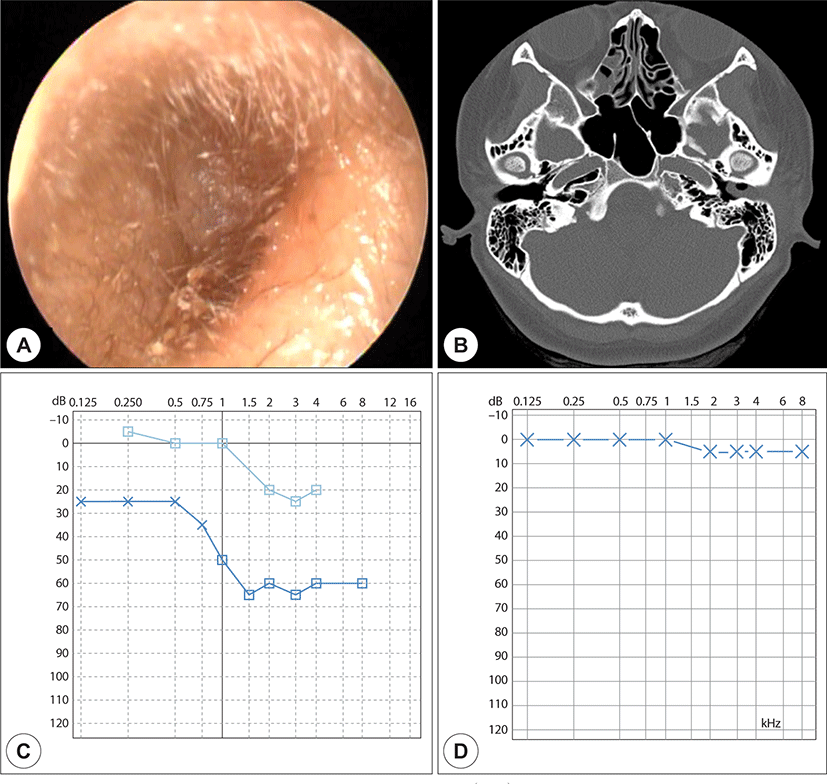
Her conductive hearing loss in 40 dB air-bone gaps was assessed using pure tone audiometry (Fig. 1C, D). High-resolution temporal bone computed tomography (TBCT) scan showed soft tissue density in the EAC (Fig. 1B). She underwent meatoplasty and canaloplasty with a retroauricular approach to resolve the EAC stenosis. The EAC was blocked with granulation tissue, and we widened the bony EAC using a drill after removing all granulation tissue. Split-thickness skin of the retroauricular lesion was harvested and grafted onto the EAC to correct a skin defect. The tympanic membrane was intact, and histological examination of the resected tissue revealed chronic inflammation with fibrosis. We tried to prevent synechia formation using Vaseline gauze packing for 1 month after surgery. The vaseline gauze was changed 2 weeks after surgery, and there were no other signs of infection during 1 month. However, after removal of the Vaseline gauze packing, the EAC narrowed again due to restenosis. We tried to maintain EAC patency using Merocel (Medtronic, Minneapolis, MN, USA) packing, but again, gradual narrowing occurred (Fig. 2A).
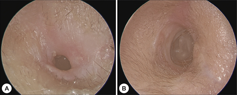
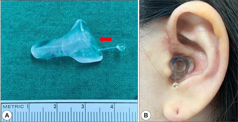
Finally, one year after the first surgery, meatoplasty was performed to resolve the restenosis. Vaseline gauze packing was maintained for 1 month after the second surgery, and an earmold stent was fabricated as an additional treatment plan to prevent restenosis. The earmold stent was accomplished using a custom-made heat-cured acryl resin with a large bore (Fig. 3). The patient wore an earmold stent for 2 months, and the EAC patency was well maintained for more than 1 year without restenosis or complications (Fig. 2B).
Discussion
Acquired EAC stenosis is very rare, with an incidence of only 0.6 cases per 100,000 people.2) EAC stenosis has a number of different causes, including trauma, infection, neoplasia, inflammation, and radiation therapy. Posttraumatic stenosis is extremely rare, accounting for approximately 10% of cases.6) There is no gold standard for maintaining patency of medical and surgical treatment of EAC stenosis. Although various methods have been reported, the progression of stenosis or restenosis remains a major problem. The etiology of EAC stenosis is diverse, but a common feature of pathogenesis is the fibroproliferative inflammatory response.7) Medical treatment is recommended when the inflammatory response is actively progressing. However, when the inflammatory response is terminated and the stenosis region is mature, surgery should be performed.6) During surgery, removal of all fibrotic tissue in the stenosis region is very important, and additional canaloplasty, meatoplasty, and split-thickness skin grafting are often performed.8) After surgery, otologists need to make efforts to reduce the incidence of restenosis. Often, surgery alone cannot prevent restenosis, and in these cases, postoperative stent maintenance can help to reduce the incidence of restenosis. In a recently study, long-term maintenance of the stent after surgery reduced the recurrence rate of restenosis,9) and new materials that improved the problem of traditional stent materials were introduced. Various stents such as cotton balls, gelatin sponge (Gelform), Vaseline gauze, expending cellulose wick, silicon dental impression, and acryl resin are used to maintain EAC patency. Materials such as cotton balls, Gelform, and gauze must be tightly packed to maintain EAC patency, and periodic changes are required during maintenance. It is also accompanied by conductive hearing loss, obstructive effects, and ventilation disorders. A customized earmold stent made with a dental impression can be manufactured quickly and inexpensively, but is soft and difficult to maintain for a long time.
This report introduces an interesting case in which EAC stenosis caused by trauma was successfully treated using a customized acryl resin earmold stent after surgery. This acryl resin earmold stent is inexpensive and easy to manufacture, and it is sufficiently firm to maintain EAC patency. The presence of a hole allows the reduction of conductive hearing loss, obstruction effect, and ventilation disturbance. Moreover, it can be used for a long time without discoloration, because the patient can insert and remove the stent for easy hygiene management. Although the available literature on the use of acrylic resin earmold stent is very limited, there are no reports of serious complications. Therefore, the use of a customized acryl resin earmold stent after surgical treatment of EAC stenosis is a treatment option for successful surgical outcomes.