증례

비중격의 연소성 황색육아종: 증례 보고

김효준1, 안유영1, 김민주2, 최지호1,*
Hyo Jun Kim1, You Young An1, Min Ju Kim2, Ji Ho Choi1,*
Author Information & Copyright
1순천향대학교 의과대학 부천병원 이비인후-두경부외과
2병리과
1Department of Otorhinolaryngology-Head and Neck Surgery, Bucheon, South Korea
2Pathology, Soonchunhyang University College of Medicine, Bucheon Hospital, Bucheon, South Korea
*교신저자: 최지호, 14584 경기도 부천시 조마루로 170, 순천향대학교 부천병원 이비인후-두경부외과 전화: (032) 621-5015·전송: (032) 621-5016·E-mail: handsomemd@hanmail.net

© Copyright 2021 The Busan, Ulsan, Gyeoungnam Branch of Korean Society of Otolaryngology-Head and Neck Surgery. This is an Open-Access article distributed under the terms of the Creative Commons Attribution Non-Commercial License (http://creativecommons.org/licenses/by-nc/4.0/) which permits unrestricted non-commercial use, distribution, and reproduction in any medium, provided the original work is properly cited.

Received: Sep 14, 2020; Revised: Oct 20, 2020; Accepted: Dec 11, 2020

Published Online: Jun 30, 2021

ABSTRACT

Juvenile xanthogranuloma is a rare benign lesion that occurs most frequently in infants and children. Histologically, juvenile xanthogranuloma represents an accumulation of non-Langerhans cell histiocytes. Its clinical condition is various but usually presented with cutaneous lesions. The treatment of juvenile xanthogranuloma is usually the observation due to the self-limiting nature but sometimes surgical excision is needed to treat cosmetic or symptomatic problems. We report a case of juvenile xanthogranuloma arising from the nasal vestibule. (J Clinical Otolaryngol 2021;32:75-79)

Keywords: 연소성 황색육아종; 비전정; 병리
Keywords: Juvenile xanthogranuloma; Nasal vestibule; Pathology

서론

연소성 황색육아종(Juvenile xanthogranuloma)은 드물게 발생하는 양성의 조직구 증식성 질환으로 대부분 유소아에서 발생하며, 자연 퇴화하는 특징을 가지고 있다.1) 주요 장기의 침범 없이 두경부, 체간의 피부에서 주로 호발하나, 드물게 중추신경계, 간, 비장, 심장, 근육 등 피부외 기관에서도 발생이 된다.2,3) 그 중에서도 비전정에서 기원한 연소성 황색육아종은 국내 논문에서는 소수의 보고만 있을 정도로 매우 드문 종양이다.4) 최근 저자들은 2세 남아에서 아주 드문 부위인 비전정에서 발생한 연소성 황색육아종의 진단 및 치료를 경험하였기에 문헌 고찰과 함께 이에 대해 보고하고자 한다.

증례

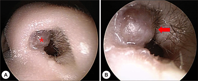
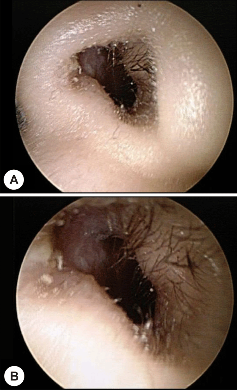
2세 남아가 내원 2개월 전부터 발견된 좌측 비강 종물을 주소로 본원 이비인후과에 내원하였다. 특이 과거력, 가족력, 두부외상 및 비수술의 병력은 없었다. 비강 종물 이외에 증상은 없었으며, 비강 내시경 검사에서 좌측 비강 입구 부위에 무통성의 부드럽고 경계가 분명한 0.5×0.5 cm 크기의 융기된 종물이 관찰되었다(Fig. 1A, B).

jcohns-32-1-75-g1
Fig. 1. Preoperative endoscopic findings. Endoscopic finding of left nostril shows bulging contoured mass like lesion originating from left nasal vestibule (marked with asterisk) (A). Smooth surface was observed and it was confirmed as juvenile xanthogranuloma (marked with arrow head) (B).
Download Original Figure

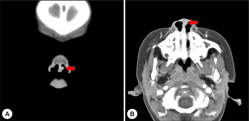
부비동 전산화 단층 촬영에서 좌측 비전정 부분에 명확한 경계를 가지며, 균질하게 조영 증강되는 폴립모양의 0.5×0.5 cm의 종물이 관찰되었다(Fig. 2A, B). 부비동 및 비갑개 등에서 특별한 이상 소견은 관찰되지 않았고, 혈액검사도 정상 범위였다.

jcohns-32-1-75-g2
Fig. 2. Contrast enhanced coronal paranasal CT image shows the left nasal vestibular mass. There is about 0.5×0.5 cm sized protruding mass like lesion originates from nasal vestibule (marked with arrow head) (A). Contrast enhanced axial paranasal CT image shows enhancing polypoid mass (marked with arrow head) (B).
Download Original Figure

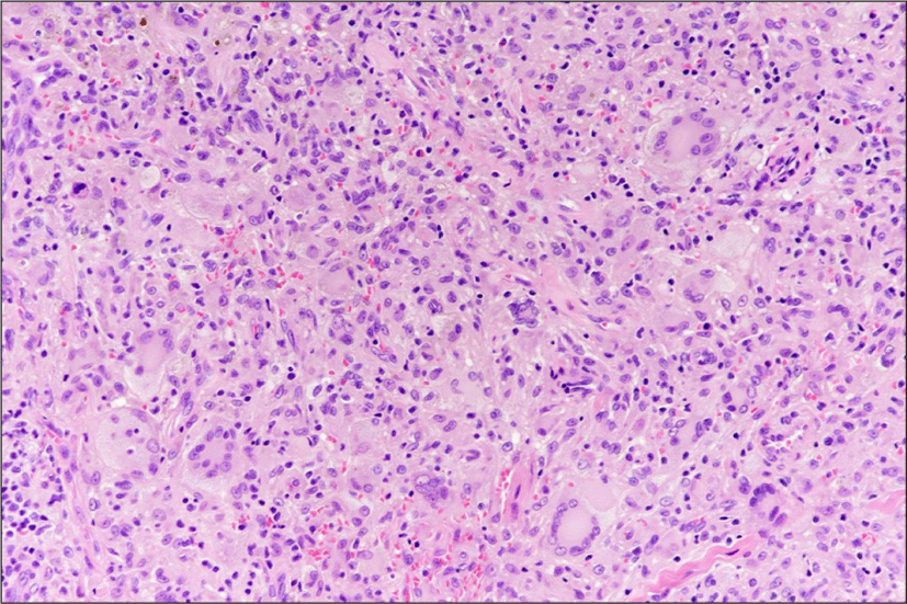
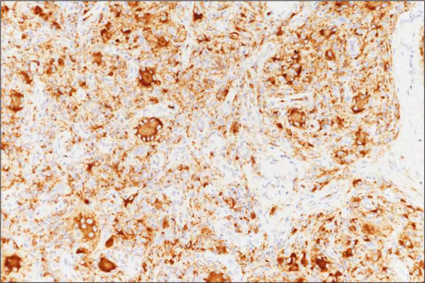
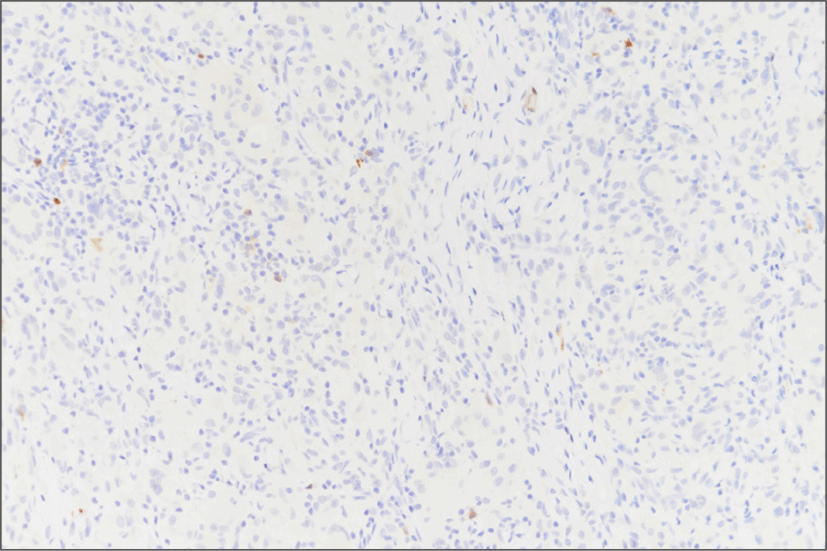
신체 진찰과 영상 소견을 통해 양성 종양이 의심되어 전신마취 하에 비내시경을 이용하여 절제 생검을 시행하였다. 수술 시작 후 좌측 비전정 전단에 직경 약 0.5×0.5 cm 크기의 부드러운 표면을 가진 종물이 보였고, 비전정 점막에서 기원하는 모습이 관찰되어 조심스럽게 종물을 제거하었다. 술 후 확인한 종물의 내부는 지방종과 유사한 소견을 보였다. 술 후 병리조직학적 검사에서 점막 상피하에 미만성으로 염증세포의 침윤이 있고, 특징적으로 대식세포와 뭇핵대식세포가 관찰되었으며, 림프구와 호산구가 동반되어 있었다(Fig. 3). 면역조직화학염색에서 CD68 양성, S-100 protein 음성의 소견을 보여 연소성 황색육아종으로 최종 진단하였다(Fig. 4, Fig. 5).

jcohns-32-1-75-g3
Fig. 3. A diffuse infiltrate of macrophages and multinucleated giant cells, is accompanied by a mixed infiltrate of lymphocytes and eosinophils (H&E, x200). Tuoton giant cells are marked in round red circles.
Download Original Figure
jcohns-32-1-75-g4
Fig. 4. Immunohistochemistry (IHC) images for CD68 positive finding at ×200 magnification.
Download Original Figure
jcohns-32-1-75-g5
Fig. 5. Immunohistochemistry (IHC) images for S100 negative finding at ×200 magnification.
Download Original Figure

3개월 동안 외래 경과관찰한 결과, 환자는 특별한 증상을 호소하지 않았으며, 국소 재발 소견은 관찰되지 않았다(Fig. 6).

jcohns-32-1-75-g6
Fig. 6. Postoperative endoscopic findings. Vestibular mass is completely removed (A). There is no deformity after opearation (B).
Download Original Figure

고찰

연소성 황색 육아종은 드문 양성 조직구증으로서 1905년 Adamson에 의해 선천성 다형성 황색육아종 (Congenital xanthoma multiplex)으로 최초로 기술된 후 종양의 기원이 지방포함 조직구라는 소견을 바탕으로 연소성 황색 육아종(juvenile xanthogranuloma)으로 최종 명명되었다.5,6) 비랑거한스세포 조직구증(non-Langerhans cell histiocytosis)인 class II histiocytosis로 분류되며, 유병률은 명확히 알려져 있지 않으나, 주로 영유아, 특히 1세 미만의 남아에서 호발하는 경향을 보인다.1,4)

정확한 병리 기전에 대해 밝혀진 바가 없으나, 원인 미상의 외상이나 염증에 의한 대식세포와 조직구의 육아종성 반응으로 고려되고 있으며, 제 1형 신경섬유종증 인슐린 의존성 당뇨병, 연소성 만성 골수성 백혈병과의 연관성도 보고되고 있다.7) 다른 황색종과는 다르게 과다지방증이나 요붕증과 같은 전신적인 질환과는 관련이 없는 것으로 알려져 있다.1)

가장 흔하게 나타나는 임상 증상은 두경부, 체간과 사지의 피부에 경계가 명확하고, 둥근 단발성 혹은 다발성으로 관찰되는 다양한 크기의 무통성 결절이다.6,8) 피부 이외의 장기로는 안구에서 가장 호발하며, 폐, 심장, 소화기, 중추신경계, 신장 등에도 발생한다.2,3) 두경부 영역에서는 드물게 비인두, 구강 등에서 발견되기도 하나, 비전정에서의 발생은 아주 드문 것으로 알려져 있다.4)

연소성 황색육아종은 감별 진단 및 미용적 치료를 위해 절제 생검 후 병리조직학적 검사와 면역조직화학염색으로 확진한다.1,5,6) 조직학적으로는 초기에는 염증세포의 침윤이 많이 관찰되지 않으나, 시간이 흐를수록 조직구, 호산구 및 림프구 등의 침윤을 보이며, 거품이 많은(foamy) 또는 호산성 세포질에 의해 둘러싸인 소용돌이 모양의 핵을 보이는 Touton형 거대세포가 특징적으로 관찰된다.5,6,9) 면역조직화학염색상 침윤된 조직구는 랑거한스세포의 특이성이 있는 marker인 OKT6(CD1), S-100 단백은 음성이며 조직 대식세포의 marker인 HAM56, factor XIIIa, CD 68에는 양성이다.6,10)

감별해야 할 질환들로는 황색종(xanthoma), 혈관종(hemangioma), 전염성 연속종(molluscum contagiosum), 랑거한스세포 조직구증(Langerhans cell histiocytosis), 신경섬유종(neurofibromatosis) 등이 있으며, 이러한 질환들의 감별은 면역조직화학염색으로 가능하다.3,6)

연소성 황색육아종은 자연관해되는 것이 대부분으로 주기적인 경과관찰로 충분하나, 병변이 오래 지속되거나 미용적으로 문제가 될 경우, 악성을 감별하고 정확한 진단과 치료를 위해 수술을 시행할 수 있다.1,4) 안구나 중추신경계 등의 주요 장기를 침범한 경우에는 항암치료나 방사선 치료 또는 병합 요법을 시행하기도 하나, 그 효과에 대해서는 아직 불분명하다.10) 재발은 드물고 예후도 좋은 편이나, 전신적으로 발생할 경우에는 치명적일 수 있으므로 주기적인 경과관찰이 필요하다.4,7)

본 증례에서는 좌측 비전정의 전반부 종물을 주소로 내원한 2세 남아에서 신체 진찰, 영상 검사 및 혈액 검사상 특이 소견이 없는 것을 확인하였으며, 진단 및 치료를 목적으로 시행한 절제 생검 후 병리조직학적 검사와 면역조직화학 염색을 통해 연소성 황색육아종을 확진할 수 있었다. 수술 후 외래에서 시행한 내시경 검사 결과, 좌측 비전정의 종물은 관찰되지 않아 더 이상의 검사나 치료를 시행하지 않았으며, 현재 정기적으로 경과관찰 중이다. 따라서, 비전정 전방 부위에서 발생한 종물을 주소로 내원한 소아의 경우 드물지만 연소성 황색육아종의 가능성을 염두에 두어야 할 것으로 사료된다.

REFERENCES

1).

Lee YW, Park YH. Juvenile xanthogranuloma in the external auditory canal. Korean J Otorhinolaryngol-Head Neck Surg 2010;53(9):571-3.

2).

Cichewicz A, Białecka A, Męcińska-Jundziłł K, Adamska U, Białecki M, Neska-Długosz I, et al. Congenital multiple juvenile xanthogranuloma. Postepy Dermatol Alergol 2019;36(3):365-8.

3).

Cypel TKS, Zuker RM. Juvenile xanthogranuloma: case report and review of the literature. Can J Plast Surg 2008;16(3):175-7.

4).

Suh SH, Son JY, Kim BM, Kim CK, Je JS. A case of the juvenile xanthogranuloma of the nasal cavity and testis. Korean J Otorhinolaryngol-Head Neck Surg 2001;44(9):997-1000.

5).

Sonoda T, Hashimoto H, Enjoji M. Juvenile xanthogranuloma. Clinicopathologic analysis and immunohistochemical study of 57 patients. Cancer 1985;56(9):2280-6.

6).

Raj D, Rathy R. Juvenile xanthogranuloma: a case report and review of the literature. Int J Odontostomat 2018;12(3):327-31.

7).

Lee SY, Jang YS, Kim MS, Yang YJ, Jung MJ, Hwang PH. A case of multiple juvenile xanthogranuloma involving skin and systemic organs. Clin Exp Pediatr 2004;47(2):227-30.

8).

Margulis A, Melin-Aldana H, Bauer BS. Juvenile xanthogranuloma invading the muscles in the head and neck: clinicopathological case report. Ann Plast Surg 2003; 50(4):425-8.

9).

Kim SW, Han TY, Lee JH, Son SJ. A case of juvenile xanthogranuloma in the nasal cavity of a neonate. Indian J Dermatol 2013;58(5):411.

10).

Freyer DR, Kennedy R, Bostrom BC, Kohut G, Dehner LP. Juvenile xanthogranuloma: forms of systemic disease and their clinical implications. J Pediatr 1996;129(2):227-37.