증례

비익에 발생한 망상조직구증 1예

문지승1,*, 구수권1, 박근형1, 이화선2
Ji Seung Moon1,*, Soo Kweon Koo1, Geun Hyung Park1, Hwa Sun Lee2
Author Information & Copyright
1부산성모병원 이비인후과
2병리과
1Department of Otorhinolaryngology-Head and Neck Surgery, Busan, Korea
2Department of Pathology, Busan Saint Mary’s Hospital, Busan, Korea
*교신저자: 문지승, 48575 부산광역시 남구 용호로 232번길 25-14, 부산성모병원 이비인후과 전화: (051) 933-7214·전송: (051) 956-1956·E-mail: zslune@naver.com

© Copyright 2020 The Busan, Ulsan, Gyeoungnam Branch of Korean Society of Otolaryngology-Head and Neck Surgery. This is an Open-Access article distributed under the terms of the Creative Commons Attribution Non-Commercial License (http://creativecommons.org/licenses/by-nc/4.0/) which permits unrestricted non-commercial use, distribution, and reproduction in any medium, provided the original work is properly cited.

Received: Sep 12, 2020; Revised: Oct 12, 2020; Accepted: Nov 11, 2020

Published Online: Dec 31, 2020

ABSTRACT

Reticulohistiocytosis is a rare form of histiocytosis that can affect the skin and other organs. There are two distinct forms of reticulohistiocytosis, reticulohistiocytoma and multicentric reticulohistiocytosis. Reticulohistiocytoma is a cutaneous condition characterized by a solitary, firm, dermal skin lesion of less than 1cm in diameter. It can be diagnosed by histopathologic examination and immunohistochemical stains. It can be treated with simple resection, and recurrence is rare. In this article, the authors experienced a case of single reticulohistiocytoma that occurred in the nasal vestibule and report it with literature review. (J Clinical Otolaryngol 2020;31:238-242)

Keywords: 망상조직구증; 비전정
Keywords: Reticulohistiocytoma; Nasal vestibule

서론

망상조직구증(Reticulohistiocytosis)은 드물게 발생하는 피부 또는 연조직의 양성 조직구 증식 질환으로, 1950년 Zak에 의해 처음 기술되었다.1) 피부에 발생하는 피부 망상조직구증은 주로 단발성으로 두경부 영역에 호발하며, 무증상인 경우가 많다. 피부 망상조직구증의 병인은 잘 알려져 있지 않지만, 사이토카인 유발 조직구(cytokine-induced histiocytes)의 국소화를 초래하는 알려지지 않은 염증 과정으로 인해 발생할 수 있다고 제안되고 있다.2) 대부분 직경이 1 cm를 넘지 않으며, 황색 또는 적갈색의 결절이나 구진 형태로 나타난다.2) 병리조직학적 검사와 면역조직화학 검사를 통해 진단할 수 있으며, 단순 절제술 만으로 치료가 가능하고, 재발하는 경우는 흔하지 않다. 국내 문헌에는 7례가 보고되었으며, 손가락3), 두피4), 흉부5), 이주6), 상순부7), 비전정8), 손바닥9) 순으로 보고되었다. 이에 저자들은 비전정에서 발생한 단발성 피부 망상조직구증 1예를 경험하였기에 문헌고찰과 함께 보고하는 바이다.

증례

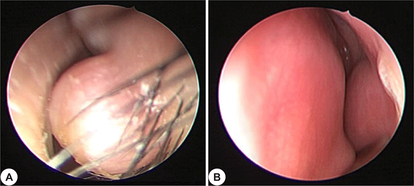
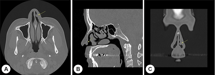
45세 여자 환자가 코막힘을 주소로 내원하였다. 당뇨 이외의 과거력 및 가족력상 특이 소견은 없었다. 이학적 검사상 좌측 내측 비전정의 피부에 둥그런 모양의 단단한 양상의 종괴가 관찰되었고(Fig. 1A), 코막힘 호소 외에 통증 등의 자각증상은 없었다. 혈액검사, 요검사, 흉부 X-ray 검사에서 이상 소견은 없었다. 부비동 컴퓨터 단층 촬영(O.M.U CT)상 비전정에서 0.6×0.5×0.6 cm 크기의 경계가 뚜렷한 종괴를 확인할 수 있었으며(Fig. 2), 국소마취하 절제생검을 시행하였다.
jcohns-31-2-238-g1
Fig. 1. Pre- and post-operative (after 6 months) nasoscopic findings : (A) round, well demarcated and solid mass on left nasal vestibule, (B) there was no recurrence and excision site healed well.
Download Original Figure
jcohns-31-2-238-g2
Fig. 2. Non-enhanced computed topographic findings of (A) axial, (B) sagittal and (C) coronal view : about 6×5×6 mm sized, well-defined, isodense nodule at lateral side of left nasal vestibule.
Download Original Figure

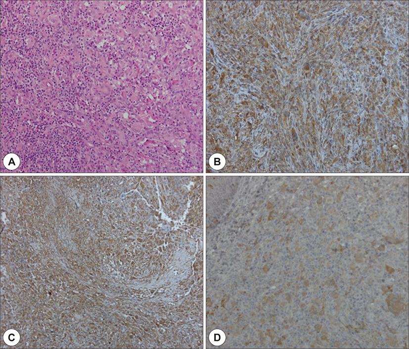
현미경 소견상 비교적 경계가 좋은 진피에 국한된 결절성 병변이 관찰되었다. 소포성(vesicular)의 단일 핵을 가진 다수의 조직구들과 소수의 다핵 거대세포가 존재하는 것을 확인할 수 있었고, 일부에서는 중성구, 호산구, 림프구 등의 염증세포가 침윤되어 있는 소견을 보였다(Fig. 3A). 조직구는 호산성을 띠는 젖빛유리(ground glass) 양상의 풍부한 세포질을 보였고, 이형성(atypia)이나 분열기세포(mitotic cell)는 확인되지 않았다. 면역조직화학염색 시행 결과, CD68(Fig. 3B), vimentin(Fig. 3C), S-100(Fig. 3D)에 양성 소견을 보였다.
jcohns-31-2-238-g3
Fig. 3. Microscopic findings of the lesion (×200 magnification) : (A) H&E staining shows a nodular infiltration of numerous large mononucleated or multinucleated histiocytes, admixed with lymphocytes, neutrophils and eosinophils in dermis. histiocytes show no nuclear atypia, no mitotic activity and dense ground glass pink cytoplasm. Immunohistochemical staining shows (B) CD 68, (C) Vimentin and (D) S-100 all positive.
Download Original Figure

조직학적 소견상 망상조직구증으로 진단 받았으며, 절제생검을 시행한지 6개월이 지나기까지 재발 및 합병증 등의 특이소견을 보이지 않고 있다(Fig. 1B)

고찰

망상조직구증은 비랑게르한스세포성 조직구증(non-langerhans cell histiocytosis)에 속하는 질환군으로, 피부와 다른 기관에 영향을 줄 수 있는 흔하지 않은 형태의 조직구증이다. 피부에만 국한하여 발생하는 단발성 및 다발성 피부 망상조직구증과, 내부장기를 비롯한 온몸을 침범하는 다중심성 망상조직구증의 2가지 형태가 있다.7) 피부 망상조직구증은 대부분 단발성으로 발생하며, 신체의 어느 부분에도 발생할 수 있지만, 두경부 영역을 포함한 신체의 상부에 주로 발생한다. 다중심성 망상조직구증이 전신적인 질환 또는 관절염 등을 동반하여 얼굴이나 신체의 말단 부위에 다발성 피부 조직구성 병변으로 나타나는데 반해, 피부 망상조직구증은 전신적인 질병을 동반하지 않는다.10) 피부 망상조직구증은 대개 경계가 뚜렷하고 융기된 1.5~11 mm 정도의 피부 표면의 결절로 나타나며, 보통 무증상이나 작열감 등을 보이기도 한다.11) 피부 망상조직구증의 병인은 아직 불분명하지만, 조직구가 알려지지 않은 염증 자극에 반응하는 비신생물 반응성 과정에 의한 것으로 생각되며,10) 국소 외상 등 물리적 자극에 대한 과민 반응이 발병에 관여하는 것으로 추정된다.7)

피부 망상조직구증의 병변은 다양한 수의 림프구 및 호중구와 함께 큰 상피성 조직구(epitheloid histiocytes)로 구성되어 있다.11) 병리조직학적으로 젖빛유리 양상의 풍부한 호산성 세포질을 가진 거대 다핵세포와 크고 균일한 조직구의 진피 침윤을 특징으로 하며, 관련된 림프구, 호산구, 호중구, 형질세포 등의 염증성 침윤 또한 동반될 수 있다.12) 조직구의 유사 분열 활성(mitotic activity)은 낮으며(넓은 고배율 시야 10개당 0~4개의 유사 분열), 병변은 다양한 수의 CD3-양성 T 세포를 포함한 반면, B 림프구, 혈장 세포, 비만 세포 등은 거의 존재하지 않는다.12)

피부 망상조직구증은 면역조직화학 검사에서 대개 중간엽 표지자(mesenchymal differentiation marker)인 vimentin 및 조직구 표지자인 CD68, CD163에는 양성 반응을 보이며, 일부에선 진피 수지상 세포의 표지자인 S-100, Factor XIIIa에도 양성을 보이고, 랑게르한스세포 표지자인 CD1a에는 음성을 나타낸다.11) 각질형성세포나 특정 멜라닌세포 표지자인 cytokeratin 또는 Melan-A(MART-1), HMB-45, and tyrosinase의 경우 일률적으로 표현되지는 않는다.13)

조직학적으로 로사이-돌프만병(Rosai-Dorfman disease), 연소성 황색육아종(juvenile xanthogranuloma), 다양한 육아종성 질환 등의 양성 질환과 조직구 육종(histiocytic sarcoma), 흑색종(melanoma), 상피모양육종(epithelioid sarcoma) 등의 악성질환들과 감별이 필요하다.11) 로사이-돌프만병의 경우, 다발성 림프절 종대를 보이는 원인 미상의 조직구 증식 질환이다. 조직학적으로 림프구, 적혈구, 형질세포 등이 조직구에 탐식되어 조직구의 세포질 내에서 이들이 관찰되는 소견인 ‘emperiopolesis’가 특징적이며, H&E stain으로도 확인 가능하지만 S-100 염색을 통해 조직구의 세포질과 핵을 염색하면 더 두드러지게 관찰된다. 또한 면역조직화학염색상 vimentin, CD68에는 양성을 보이며, CD1a에는 음성을 보이기 때문에 랑게르한스세포성 조직구증과는 구별된다.14,15) 연소성 황색육아종의 경우, 비랑게르한스세포성 조직구증 중에서 가장 흔하며 두경부나 체간에 주로 발생한다. 대개 1세 미만의 남아에서 나타나고, 두경부와 상지를 포함하여 모든 피부에서 발생 가능하고, 피부외 병변과 동반될 수도 있다.16) 조직학적으로는 다수의 조직구들이 진피에 침윤하여 뚜렷한 경계를 이루며, 주변에 림프구, 형질세포, 호산구를 동반한 소견을 보인다. 특징적인 다핵의 거대 세포(multinucleated giant cell; Touton cell)의 침윤이 85% 가량에서 확인된다. 연소성 황색 육아종 또한 비랑게르한스성 조직구증으로써, 면역조직화학염색상 CD68, vimentin, Ki-M1P, anti F XIIIa, anti-CD4 등에 양성소견을 보인다.

피부 망상조직구증은 전신 침범이 없으며, 양성 질병의 경과를 보이는 질환으로, 단순 외과 절제술 만으로도 재발 없이 치료가 가능하며, 드물게 자연적으로 치유되기도 한다. 본 환자도 단순 절제술 시행 후, 재발 없이 경과관찰 중이다.11)

REFERENCES

1).

ZAK FG. Reticulohistiocytoma (‘ganglioneuromma’) of the skin. Br J Dermatol Syph 1950;62(9):351-5.

2).

Otchere E, Wang CX, Compton LA, Nahmias Z. Eruption of solitary reticulohistiocytoma after intense pulsed light burn. JAAD Case Rep. 2018;4(10):1039-41.

3).

Kwon HC, Kim HU, Ihm CW. A case of solitary cutaneous reticulohistiocytoma. Korean J Dermatol 1989;27(6):777-80.

4).

Cho SY, Lim YS, Choi HY, Myung KB. A case of reticulohistiocytoma. Ann Dermatol 1999;11(4):255-9.

5).

Kang H, Park YM, Son SJ. Two cases of solitary cutaneous reticulohistiocytoma. Korean J Dermatol 2000;38(5):681-3.

6).

Mo HJ, Park HJ, Lee JY, Cho BK. A case of solitary cutaneous reticulohistiocytoma occurring on the tragus. Korean J Dermatol 2003;41(8):1094-6.

7).

Kim JW, Kang HW. Solitary cutaneous reticulohistiocytoma on the upper lip. Korean J Pathol 2006;40(5):373-6.

8).

Kim JH, Eom DW, Ko HS, Song YJ. Solitary cutaneous reticulohistiocytoma on the nasal vestibule. Korean J Otorhinolaryngol-Head Neck Surg 2008;51(5):484-6

9).

Yoon LR, Jeon SE, Seo JH, Lim YT. A case of reticulohistiocytoma mimicking primary cutaneous CD 4 positive small/medium T-cell lymphoma. Clin Pediatr Hematol Oncol 2014;21(2):153-6.

10).

Shibuya R, Tanizaki H, Kaku Y, Yonezawa M, Ryu Y, Otsuka A, et al. A plaque-type dolitary reticulohistiocytoma in a two-year-old boy. Case Rep Dermatol 2015;7(1):7-9.

11).

Miettinen M, Fetsch JF. Reticulohistiocytoma (solitary epithelioid histiocytoma): a clinicopathologic and immunohistochemical study of 44 cases. Am J Surg Pathol 2006;30(4):521-8.

12).

Kieselova K, Santiago F, Amado C, Henrique M. Case for diagnosis. Solitary violaceous nodule on the toe. An Bras Dermatol 2018;93(4): 595-7.

13).

Cohen PR, Lee RA. Adult-onset reticulohistiocytoma presenting as a solitary asymptomatic red knee nodule: report and review of clinical presentations and immunohistochemistry staining features of reticulohistiocytosis. Dermatol Online J 2014;17;20(3):
.

14).

Rosai J. Rosai and Ackerman’s surgical pathology, 10th ed Edinburgh; Mosby Elsevier;2011.

15).

Finger PT, Perry HD, Kempin S, Iacob CE. High-frequency ultrasound of extranodal limbal Rosai-Dorfman disease: affecting the conjunctiva, sclera, and cornea. Cornea 2007;26(7):888-90.

16).

Sonoda T, Hashimoto H, Enjoji M. Juvenile xanthogranloma clinicopathologic analysis and immunohistochemistrical study of 57 patients. Cancer 1985;56(9):2280-6.