Introduction
Nasal septal deviation causes symptoms such as nasal obstruction and is one of the most common reasons for outpatient visits to otorhinolaryngology clinics.1) Rhinosurgery such as septoplasty or even septorhinoplasty is required for correcting nasal septal deviation. However, straightening a caudal septum is difficult, which mostly requires an external approach such as extracorporeal septoplasty for complete correction.2) An L-shaped strut is included because the caudal portion of the nasal septum is critical for external nasal support. The classical endonasal approach is mostly insufficient for complete correction of the caudal nasal septum because of the intrinsic memory of cartilage and is the main reason for revision septoplasty.3,4) Thus, complete caudal septal deviation is always a challenge for rhinoplastic surgeons. Previous studies reported several surgical methods including the suture technique, cartilaginous batten grafting, and bony batten grafting.5,6) Batten grafting is an excellent method for caudal septal correction. However, autologous graft materials would be lack in most surgical field and also needed more surgical time for the harvest. There could be the possibility of donor site morbidity including inflammation and pain if graft tissue is harvested from other sites such as the auricular and costal areas. Recently, several studies have reported the effectiveness of alloplastic implants for nasal septal correction.7,8) TnR Nasal Mesh® (T&R Biofab Co. Ltd, Siheung, Korea) is new bioabsorbable implant, which is composed of polycaprolactone (PCL). We reported three cases of caudal septal correction with septal batten grafting using the TnR Nasal Mesh®. Surgical procedures were performed using endonasal septoplasty via bilateral flap elevation.
This study aimed to introduce the effectiveness of TnR Nasal Mesh® via the endonasal approach and reported the clinical progress of patients treated with this method.
Case Report
The effectiveness of the TnR Nasal Mesh® was evaluated clinically in three patients with caudal nasal deviation in 2019. This study was approved by the institutional review board of Konyang university hospital (KYUH2018-11-006). The patients provided informed consent after receiving a complete description of the study protocol. The Visual Analog Scale (VAS) was used as an indicator of the subjective outcome of nasal obstruction (0 : no nasal obstruction, 10 : complete nasal obstruction). Endoscopic findings were scored on semi-quantitative scale of 0 to 3 (0 : no deviation of nasal septum, 1 : mild deviation, less than half of the nasal floor width; 2 : moderate deviation, adjacent to half of the nasal floor width; and 3 : severe deviation, more than half of the nasal floor width). These scores were measured by using endoscopic photos. The minimal cross-sectional area (MCA) of the convex side of nasal cavity was also evaluated for determining the objective outcome. Acoustic rhinometric measurements were performed using the Eccovision Acoustic Rhinometer (Model AR-1003, Hood Laboratories, Pembroke, MA). VAS and acoustic rhinometry outcomes before and on the last day of follow-up in the out-patient clinic were used to validate the feasibility of correcting caudal septal deviation with the TnR Nasal Mesh®.
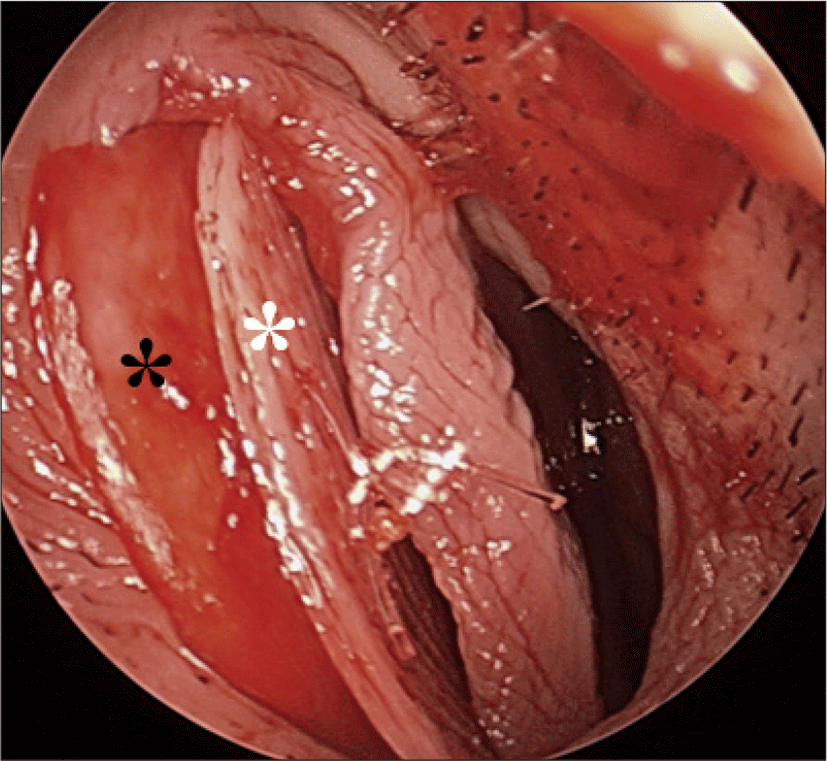
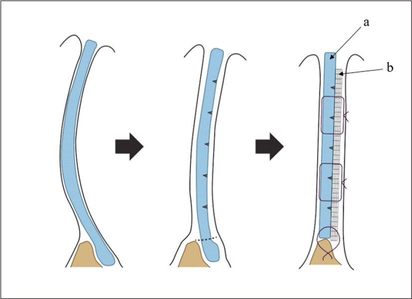
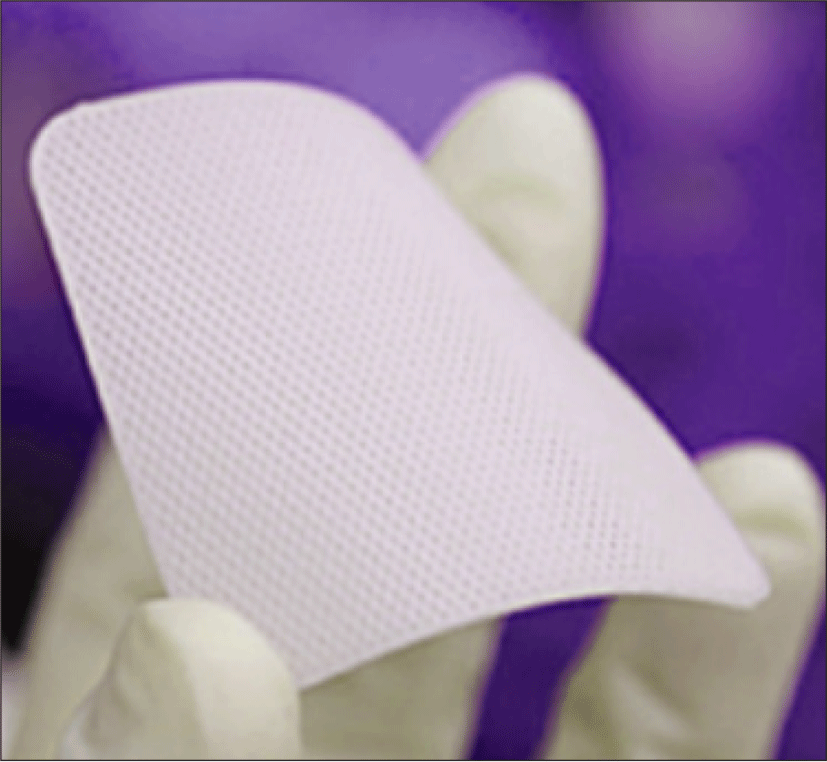
A 27-year-old male patient visited our department with a complaint of unilateral nasal obstruction caused by caudal septal deviation. The right nostril appeared narrow enough (caused by caudal septal deviation) to block the nasal passage on nasal endoscopy. Computed tomography (CT) revealed caudal nasal septal deviation, hypertrophy of the inferior turbinates, and clear sinuses. A hemitransfixion incision was performed in the caudal nasal septum and a mucoperichondrial/mucoperiosteal flap was elevated bilaterally through the incision. The caudal septum was repositioned over the nasal spine along the midline and straightened with wedge resection after resecting the deviated part of the septal cartilage and bony septum. The excessive cartilage was removed and fixed using the figure of eight anchoring suture technique. The TnR Nasal Mesh® was carved to fit the desired location and placed on the concave side (Fig. 1 and 2). The average size of the implant was 1.3×2 cm. The carved mesh was secured adjacent to the weakened caudal segment using 5-0 polydiaxanone (PDS) sutures (Fig. 3). The incision site was sutured after ensuring that the septum was straight. Silastic sheets were placed bilaterally and absorbable packing was placed for 2 days.


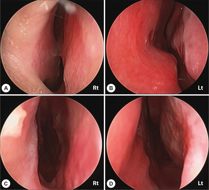
The patient showed improvement in the subjective symptoms of nasal obstruction and the objective outcomes determined using endoscopic scores and MCA and NCV scores (obtained by acoustic rhinometry) (Table 1). The VAS scores were improved from 6 (preoperative), to 3 (postoperative 2-weeks) and 1 (postoperative 1month). The endoscopic scores also improved from 3 to 1 (Fig. 4). The preoperative MCA values of the concave side improved from 0.06 cm2 (preoperative) to 0.80 cm2 (postoperative). The postoperative nasal volume of the convex side increased significantly to 6.63 cm3 from pre-operative values of 5.67 cm3. The nasal volume of the concave side also increased from 1.27 cm3 to 8.76 cm3. No significant complications were observed during or after the surgical procedure with TnR Nasal Mesh® without transient partial exposure of the implant. The patient has been followed for 18 months with no recurrence or complication.

A 53-year-old male patient came to the clinic with a complaint of unilateral nasal obstruction caused by caudal septal deviation. He had symptoms of chronic rhinitis such as sneezing, rhinorrhea, and frequent itching sensation and also complained of sleep disorders and decreased sense of smell caused by nasal congestion. Nasal endoscope showed severe caudal septal deviation with unilaterally blocked nostril and CT finding also showed caudal nasal septal deviation, hypertrophy of the inferior turbinates with clear sinuses. The surgical procedure was performed with the TnR Nasal Mesh® as previous fashioned (Patient 1). The VAS scores were decreased from 10 (preoperative), to 2 (postoperative 2 weeks) and 2 (postoperative 1 month). The endoscopic scores revealed from 3 to 1 and MCA values of the concave side improved from 0.27 cm2 to 1.01 cm2, respectively. The postoperative nasal volume of the convex side increased significantly to 8.56 cm3 from pre-operative values of 3.41 cm3, respectively. The postoperative nasal volume of the concave side also increased from 3.11 cm3 to 10.70 cm3. There was no surgical complication within 18 months of follow-up periods.
A 57-year-old male patient came to our clinic with nasal congestion that worsened a month ago. He complained of rhinitis symptoms and also discomfort due to nasal congestion, while eating or undergoing dental treatment. The endoscope and CT findings showed caudal nasal septal deviation and turbinate hypertrophy without sinusitis. The surgery was finished via previous same method (Patient 1). The subjective VAS scores about nasal obstruction were improved from 5 (preoperative), to 1 (postoperative 2 weeks) and 0 (postoperative 1 month). The endoscopic findings also improved from 3 to 1. The preoperative MCA values of the concave side improved from 0.26 cm2 to post-operative values of 0.78 cm2. The postoperative nasal volume of the convex side increased significantly to 9.08 cm3 from pre-operative values of 6.38 cm3. The postoperative nasal volume of the concave side also increased from 4.53 cm3 to 8.64 cm3. No significant complications were reported during or after the surgical procedure within 7 months of follow-up periods
Discussion
The frequency of caudal deviation is approximately 5 to 10% among all cases of nasal septal deviation.9,10) The caudal septum is critical supporting structure for the nasal shape, which involves an L-shaped strut. Several previous studies have reported techniques for the correction of caudal nasal septum deviation.3,6,11) Seo et al.11) reported the septal cartilage traction suture technique for caudal septal deviation correction. They reported good outcomes with the subjective and objective findings, which suggested a simple, safe, and effective method for correcting caudal septal deviation. Other techniques, such as cross-hatching, batten grafting, swinging door technique, and cutting sutures, have been reported previously, to facilitate better outcomes of caudal nasal septal deviation correction.3-6) In contrast, it is difficult to correct caudal septal deviation completely with the conventional septoplasty technique due to the possibility of weakening the supporting structure. The batten graft is one of the most powerful methods for straightening caudal nasal septal deviation. Dingman et al.12) first reported the utility of batten grafting for correcting septal deformity. Since then, batten grafting has served as a versatile surgical method for caudal septal correction. Most surgical materials for batten grafting would be autologous tissues such as septal cartilage, septal bone, auricular cartilage, and costal cartilage. Previous studies reported the utility of alloplastic implants for the batten grafting for septal correction.7) They provided verification for the use of the PDS-cartilage composite graft in septorhinoplasty. Boenisch et al.13) reported extracorporeal septal reconstruction with a combination of nasal septal cartilage and auricular conchal cartilage on PDS. They reported that the functional and cosmetic results were satisfactory without any complications and that the use of a PDS plate during septal reconstruction facilitated external septoplasty and corrected severe nasal deformity. TnR Nasal mesh® is absorbable, three-dimensionally printed, homogeneous, and composite nasal implant, which demonstrated proper mechanical support, which was adequately thin with excellent biocompatibility.14) TnR Nasal Mesh® is composed of microporous PCL. This polymer is ultimately hydrolyzed into carbon dioxide (CO2) and water (H2O). After being grafted on the body and filling the space the porous structural characteristics of the products allow the various cells normally present in the surrounding tissues to migrate into the porous scaffold. Consequently, it promotes the generation of extracellular matrix material such as collagen after a certain amount of time, while contributing to the reconstruction of the damaged tissue. These characteristics could prove the biocompatibility of the surgical material. On the other hand, most previous studies with alloplastic implants for treating caudal septal deviation used the extracorporeal septoplasty or rhinoplasty approach, combined with the caudal septal extension graft or cutting suture technique.4,15) There is a paucity of informations using alloplastic implants via pure endonasal septoplasty for correcting causal septal deviations. Recently, there was a previous similar study of this three-dimensional printing mesh.16) Our study emphasized that TnR Nasal Mesh® can be used for pure endonasal septoplasty via bilateral flap elevation and facilitates complete caudal septal correction without any external scar or extracorporeal manipulation, as previous reported. The possible complications of the alloplastic implant include infection, migration, and cosmetic changes. We did not observe any immediate (bleeding, hematoma formation, infection, or necrosis) or long-term (septal perforation, fibrosis of adjacent soft tissue, or immunologic rejection of the implant) complications in our patients in one and half year follow-up period.
The key finding of this brief case study is that surgical method described here can be used easily for endonasal septoplasty via bilateral flap elevation. Classically, the septorhinoplasty approach is often required for flawless correction of caudal septal deviation. Pure endonasal approach without open rhinoplasty was preferred in our patients because of the shorter operation time, lesser external scar tissue formation, lower rates of flap-related complications, faster healing process, and low cost. Using the TnR Nasal mesh® endonasal approach alone with bilateral flaps could provide alternative option for the correction of caudal septal deviation.
Conclusion
We used the TnR Nasal Mesh® for caudal septal correction in three patients. Significant improvements were observed in the subjective measurement of nasal obstruction (determined by VAS) and objective measurement of the MCA on acoustic rhinometry and endoscopy. In present case series, it produced satisfactory results without major complications such as recurrence of septal deviation, necrosis of the septal mucosa, and inflammation of the recipient site. Further studies are needed to evaluate larger samples and long-term effects.


