증례

72세 고령 환자에서 비인두 새열 낭종 1예

김도연1, 박준욱1, 김병국1, 김지선1,*
Doyeon Kim1, Jun-Ook Park1, Byung Guk Kim1, Ji-Sun Kim1,*
Author Information & Copyright
1가톨릭대학교 의과대학 은평성모병원 이비인후-두경부외과학교실
1Department of Otorhinolaryngology-Head and Neck Surgery, Eunpyeong St. Mary’s Hospital, College of Medicine, The Catholic University of Korea, Seoul, Korea
*교신저자: 김지선, 03312 서울 은평구 통일로 1021 가톨릭대학교 의과대학 은평성모병원 이비인후-두경부외 과학교실 전화: (02) 2030-4631·전송: (02) 2030-4631 E-mail:skswltjs23@hanmail.net

© Copyright 2020 The Busan, Ulsan, Gyeoungnam Branch of Korean Society of Otolaryngology-Head and Neck Surgery. This is an Open-Access article distributed under the terms of the Creative Commons Attribution Non-Commercial License (http://creativecommons.org/licenses/by-nc/4.0/) which permits unrestricted non-commercial use, distribution, and reproduction in any medium, provided the original work is properly cited.

Received: Feb 06, 2020; Revised: Apr 08, 2020; Accepted: May 12, 2020

Published Online: Jun 30, 2020

ABSTRACT

Second branchial cleft cysts are almost located in the neck; thus, their presence in the nasopharynx is extremely rare. A 72-year-old woman was referred to our department for a mass found in the left lateral nasopharyngeal wall incidentally on a neck magnetic resonance imaging. The location and pathological feature of the cyst indicated that this was of branchial origin. When a cystic lesion presents in the lateral nasopharynx, branchial cleft cyst should be considered in the differential diagnosis. Here, we report a case of nasopharyngeal branchial cleft cyst with a literature review. (J Clinical Otolaryngol 2020;31:94-97)

Keywords: 비인두 낭종; 새열낭종; 조대술
Keywords: Nasopharyngeal cyst; Branchial cleft cyst; Marsupialization

서 론

새성기형은 태생기 2주경에 발생하여 6주경에 분화를 마치는 새성기관(branchial apparatus)의 불완전 폐쇄에 의해 발생하며, 피부 또는 점막과의 개통 여부에 따라 낭 종(cyst), 동(sinus) 또는 누공(fistula)의 형태로 나타나고 지칭된다.1-3) 새열낭종(branchial cleft cyst)은 일반적으 로 경부의 측면에서 단발성 무통성 종물의 형태로 나타 나게 되며,4) 비인두에서 발견되는 경우는 극히 드물다.5)

비인두의 낭성 병변은 대게 양성이나, 악성 병변과 구 별하기 위해선 주의를 기울여야 한다. 비인두 낭종은 중 앙에서 발생하는 것과 측면에서 발생하는 것으로 분류할 수 있으며, 선천적 병변과 후천적 병변으로 분류할 수 있 다. 비인두의 측면에 발생하는 낭종의 원인으로 감별해야 할 진단으로는, 선천적 병변으로 새열낭종과 후천적 병변 으로 점액 저류 낭종(mucous retention cyst)이 있다.6)

본 저자들은 극히 드문 질환인 비인두 새열낭종을 경험 하였기에 이에 대해 보고하고자 한다. 저자들이 아는 한, 본 증례는 가장 고령의 비인두 새열낭종에 대한 보고이다.

증 례

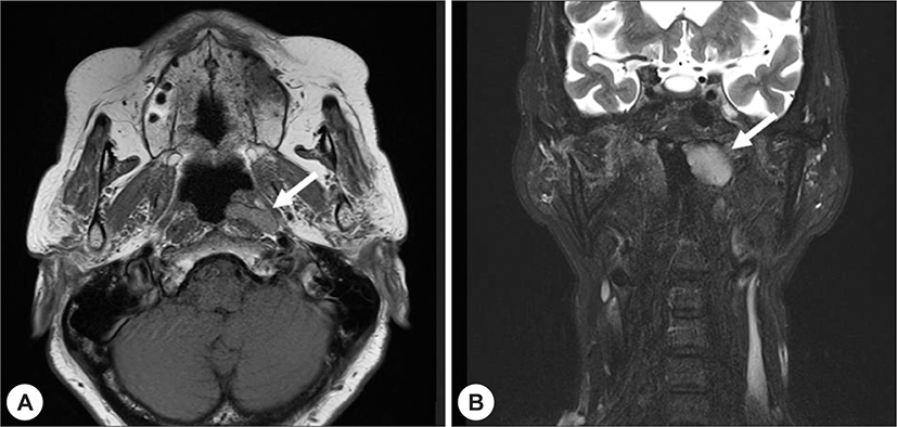
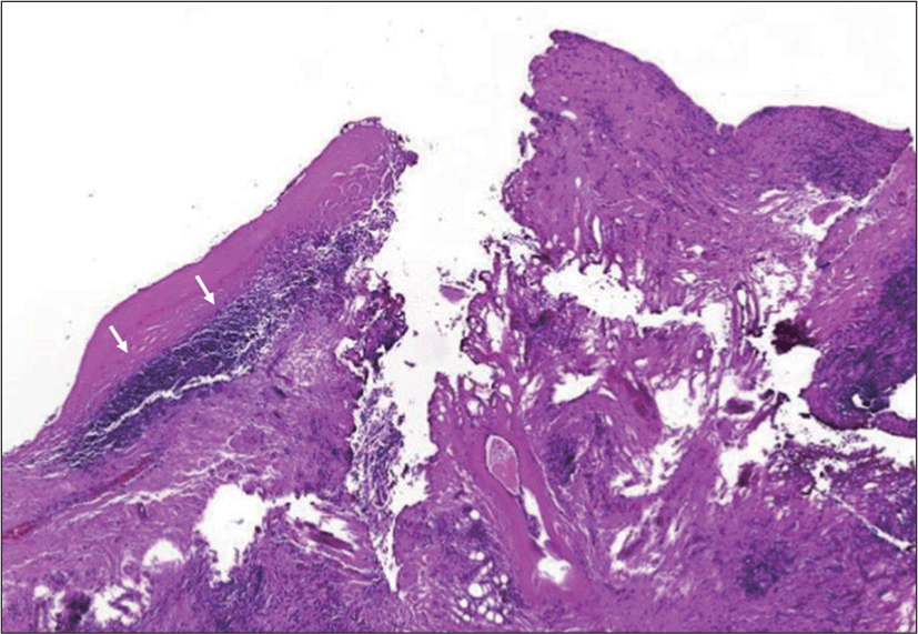
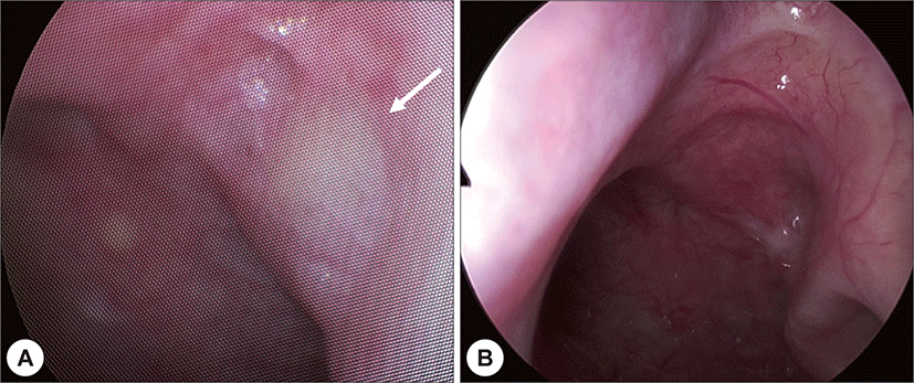
72세 여자환자가 내원 6개월전부터 발생한 두통을 주 소로 신경외과에 내원하여 시행한 뇌 자기공명영상에서 우연히 발견된 좌측 비인두 종괴로 본과에 의뢰되었다. 과거력에서 특이 사항은 없었고, 계통 문진상에서 비폐 색이나 이충만감과 같은 본과적 특이 호소증상은 없었 다. 비인두 내시경에서 좌측 귀인두관 융기(torus tubarius) 후방의 비인두 표면에서 융기된 매끄러운 종괴가 확 인되었고, 후비공이나 유스타키오관의 폐색은 없었으며, 양측 고막 또한 정상소견이었다. 자기공명영상의 축상면 에서 좌측 비인두로 돌출된 아령 모양(dumbbell shape) 의 종괴를 확인 할 수 있었으며, 이는 축상과 관상면을 종합하였을 때 약 0.7×2.2×2 cm 크기로 확인되었다 (Fig. 1). 진단 감별을 위해 비인두 종괴에 대한 비내시경 하 조직검사를 계획하였고, 수술 시 종괴의 벽이 제거되 면서 갈색의 점액성 분비물이 배출되는 것을 확인하였 다. 수술 소견 및 동결절편 조직검사를 종합하였을 때 전 형적인 낭종 소견으로 판단되어 미세흡입분쇄기를 이용 한 조대술을 시행하였다. 수술 후 영구검체의 병리조직 학적 소견은 원주 상피 세포 하에 림프계 세포가 산재해 있는 것으로 나타났다(Fig. 2) 낭종의 위치와 병리학적 특징은 제2 새열낭종과 일치하였다. 환자는 수술 다음날 특이 사항 없이 퇴원하였고, 수술 후 6개월동안 비인두 내시경 검사에서 재발 소견 없이 추적관찰 중이다(Fig. 3).

jcohns-31-1-94-g1
Fig. 1. Magnetic resonance imaging shows iso-intense signal on (A) T1-weighted image, axial view, and hyper-intense signal (B) T2-weighted image, coronal view. The mass is homogeneous and involves the left lateral aspect of the nasopharynx (white arrow). The boundaries were clear without invasion into the surrounding structures.
Download Original Figure
jcohns-31-1-94-g2
Fig. 2. The histopathology reveals columnar epithelium with lymphoid aggregates (white arrow)(original magnification ×100).
Download Original Figure
jcohns-31-1-94-g3
Fig. 3. The endoscopic findings of the left nasopharynx. A: Preoperative clinical photograph; nasal endoscopy showed a mass with a smooth surface at the left nasopharynx (white arrow). B: Postoperative clinical photograph.
Download Original Figure

고 찰

새성기형은 선천성 경부 종물 중 갑상설관 낭종 다음 으로 흔하며, 그 중 제2 새성기형이 90% 이상으로 가장 흔한 것으로 알려져 있다.7) 제2 새성누공은 흉쇄유돌근 하방 1/3 전방에서 경동맥의 외측 상부로 주행하여 내경 동맥과 외경동맥의 사이에서 내측으로 들어가며 설인신 경 및 설하신경의 상부와 경설골인대의 하부로 주행하 여 편도와(tonsillar fossa) 부근에서 인두벽으로 연결된 다.8) 낭종은 이 누공 경로의 어느 곳에서나 발생할 수 있 다. 제2 새열낭종은 Proctor에 의해 네 가지 유형으로 분 류되었다.9) 제1형은 흉쇄유돌근의 앞쪽 경계와 경부 근 막 아래에 놓여있고, 가장 흔한 제2형은 심경부 근막아래 에 있으며 큰 혈관과 맞닿아 있다. 제3형은 내경동맥과 외경동맥 사이를 통과하며 인두벽까지 뻗어있고, 제4형 낭동은 인두벽에 인접하여 큰 혈관들의 안쪽으로 위치 해 있다.9)

새열낭종은 일반적으로 층화된 평편상피(stratified squamous epithelium), 가성 중층섬모원주 상피(pseudostratified ciliated columnar epithelium) 또는 두 가 지 모두로 구성될 수 있으며, 상피아래에는 다양한 림프 조직으로 구성되어 있다.10) 새궁 기원의 진단은 해부학 적 위치와 조직학적 특성에 기초한다. 이러한 결과를 종 합해 보았을 때, 본 증례의 비인두 낭종은 제2 새열낭종 의 제4형에서 파생되었음을 알 수 있었다.11)

비인두 종양의 감별진단에는 라트케 낭(Rathke’s pouch), Tornwaldt 낭종, 아데노이드내 낭종, 새열낭종, 점액 저 류 낭종, 유피낭종, 비인두암 등이 있다.6) 비인두암과의 감별진단이 중요하며, 비인두암은 일반적으로 성인에서 Rosenmuller’s fossa의 궤양성 종괴 형태로 주로 나타나 며, 경부 림프절 비대 및 이관 기능장애 및 혈성 후비루 를 동반할 수 있다.12) Tornwaldt 낭종은 비인두에 발생하 는 선천성 낭종으로, 비인두의 중앙선에 가깝게 위치하 며 조직학적으로 림프 조직이 없이 상피로 구성되어 있 어 새열낭종과 감별이 가능하다.13) 점액 저류 낭종은 새 열낭종과 비슷한 위치인 비인두의 측면에서 발생할 수 있는 후천적 낭종으로, 상피 세포층이 없이 결합 조직 내 에 점액물질을 가진 병리학적 특성으로 새열낭종과 감 별할 수 있다.14)

비인두 새열낭종은 국내에선 보고된 증례가 없으며, 국외에서도 20건 이하의 보기 드문 증례이다. 또한 비인 두 낭종의 표준 치료 방법은 완전 절제로 알려져 왔으 나, 최근 조대술만으로 효과적으로 치료되는 사례들이 보고되고 있다.15,16) 내시경하 비내 접근법은 조직 손상과 관련된 합병증을 감소시킬 수 있고 재원 기간이 짧은 장 점이 있어, 본 증례와 같은 비인두 종괴에 대한 수술법 으로 조대술을 시행하는 것이 가장 효과적인 것으로 생 각되어 이 증례를 보고한다.

REFERENCES

1).

Doi O, Hutson JM, Myers NA, McKelvie PA. Branchial remnants: a review of 58 cases. J Pediatr Surg 1988;23(9):789-92.

2).

Tae K, Jeong SW, Lee EJ, Lee SH, Kim KR, Park CW, et al. A clinical study of branchial anomaly. Korean J Otorhinolaryngol-Head Neck Surg 2002;45(10):998-1003.

3).

Chandler JR, Mitchell B. Branchial cleft cysts, sinuses, and fistulas. Otolaryngol Clin North Am 1981;14(1):175-86.

4).

Panchbhai AS, Choudhary MS. Branchial cleft cyst at an unusual location: a rare case with a brief review. Dentomaxillofac Radiol 2012;41(8):696-702.

5).

Boysen ME, de Besche A, Djupesland G, Thorud E. Internal cysts and fistulae of branchial origin. J Laryngol Otol 1979;93(5):533-9.

6).

Marom T, Russo E, Ben Salem D, Roth Y. Nasopharyngeal cysts. Int J Pediatr Otorhinolaryngol 2009;73(8):1063-70.

7).

Taylor JN, Burwell RG. Branchiogenic nasopharyngeal cysts. J Laryngol Otol 1954;68(10):667-79.

8).

Benson MT, Dalen K, Mancuso AA, Kerr HH, Cacciarelli AA, Mafee MF. Congenital anomalies of the branchial apparatus: embryology and pathologic anatomy. Radiographics 1992;12(5):943-60.

9).

Proctor B. Lateral vestigial cysts and fistulas of the neck. Laryngoscope 1955;65(6):355-401.

10).

Zaifullah S, Yunus MR, See GB. Diagnosis and treatment of branchial cleft anomalies in UKMMC: a 10-year retrospective study. Eur Arch Otorhinolaryngol 2013;270(4):1501-6.

11).

Heo CY, Kim BG, Kim YB, Park IS. A second branchial cleft cyst of palatine tonsil. J Clinical Otolaryngol 2010;21(1):123-6.

12).

Chen YA, Su JL, Hao SP. Nasopharyngeal branchial cleft cyst. Otolaryngol Head Neck Surg 2007;136(1):144-6.

13).

Kwok P, Hawke M, Jahn AF, Mehta M. Tornwaldt’s cyst: clinical and radiological aspects. J Otolaryngol 1987;16(2): 104-7.

14).

Nahata V. Branchial Cleft Cyst. Indian J Dermatol 2016;61(6): 701.

15).

Kim YW, Baek MJ, Jung KH, Park SK. Two cases of nasopharyngeal branchial cleft cyst treated by powered instrument assisted marsupialisation. J Laryngol Otol 2013;127(6): 614-8.

16).

Flis DW, Wein RO. Nasopharyngeal branchial cysts-diagnosis and management: a case series. J Neurol Surg B Skull Base 2013;74(1):50-3.