증례

비인두에 발생한 Melanotic Oncocytic Metaplasia(MOM) 1예

박근형1, 지창록1, 고태경1,*, 이화선2
Geun Hyung Park1, Chang Lok Ji1, Tae Kyung Koh1,*, Hwa Sun Lee2
Author Information & Copyright
1부산성모병원 이비인후과
2병리과
1Department of Otorhinolaryngology-Head and Neck Surgery, Busan, Korea
2Pathology, Busan Saint Mary’s Hospital, Busan, Korea
*교신저자: 고태경, 48757 부산광역시 남구 용호로 232번길 25-14 부산성모병원 이비인후과 전화: (051) 933-7214· 전송: (051) 956-1956 E-mail:ktk0305@hotmail.com

© Copyright 2020 The Busan, Ulsan, Gyeoungnam Branch of Korean Society of Otolaryngology-Head and Neck Surgery. This is an Open-Access article distributed under the terms of the Creative Commons Attribution Non-Commercial License (http://creativecommons.org/licenses/by-nc/4.0/) which permits unrestricted non-commercial use, distribution, and reproduction in any medium, provided the original work is properly cited.

Received: Jan 14, 2020; Revised: Apr 16, 2020; Accepted: May 30, 2020

Published Online: Jun 30, 2020

ABSTRACT

Melanotic oncocytic metaplasia (MOM) of the nasopharynx is a rare lesion that occurs mostly in Asian men in their 60s to 70s who are heavy smokers. Although it is a benign lesion, it’s sometimes mistaken for malignant melanoma, nevus or early nasopharyngeal carcinoma. A 75 year-old male visited our outpatient clinic with hearing loss that occurred a week earlier. Otoscopic findings revealed left serous otitis media. A nasopharyngoscopic examination to rule out a malignant tumor in the nasopharynx revealed multiple black nodules in the left torus tubarius and left nasal floor. Under local anesthesia, some of nodules were excised for histopathology. The histological examination revealed oncocytic metaplasia of mucous glands and the presence of numerous brown-colored melanin pigments. Immunohistochemically, S 100-positive and HMB 45-negative dendritic melanocytes were identified. During 3 months of regular follow up, there was no recurrence at the excision site and no expansion of the remaining lesion. (J Clinical Otolaryngol 2020;31:88-93)

Keywords: 멜라닌; 화생; 비인두
Keywords: Metaplasia; Melanins; Nasopharynx

서 론

비인두에 발생하는 Melanotic Oncocytic Metaplasia (MOM)는 매우 드문 병변으로, 대게 아시아계의 고령 남성에서 발견된다. 병변은 수 밀리미터 정도로 크기가 작으며 갈색에서 흑색을 띄는 다수의 결절이 이관 개구 부 주변에 발생하는 것이 특징이고 일측성 혹은 양측성 을 보인다. 대게 무증상이기 때문에 우연히 발견되나 때때로 삼출성 중이염, 이명, 목불편감, 쉰목소리 코피, 객혈, 비루 등을 호소하기도 한다. 현재까지 보고된 MOM은 모두 양성이나 외래에서 간혹 악성 흑색종 (malignant melanoma)으로 오인되기도 한다.

비인두에 발생하는 MOM은 Shek 등1)이 1995년 처 음 보고하였으며, 현재까지 28개의 증례가 영문 문헌에 보고되었고 우리나라에서는 4건이 보고되었다.

저자들은 내원 일주일 전부터의 청력저하를 주소로 내원하였던 75세 남자 환자에게 발견된 이관융기부 (torus tubarius)와 비강저부(nasal floor)의 MOM에 대 한 증례 1예를 문헌 고찰과 함께 보고하는 바이다.

증 례

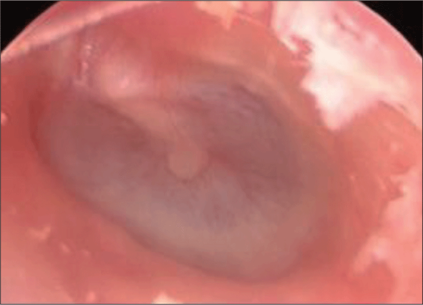
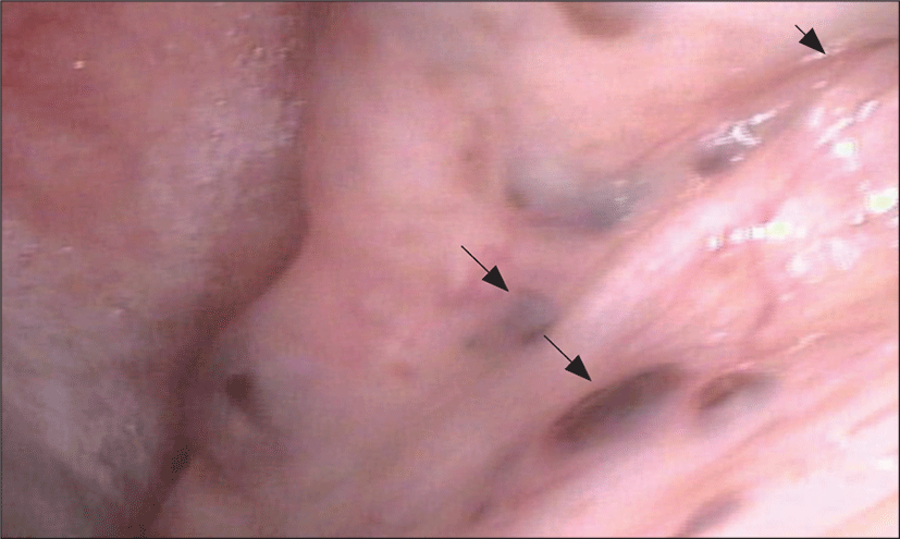
75세 남자 환자가 약 일주일 전부터 발생한 청력저하 와 이충만감으로 내원하였다. 과거력상 당뇨, 대장 용종 및 사회력상 흡연 25갑년(0.5갑/일×50년)이 확인되었 다. 이내시경상 좌측에 경도로 함몰된 호박색 고막이 확인되었고(Fig. 1), 순음청력검사상 좌측 공기뼈전도 차이(air-bone gap)가 확인되어 좌측 삼출성 중이염으 로 진단 되었다. 고령의 삼출성 중이염 환자는 반드시 비 인두 악성 종물 유무를 확인해야 하므로 비내시경 검사 를 시행하였다. 비내시경상 좌측 이관융기(torus tubarius)와 좌측 비강저부(nasal floor)에 다발성 흑색 결절이 확인되었으며(Fig. 2), 후두 내시경 및 경부 촉진상 다른 이상 소견은 확인되지 않았다.

jcohns-31-1-88-g1
Fig. 1. Otoscopic findings: retracted and ambor colored tympanic membrane.
Download Original Figure
jcohns-31-1-88-g2
Fig. 2. Nasoscopic findings of the lesion: multiple, dark blue colored small nodules (arrow) around eustachian tube orifice (arrowhead).
Download Original Figure

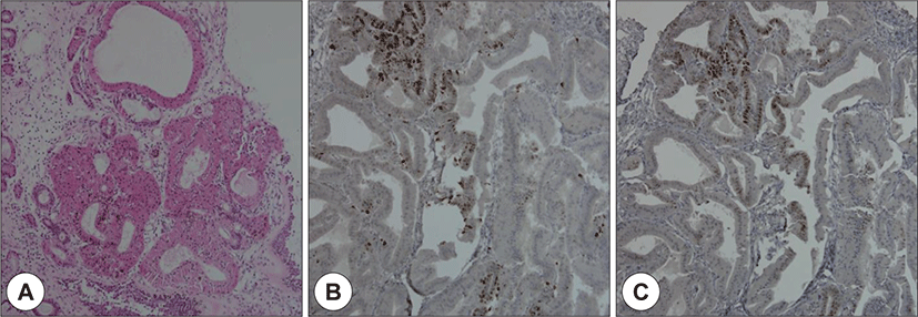
국소 마취하에 후두 가위(larygneal scissor), 후두 겸자 (laryngeal forcep) 등으로 이관융기에서 2건, 비강저부에 서 1건 절제 생검(excisional biopsy)하였고 출혈은 경미 하였다. 수술 후 좌측 이관의 개구부는 정상 소견을 보였 다. 절제한 결절은 육안상 약 0.3 cm이었으며, H&E stain 하에 현미경 병리조직소견상 바깥층에 정상 호흡 상피세 포가 확인되었고, 상피하 점액샘(subepithelial mucous gland)의 상피세포에서 호산성 화생(oncocytic metaplasia)이 관찰되었고 호산성 화생을 보이는 상피세포 내에 수많은 갈색의 멜라닌 색소 침착을 동반하고 있었다(Fig.3). 분열기세포(mitotic cell)나 비정형성(atypia)은 관찰되 지 않았다. 면역조직화학염색법상, 분비샘(mucous gland)의 기저층(basal layer)에 위치한 수지상세포(dendritic cell)에서 S-100 단백질 양성, Human Melanoma Black(HMB)-45 음성(Fig. 3) 소견을 보여 멜라닌세포 (melanocyte)의 존재가 확인되었다. 따라서 멜라닌 침착 을 동반한 호산성 화생(oncocytic metaplasia)인 Melanotic Oncocytic Metaplasia(MOM)로 진단되었다.

jcohns-31-1-88-g3
Fig. 3. Microscopic findings of the lesion (hematoxylin and eosin stain, ×100 magnification): subepithelial mucous glands show oncocytic metaplasia with numerous brown colored melanin pigments (A). Immunohistochemical staining of the lesion (S-100): dendritic melanocytes are positive for S-100 (B). Immunohistochemical staining of the lesion (HMB-45): dendritic melanocytes are negative for HMB-45 (C).
Download Original Figure

수술 2주, 1개월 그리고 3개월 뒤에도 남아있는 흑색 결절의 크기는 변하지 않았다. 수술 1개월 뒤에도 좌측 삼출성 중이염은 자연 관해 되지 않아 고막절개술 시행 하였으며 고막 절개시 장액성 이루가 배출되었고 청력 도 호전되었으나 이후 중이염은 반복적으로 재발하였다.

고 찰

호산성 화생(oncocytic metaplasia)이란, 분비샘 (gland)의 정상 상피세포에 미토콘드리아가 축적 되어 풍부한 호산성(eosinophilic) 및 과립성(granular) 세포 질을 가지는 비대한 세포로 변하는 것을 뜻한다.1) 호산 성 화생은 주타액선(major salivary gland)과 상기도 점 막의 소타액선(minor salivary gland) 모두에서 나타날 수 있으며, 나이가 들수록 증가하는 양성의 퇴행성 변 화로 알려져 있다.2) 비인두에서의 호산성 화생은 내시 경상 작은 결절, 종괴, 낭종, 비정상 점막 등으로 보이기 도 하나, 대게 선별을 위한 임의 생검에서 우연히 발견 된다.3)

호산성 화생에 멜라닌 침착이 동반되는 경우는 더 드 물며, 영문 문헌상 현재까지 총 28건만이 보고되었다. 호산성 세포(oncocyte)는 스스로 멜라닌을 생성하지 못 하며, 이 멜라닌의 정확한 기원은 밝혀지지 않았다.4) 정 상 비강 점막에서 멜라닌세포(melanocyte)가 관찰되는 경우는 매우 드문데, MOM을 최초로 보고한 Shek et al. 은, 조직 소견상 호산성 세포와 멜라닌세포 군집이 매우 인접하며, 이러한 두 가지 소견의 조합이 흔하지 않은 것을 고려해 볼 때 호산성 세포가, 잠복된 멜라닌 세포(dormant melanocyte)를 활성화 시키는 인자를 분 비한다는 가설을 제시했다.1)

한편 피부의 멜라닌 세포는 이웃한 각질세포(keratinocyte)에 멜라닌소체(melanosome)를 이동시킴으로 써, 이웃한 각질세포에 멜라닌을 전달할 수 있다는 사 실이 알려져 있는데,5) Hirakawa 등6)은 전자현미경을 이용하여, 호산성 세포내에 멜라닌소체가 존재함을 확 인하였으며, 멜라닌소체의 전구체인 전멜라닌소체 (premelanosome)는 발견되지 않는 다는 사실을 확인 하였다. 이를 통해 호산성 세포(oncocyte)내의 멜라닌 침착도, 각질세포의 멜라닌과 같이 스스로 생산한 것이 아니라, 멜라닌세포(melanocyte)에서 전달받은 것이라 추정하였으며, 분비샘 상피세포들 사이로 뻗어있는 멜 라닌세포(melanocyte)의 가지돌기(dendrite process)들 을 통해 멜라닌이 전달된다고 추정하였다.

멜라닌 침착이 흡연과 관련이 있다는 가설이 있는데, 태국과 말레이시아에서 치과 외래 환자 467명을 대상으 로 한 연구에서, 흡연자의 약 30%에서 구강 내 멜라닌 침 착 소견을 보였다.7) 이외에도 터키와 스웨덴에서도 흡연 과 구강 내 멜라닌 침착의 연관성에 대해 보고하였다.8,9) 처음으로 다수의 MOM 증례(7건)를 보고한 Sakaki 등10) 의 보고에서 2명이 흡연자로 확인되었으며(Table 1), 비 인두 MOM의 선행요인으로 흡연의 가능성을 제시하였 다. 본 증례의 환자도 약 25갑년의 흡연력을 갖고 있었으 며, 현재까지 보고된 28건의 증례 보고에서도 총 12명이 흡연자였다(1명은 비흡연자, 15명은 흡연력이 확인되지 않았다). 특히 흡연자 12명은 1명을 제외하고 모두 25갑 년 이상의 흡연력을 가진 heavy smoker였다.

Table 1. Clinical features of 28 cases (including our case) with melanotic oncocytic metaplasia
Case Author Year Sex/Age Site Number Clinical Impression Symptom Smoking history
1 Shek et al. 1995 M/67 Eustachian opening (ND) Single Early carcinoma Otitis media ND
2 Shek et al. 1995 M/63 Eustachian opening (L) Single Early carcinoma Tinnitus ND
3 Kurihara et al. 1997 M/69 Nasopharynx (L) Multiple ND None ND
4 Hirakawa et al. 1999 M/64 Eustachian opening (B), nasopharynx and nasal cavity (L) Multiple Malignant tumor Discomfort in throat ND
5 Xue et al. 1999 M/70 Eustachian opening (B) Multiple Nevus Tinnitus ND
6 Sakaki et al. 2004 M/80 Nasophraynx and nasal cavity (L) Multiple Melanoma Hoarseness 10/day×50yr
7 Sakaki et al. 2004 M/69 Eustachian opening (L) Single Melanoma Rhinorrhea 40/day×60yr
8 Sakaki et al. 2004 M/74 Nasopharynx (L) Single Tumor Epistaxis ND
9 Sakaki et al. 2004 M/74 Eustachian opening (R) Multiple Melanoma Discomfort in throat ND
10 Sakaki et al. 2004 M/68 Nasopharynx (ND) Single Tumor ND ND
11 Sakaki et al. 2004 M/65 Eustachian opening (R) Single ND Hemoptysis ND
12 Sakaki et al. 2004 M/63 Eustachian opening (L) Single Melanoma Epistaxis ND
13 Takano et at. 2004 M/64 Eustachian opening (L), torus tubarius (B) Multiple Melanoma Discomfort in the ear ND
14 Lui et al. 2004 M/79 Eustachian opening (R) Single ND Otitis media ND
15 Liao et al. 2005 M/ND Nasopharynx (B) Multiple ND None ND
16 Kondo et al. 2010 M/73 Torus tubarius (B) Multiple Melanoma or hemangioma Nasal congestion Non-smoker
17 Li et al. 2010 M/58 Nasopharynx (B) Multiple ND Epistaxis 20/day×40yr
18 Na et al. 2012 M/72 Torus tubarius (B) Torus tubarius (L), Multiple Melanotic dysplasia Nasopharynqeal Hearing impairment 40/day×50yr
19 Na et al. 2012 M/71 soft palate (ND) Multiple pigmented lesion Hoarseness 20/day×40yr
20 Na et al. 2012 M/51 Torus tubarius (B) Multiple Melanotic oncocytic metaplasia Pain of the tongue ND
21 Chang et al. 2014 M/63 Nasopharynx (ND) Multiple Melanoma Ep it axis 40/day×40yr
22 Tajima et al. 2014 M/57 Torus tubarius (R) Multiple Melanoma None 30/day×40yr
23 Uehara et at. 2015 F/70 Eustachian opening (ND) Multiple Melanosis None 5/day×50yr
24 Uehara et at. 2015 F/61 Eustachian opening (L) Single Melanosis Hoarsness 23/day×40yr
25 Uehara et at. 2015 M/74 Nasopharynx (B) Multiple Melanosis Hoarsness 20/day×50yr
26 Lin at el. 2015 M/60 Torus tubarius (L) Single Nevus None ND
27 Chen at el. 2020 M/75 Torus tubarius (R) Multiple Nevus or melanoma Epistaxis 30/day×50yr
28 Present case 2020 M/75 Torus tubarius and nasal cavity (L) Multiple Melanosis Otitis media 10/day×50yr

M: male, F: female, B: both, R: right, L: left, ND: non-documented

Download Excel Table

현재까지 보고된 28건의 MOM의 역학, 발생 위치, 개 수, 증상 등을 Table 1에 정리하였다. 28명 모두 동아시아 인(일본 15명, 중국계 9명, 한국4명)이었으며, 남자 26명, 여자 2명으로 남자가 대다수였으며, 평균나이는 68세였 다. 발생 위치는 이관 개구부(Eustachian opening)가 12 명, 이관융기(torus tubarius)가 8명으로 보고되어 이관에 서 발생하는 경우가 대부분이었고, 비인두(nasopharynx), 비강(nasal cavity), 연구개(soft palate)등의 명칭으 로도 보고 되었다. 병변의 개수는 1개인 경우가 10명, 여 러 개인 경우가 16명이었다. 증상은 무증상이 6명, 삼출 성 중이염이 3명이었으며 이외에도 이명, 쉰목소리, 목불 편감, 코피, 객혈, 콧물 등의 비특이적인 증상이 보고되었 다. 삼출성 중이염의 경우, 이관개구부 주변의 조직 부종 에 의한 이관기능장애로 인해 발생할 것으로 추정되며,2) 본 증례를 포함한 3명의 삼출성 중이염 환자 모두 이관에 병변이 있었다. 내시경 소견을 바탕으로 한 임상의의 추 정진단은 조기 비인두암(early nasopharyngeal carcinoma), 악성 흑색종(melanoma), 모반(nevus), 멜라닌증 (melanosis) 등으로 보고되었다.

MOM과 악성 흑색종은 멜라닌을 포함한 색소성 병 변을 갖는다는 점에서 비슷하여 감별이 필요하다. 점막 흑색종(mucosal melanoma)은 피부 흑색종(cutaneous melanoma)에 비해 임상 양상이 공격적이며 예후가 나 쁘다. 악성 흑색종의 경우 초기에는 반복되는 코피와 진 행성 코막힘이 주된 증상이며 무증상일 수도 있고, 병변 이 점차 진행되면 통증, 안면비대칭, 안구돌출, 복시 등 이 나타나기도 한다.11) 내시경 소견상 종괴가 비교적 크 고, 폴립모양(polypoid)이며, 부스러지기 쉬우며(friable), 혈성(bloody)을 띄는 특징을 보이고,12) 검정에서 푸른색(blackish to blue), 창백한 노란색(pale yellow) 또는 멜라닌결핍 흑색종(amelanotic melanoma)인 경우 투명한색(translucent)을 띌 수 있다.13) 반면 MOM은 보 고된 28건의 증례에서, 흑색종과 마찬가지로 코피나 객 혈을 호소한 경우는 있었지만(6건), 통증(0건)이나 코막 힘(1건)을 호소한 경우는 거의 없었다. 또한 내시경 소 견상 수 밀리미터로 비교적 크기가 작고 흑갈색을 띄며 대게 납작하거나 결절 형태를 보였다. 또한 두 질환은 헤마톡실린&에오신 염색하의 병리조직소견으로 구분 된다. 보고된 모든 MOM은, 조직학적으로 악성을 시사 하는 비정상 분비샘 구조(glandular architecture), 괴사 조직, 비정형 핵, 분열기세포(mitotic cell) 등이 보고된 바가 없다. 반면 흑색종은 정상 분비샘 구조가 파괴되 고, 비정형 세포, 주변 조직 침습 등의 조직학적 악성 소 견을 보인다.14)

한편 명확한 감별을 이용하여 특수염색을 시행할 수 있는데, Fontana-Masson stain과 iron stain(Berlin blue stain)을 시행한 증례 보고에서 Fontana-Masson stain에 양성, iron stain에 음성임을 보고 하여 호산성 세포 내의 색소성 병변이 hemosiderin이 아니라 melanin임을 재확 인하기도 하였다.10,15) 면역조직화학염색은 멜라닌세포를 확인하기 위해 사용되는데, S-100은 멜라닌세포에 대해 가장 민감도가 높은 표지자이다.16) Human Melanoma Black(HMB)-45는 전멜라닌소체(premelanosome)의 당 단백질(glycoprotein)을 염색하여 멜라닌세포를 검출하며 특이도가 높다.17) 본 증례에서는 기존 증례 보고들과 마 찬가지로 S-100에 양성, HMB-45에는 음성을 보였다. 이러한 결과에 대해 Tajima 등18)은 MOM 주변의 멜라닌 세포(melanocyte)는 활발한 활성 상태가 아니기 때문에, 전멜라닌소체(premelanosome)가 검출되지 않아 HMB-45에 음성인 것으로 추정하였다. 현재 28건의 증례 중 면 역조직화학 염색을 사용한 모든 증례에서 S-100에는 양 성, HMB-45에는 음성으로 보고되었다. 한편 멜라닌세포 검출에 대한 민감도, 특이도를 높이기 위해 melanA(melanoma angigen), Melanocyte Inducing Transcription Factor(MITF) 등이 추가적으로 사용되기도 한다. Chang 등19)은 MOM에 대해 Melan A 양성으로 보고하였 으며. Tajima 등18)은 MOM에 대해 Melan A 양성, MITF 양성으로 보고하여 멜라닌세포 존재를 재입증했다. Uehara 등20)도 3건의 증례에 대해 Melan A 양성으로 보고 하였다. 반면 악성 흑생종의 경우 면역조직화학염색에서 MOM과 달리 HMB-45 양성이 자주 나타난다는 점에서 차이가 있다.21) 또한 유전학적 분석에서 KIT(tyrosine-protein kinase) 양성소견이 진단적 가치를 가진다.22)

현재 보고된 모든 MOM은 양성의 경과를 보였으며, 재발이나 병변의 진행은 없었다. 따라서 MOM의 치료 는 절제 생검(excisional biopsy)을 시행하고 남아있는 병변을 주기적으로 경과관찰하는 것 만으로 충분하다.1)본 증례의 경우도 3개월간의 경과관찰에서 재발이나 남 아있는 병변의 확장은 관찰되지 않았다. 한편 현재 보고 된 MOM 28건 중 3건에서 병변의 동측에서 삼출성 중 이염이 관찰되었으며, 본 증례의 경우 절제 생검 시행 후에도 중이염이 지속되어 고막절개술 시행하였으나 중이염이 반복해서 재발하였다.

REFERENCE

1).

Shek T, Luk I, Nicholls J, Fok K. Melanotic oncocytic metaplasia of the nasopharynx. Histopathology 1995; 26(3):273-5.

2).

Morin GV, Shank EC, Burgess LP, Heffner DK. Oncocytic metaplasia of the pharynx. Otolaryngology-Head and Neck Surgery 1991;105(1):86-91.

3).

Li JJ, Ng JK, Chan AB. Clinicopathological features of melanotic and non-melanotic oncocytic lesions of the nasopharynx. Pathology 2019;51(6):600-4.

4).

Zak FG, Lawson W. The presence of melanocytes in the nasal cavity. Ann Otol Rhinol Laryngol 1974;83(4):515-9.

5).

Murphy G. Melanocytes. Lever’s histopathology of the skin. Lippincott-Raven, Philadelphia;1997. p.12-4.

6).

Hirakawa E, Miki H, Ohmori M, Kobayashi S, Haba R, Nagai Y. Melanin pigmented oncocytic metaplasia of the nasopharynx. Virchows Archiv 1999;434(5):455-7.

7).

Hedin CA, Axell T. Oral melanin pigmentation in 467 Thai and Malaysian people with special emphasis on smoker’s melanosis. J Oral Pathol Med 1991;20(1):8-12.

8).

Salonen L, Axéll T, Helldén L. Occurrence of oral mucosal lesions, the influence of tobacco habits and an estimate of treatment time in an adult Swedish population. J Oral Pathol Med 1990;19(4):170-6.

9).

Unsal E, Paksoy C, Soykan E, Elhan AH, Sahin M. Oral Melanin Pigmentation Related to Smoking in a Turkish Population. Community. Dent Oral Epidemiol 2001;29(4):272-7.

10).

Sakaki M, Shek TW, Hirokawa M, Kashima K, Daa T, Gamachi A, et al. Melanotic oncocytic metaplasia of the nasopharynx: a report of seven cases and review of the literature. Virchows Archiv 2004;444(4):345-9.

11).

Paolino G, Didona D, Macrì G, Calvieri S, Mercuri SR. Nasopharyngeal Melanoma. In Noncutaneous Melanoma [Internet]: Codon Publications;2018.

12).

Thompson LD, Wieneke JA, Miettinen M. Sinonasal tract and nasopharyngeal melanomas: a clinicopathologic study of 115 cases with a proposed staging system. Am J Surg Pathol 2003;27(5):594-611.

13).

Alves I, Berriel L, Alves R, Pinto M, Oliveira C, Cazzotto A, et al. Sinonasal melanoma: a case report and literature review. Case Rep Oncol Med 2017;2017:8201301.

14).

Ewing E, Jorgensen J, Erhardt B, Layfield L. Malignant melanoma arising in association with sinonasa l melanosis: a case report and review of the literature. Int J Pathol Clin Res 2017;3:058.

15).

Lin YZ, Lin YH, Hung SH. A dark pigmented spot in the nasopharynx. JAMA Otolaryngol Head Neck Surg 2015; 141(7):661-2.

16).

Clarkson K, Sturdgess I, Molyneux A. The usefulness of tyrosinase in the immunohistochemical assessment of melanocytic lesions: a comparison of the novel T311 antibody (anti-tyrosinase) with S-100, HMB45, and A103 (anti-melan-A). J Clin Pathol 2001;54(3):196-200.

17).

Smoller B, McNutt N, Hsu A. HMB-45 recognizes stimulated melanocytes. J Cutan Pathol 1989;16(2):49-53.

18).

Tajima S, Ohkubo A, Yoshida M, Koda K, Nameki I. Melanotic oncocytic metaplasia of the nasopharynx: a case report with a focus on immunohistochemical analyses and literature review. Int J Clin Exp Pathol 2015;8(2):2103-10.

19).

Chang IW, Wang CC, Liu KW, Lan CH, Hung CH. Melanotic oncocytic metaplasia of the nasopharynx. Pol J Pathol 2014;65(2):162-4.

20).

Uehara K, Usami Y, Imai Y, Shimizu M. Melanotic oncocytic metaplasia of the nasopharynx. Pathol Int 2015;65(3): 144-7.

21).

Zhu W, Zou B, Wang S. Clinicopathological features and prognosis of sinonasal mucosal malignant melanoma: a retrospective study of 83 cases in a Chinese population. ORL 2016;78(2):94-104.

22).

Lazarev S, Gupta V, Hu K, Harrison LB, Bakst R. Mucosal melanoma of the head and neck: a systematic review of the literature. Int J Radiat Oncol Biol Phys 2014;90(5):1108-18.