증례

비강 외측벽에서 발생한 거대 사구 종양 1예

구본민1, 정종인1, 김동은1,*
Bon Min Koo1, Jong In Jeong1, Dong Eun Kim1,*
Author Information & Copyright
1계명대학교 의과대학 이비인후과학교실
1Department of Otorhinolaryngology, School of Medicine, Keimyung University, Daegu, Korea
*교신저자: 김동은, 41931 대구광역시 중구 달성로 56 계명대학교 의과대학 이비인후과학교실 전화 :(053) 250-7723·전송 :(053) 256-0325 E-mail:entkde@dsmc.or.kr

© Copyright 2019 The Busan, Ulsan, Gyeoungnam Branch of Korean Society of Otolaryngology-Head and Neck Surgery. This is an Open-Access article distributed under the terms of the Creative Commons Attribution Non-Commercial License (http://creativecommons.org/licenses/by-nc/4.0/) which permits unrestricted non-commercial use, distribution, and reproduction in any medium, provided the original work is properly cited.

Received: Mar 29, 2019; Revised: May 29, 2019; Accepted: Sep 02, 2019

Published Online: Dec 31, 2019

ABSTRACT

Glomus tumor of the lateral nasal wall is rare. We experienced a large glomus tumor that had developed for over 25 years before the diagnosis and showed highly enhanced vessels on computer tomography. If the tumor is large and has high bleeding tendency, angiography and embolization can be good choices before endoscopic resection of the glomus tumor. To our knowledge, only 30 cases of glomus tumor in the nasal cavity have been reported worldwide. Therefore, we report this case with a literature review. (J Clinical Otolaryngol 2019;30:243-246)

Keywords: 사구종양; 색전술; 출혈
Keywords: Glomus tumor; Embolization; Bleeding

서 론

사구종양(glomus tumor)은 사구체(glomus body)에 서 기원한 종양으로, 혈관 주위종에 속한다. 사구체는 신경-동맥 구조의 결합체로 혈류를 조절하여 체온을 조절하는 기능을 가지고 있으며, 사구체 평활근 세포의 과증식 혹은 과오종으로 인해 사구종양이 발생하게 된 다.1-3) 사구종양은 주로 사지 말단에서 발견되는 것이 대부분이나, 드물게 위장관, 종격동, 기관지, 폐, 뼈, 중 이의 고실에서도 발견된다.3) 비강에서 발생하는 사구종 양은 전세계적으로 드물어 현재까지 30예가 보고되어 있다.4,5) 일반적으로 비강 사구종양은 1 cm 내외의 크기 이며, 비중격 혹은 하·중비갑개에서 주로 발생하는 것 으로 알려져 있다.5) 저자들은 비강의 외측벽에서 발생 하여 전비강까지 확장된 거대 사구종양을 문헌 고찰과 함께 보고하는 바이다.

증 례

80세 여자 환자가 반복적인 좌측 비출혈을 주소로 본 원 응급실에 내원하였다. 내원 30년 전부터 간헐적 비 출혈이 있었고, 내원 25년 전 비출혈로 인해 본원에 내 원하였을 때 시행한 생검에서 사구종양 소견이 보였다. 당시 종양 제거술을 권유하였으나, 단순 비강 패킹 이 후 추적 관찰되지 않았다.

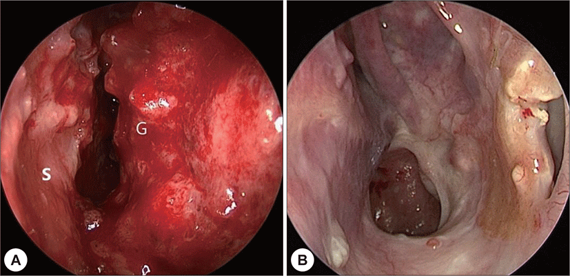
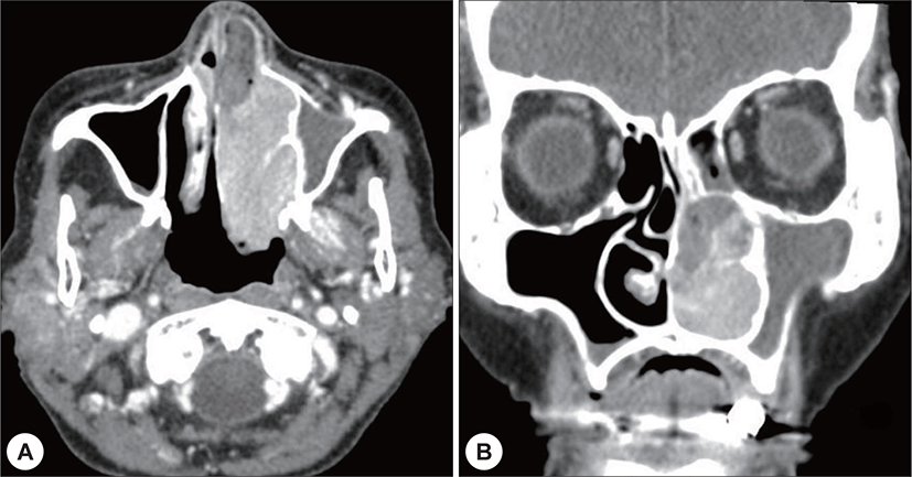
금번 응급실 내원 당시 시행한 이학적 검사에서 좌측 비강에 접촉 시 쉽게 부서지며 출혈 경향을 보이는 종물 이 발견되었다(Fig. 1). 부비동 전산화단층 촬영에서 연조 직 음영이 좌측 비강 입구에서부터 비인두까지 확장되어 있는 것이 관찰되었다. 종물은 비중격과 상악동을 압박하 고 있었고, 하·중비갑개에 골미란이 있었으며, 조영 증 강되는 혈관 조직이 종물 내에서 발견되었다(Fig. 2).

jcohns-30-2-243-g1
Fig. 1. A : Preoperative endoscopic finding of nasal cavity showing a friable large mass of left nasal cavity. S : septum, G : glomus tumor mass. B : Endoscopic finding of nasal cavity 5 months after operation showing a clear nasal cavity and no evidence of recurrence.
Download Original Figure
jcohns-30-2-243-g2
Fig. 2. Contrast enhanced CT image showing a 33×55×23 mm-sized mass. The mass had highly enhanced tubular vessels and caused bony erosions of left inferior and middle turbinates (A : axial view. B : coronal view).
Download Original Figure

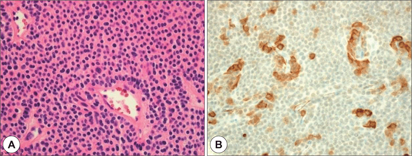
국소 마취 하에 펀치 생검을 시행한 종물은 병리조직 학적 소견 상 혈관 주위에 사구종양 세포들이 위치해 있었다. 사구종양 세포는 둥근 모양으로, 호산성의 세포 질을 가지고 있었고, 특수 염색을 한 결과 smooth muscle actin(SMA) 양성 소견이 보여 사구종양으로 확진되 었다(Fig. 3). 이에 따라 환자의 연령, 종물의 크기 및 위 치를 고려하여 수술 전 혈관 조영술 및 색전술을 계획 하였다.

jcohns-30-2-243-g3
Fig. 3. Histological findings. A : Glomus cells surrounding separate vessel tissues of variable sizes (H&E stain, ×400). Glomus cells have rounded nuclei and eosinophilic cytoplasm. B : Immunohistochemical staining showing that the tumor reacted positively (smooth muscle actin stain, ×400).
Download Original Figure

혈관 조영술 도중 접형구개동맥의 분지들을 통해 혈 액 공급을 받는 종물이 좌측 비강 내에 관찰되어 해당 혈관에 색전술을 시행하였고(Fig. 4), 15시간 후 전신마 취 하 내시경적 비강 종물 절제술을 시행하였다. 비입 구에서부터 좌측 하비갑개 후방까지 쉽게 부스러지는 종물이 있는 것을 확인하였고, 미세 분쇄기를 이용하여 종물을 제거하면서 범위를 좁혀 들어가서 종양의 기원 범위로 보이는 비강 외측벽의 출혈 부위를 전기 소작기 로 지혈하였다. 종양의 기원 부위는 미만성으로 퍼져 있었으나, 종물 제거 시 소량의 출혈만 있었다. 수술 후 5개월간 외래 관찰 중 재발 소견은 없었다(Fig. 1).

jcohns-30-2-243-g4
Fig. 4. A : Angiography of Left external carotid artery. The hypervascular tumor is supplied by the left sphenopalatine artery and branches (arrows). B : Post-procedure angiography. Superselction with microcatheter and embolization was performed. No visible hypervascular tumor after embolization was found.
Download Original Figure

고 찰

지금까지 보고된 30개의 증례들에 의하면 사구종양 이 발생한 연령대는 평균 54.4세이며, 60세 이상에서 발 생한 경우가 12예였다. 또한 남성보다 여성에서 높은 발병률을 보였다.5)

사구종양이 비강 내에서 발견되는 경우 내시경적, 조 직학적 소견이 다른 종양과 유사한 경우가 많아 감별이 중요하다. 감별에 주의해야 할 종양은 사구맥관종(glomangioma), 사구맥관근종(glomangiomyoma), 사구맥 관혈관주위세포종(glomangiopericytoma) 등이 있다. 사구종양은 병리조직학적 소견 상 사구세포가 주를 이 루며, 사구세포가 둘러싸고 있는 혈관 성분이 매우 적 으며, 평활근 성분은 거의 없는 것이 특징이다. 그에 비 해 사구맥관종은 사구종양에 비해 혈관 성분이 풍부하 며, 사구맥관근종은 사구종양에 비해 혈관과 평활근 성 분이 풍부하다.3,6) Smooth muscle actin, vimentin, collagen 등의 면역화학염색법이 감별 진단에 사용되고 있 다.6-8) 사구맥관혈관주위세포종은 일반적으로 잘 부서 지며, 출혈이 쉽게 일어나고, 전산화단층촬영 및 자기공 명영상 등의 영상학적 검사상 연부 종양 형태를 보이기 때문에 임상 소견만으로는 사구종양과 감별하기가 어 렵다. 그러나 사구체 유사 근육양 세포(perivascular glomus-like myoid cells)의 증식으로 발생하기에 조직 학적으로 감별이 된다.9)

현재까지 보고된 사구종양의 증례 중 대부분이 단순 절제로 치료하였고, 환자가 수술적 치료를 거부하여 방 사선으로 치료한 1예가 있었다.10) 수술로 치료한 증례 중 Shimono11)는 1240 mL의 출혈이 있었다고 보고하였 다. 이 증례는 사골동에서 발생한 사구종양이어서 비중 격에서 발생한 사구종양보다 지혈이 쉽지 않았을 것으 로 생각된다.

두경부 영역의 경정맥 사구종양은 수술 전 색전술을 시행하는 것이 표준치료이나, 비강의 사구종양에서 색 전술 후 수술을 시행한 것은 본 증례가 최초이다. 사구 종양이 비중격에 위치하고 크기가 작은 경우는 수술적 절제 시 대량 출혈의 위험성은 낮아 보인다. 그러나 사 구종양의 위치가 부비동 안에 있거나, 비강 측벽에 있 어서 위치상 지혈이 용이하지 않은 경우, 사구종양의 영양혈관 조직이 발달하여 종물 제거 도중 출혈의 위험 이 커 보이는 경우, 사구종양이 대혈관의 분지에서 직 접 혈액 공급을 받는 경우 등에서는 대량 출혈을 예방 하기 위해 수술 전 혈관 조영술 및 색전술을 고려해 볼 수 있겠다. 특히 본 증례와 같이 고령인 경우 출혈에 더 세심한 주의를 기울이는 것이 필요하다.

REFERENCES

1).

Ahn DB, Sin CM, Park JH, Kim JS. Glomus tumor of the nasal septum: a case report. J Rhinology 2008;15(2):156-9.

2).

Potter AJ, Khatib G, Peppard SB. Intranasal glomus tumor. Arch Otolaryngol 1984;110(11):755-6.

3).

Weiss SW, Goldbum JR. Perivascular tumors. In: Weiss Sw, Goldbum JR, editors. Soft Tissue Tumors, 4th ed. St Louis: Mosby Year Book;2001. p.985-1035.

4).

Li XQ, Hisaoka M, Morio T, Hashimoto H. Intranasal pericytic tumors (glomus tumor and sinonasal hemangiopericytoma-like tumor): report of two cases with review of the literature. Pathol Int 2003;53(5):303-8.

5).

Koh YW, Lee BJ, Cho KJ. Glomus tumor of the sinonasal tract: two case reports and a review of literature. Korean J Pathol 2010;44(3):326-9.

6).

Ahmed A, Sheehan A, Dugar J. Intranasal glomangioma. Rhinology 2003;41(1):58-60.

7).

Chu PG, Chang KL, Wu AY, Weiss LM. Nasal glomus tumors: report of two cases with emphasis on immunohisto-chemical features and differential diagnosis. Hum pathol 1999;30(10):1259-61.

8).

Shek TW, Hui Y. Glomangiomyoma of the nasal cavity. Am J Otolaryngol 2001;22(4):282-5.

9).

Byun JY, Lee YJ, Koh ES, Lee JY. A case of glomangiopericytoma of the ethmoid sinus. Korean J Otorhinolaryngol-Head Neck Surg 2013;56(4):240-3.

10).

Gaut AW, Jay AP, Robinson RA, Goh JP, Graham SM. Invasive glomus tumor of the nasal cavity. Am J Otolaryngol 2005;26(3):207-9.

11).

Shimono T, Hayakawa K, Yamaoka T, Nishimura K, Takasu K, Mimaki S. Case report: glomus tumour of the nasal cavity and paranasal sinuses. Neuroradiology 1998;40 (8):527-9.