Journal of Clinical Otolaryngology Head and Neck Surgery
The Busan, Ulsan, Gyeoungnam Branch of Korean Society of Otolaryngology-Head and Neck Surgery
Case Report

비강내시경적 누낭비강문합술 후 발생한 난치성 추정 육아종의 구제 치료: 누도 탐침 보조 비강 내 돌출 및 선택적 소작술

구형본1https://orcid.org/0000-0002-1342-9450, 김종빈1https://orcid.org/0009-0001-9015-5561, 이재훈1,*https://orcid.org/0000-0002-0856-8173
Hyung Bon Koo1https://orcid.org/0000-0002-1342-9450, Jong Bin Kim1https://orcid.org/0009-0001-9015-5561, Jae Hoon Lee1,*https://orcid.org/0000-0002-0856-8173
1원광대학교 의과대학 이비인후과학교실
1Department of Otolaryngology-Head and Neck Surgery, Wonkwang University School of Medicine, Iksan, Korea
*Corresponding author: Jae Hoon Lee, Department of Otolaryngology-Head and Neck Surgery, Wonkwang University School of Medicine, Iksan 54538, Korea, Tel: +82-63-859-1441, Fax: +82-63-841-6556, E-mail: Leejaehoon64@gmail.com

© Copyright 2026. The Busan, Ulsan, Gyeoungnam Branch of Korean Society of Otolaryngology-Head and Neck Surgery. This is an Open-Access article distributed under the terms of the Creative Commons Attribution Non-Commercial License (http://creativecommons.org/licenses/by-nc/4.0/) which permits unrestricted non-commercial use, distribution, and reproduction in any medium, provided the original work is properly cited.

Received: Jun 17, 2026; Revised: Jul 15, 2026; Accepted: Aug 05, 2026

Published Online: Sep 30, 2026

ABSTRACT

Postoperative ostium granulomas after dacryocystorhinostomy (DCR) may compromise the internal common opening (ICO) and cause functional failure. Although observation and topical therapy are appropriate for many early or nonobstructive lesions, deep inner or bang-on-ICO lesions may be difficult to expose and may recur after office-based removal. We report a patient with recurrent, clinically presumed granuloma adjacent to the ICO after endoscopic DCR. A size 00 lacrimal probe was introduced through the lower canaliculus to gently exteriorize the lesion into the nasal cavity under endoscopic visualization, and the exposed portion was selectively treated with a needle-tip monopolar electrocautery. At the 1-year follow-up, the ostium remained patent, and no recurrence of the presumed granuloma was observed. The Munk score improved from 3 preoperatively to 0 at the 1-year follow-up, and lacrimal irrigation demonstrated free passage without regurgitation; the fluorescein dye disappearance test was not performed. This single case suggests that probe-assisted exteriorization followed by selective ablation may be useful as a salvage exposure-and-ablation technique for selected mobile, refractory inner or bang-on-ICO lesions. Further cases are required to establish safety and reproducibility.

Keywords: Dacryocystorhinostomy; Granuloma; Internal common opening; Lacrimal probe; Electrocoagulation

서론

비강내시경적 누낭비강문합술(endoscopic dacryocystorhinostomy, DCR)은 비루관 폐쇄의 치료를 위해 누낭과 비강 사이에 직접적인 배액 통로를 만드는 수술이다. 충분한 골공(bony ostium), 누낭의 완전한 개방(marsupialization), 점막-점막 접합을 통해 안정적으로 상피화된 배액로를 확보하는 것이 핵심이다.1–4)

DCR 후 ostium 주변에 발생하는 육아종(granuloma)은 비정상적 창상 치유, 노출된 골, 감염, 가피, 반복적인 비강 내 조작, 실리콘관에 의한 기계적 자극 등과 관련될 수 있다.1,4–6) 특히 ostium 기저부의 inner type 또는 ICO(internal common opening)를 직접 덮는 bang-on-ICO type 육아종은 기능적 눈물 배출 경로를 침범하고, 위치가 깊어 비강 내에서 병변의 경계와 기저부를 확인하기 어렵다는 점에서 치료가 까다롭다.5,6)

초기 또는 비폐쇄성 ostium granuloma에는 관찰, 국소 비강 스테로이드, 병소 내 스테로이드 주사 등의 보존적 치료를 우선 고려할 수 있으며, 지속성·재발성 또는 기능적 폐쇄를 유발하는 병변에는 내시경적 절제나 revision DCR이 필요할 수 있다.5–8) 본 증례에서는 반복적인 외래 처치에도 재발한 ICO 인접 추정 육아종을 누도 탐침으로 비강 내로 돌출시켜 내시경 시야에 노출한 뒤 선택적으로 소작(누도 탐침 보조 비강 내 돌출 및 선택적 소작술: Probe-Assisted Intranasal Exteriorization and Selective Cauterization)하였다. 본 술식의 핵심은 소작 자체가 아니라, 비강 내 접근이 어려운 병변을 눈물소관 경로를 통해 비강 내로 돌출시켜 병변과 정상 구조를 직접 구분하도록 하는 노출 단계에 있다. 본 연구는 본원 기관심의위원회의 심의면제 승인을 받아 수행되었다(IRB No. 2026-02-016).

증례

35세 여성 환자가 유루증(epiphora)과 안구분비물(eye discharge)을 주소로 내원하였다. 우측 눈물길 실리콘관 삽입술을 받은 후에도 증상 호전이 없었다. 검사에서 우측 비루관 폐쇄가 확인되었고, 눈물길 탐침검사(lacrimal probing)에서 공통눈물소관(common canaliculus) 폐쇄는 없었으나 화농을 동반한 누낭 확장 소견이 있어 비강내시경적 DCR을 시행하였다. 상부 눈물소관을 통해 조명을 삽입한 후 비점막 절개, 골공 형성, 누낭 개방, 누낭-비점막 피판 형성 및 실리콘관 삽입을 시행하였다. 실리콘관은 DCR 2개월 후에 제거되었다.

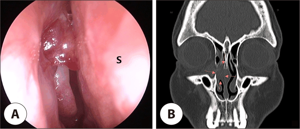
수술 6개월 후부터 누런 코와 코막힘이 지속되어 이비인후과로 의뢰되었다. 비강내시경검사와 부비동 전산화단층촬영에서 우측 DCR ostium의 ICO 인접 부위에 연조직 병변이 확인되었고(Fig. 1), 실리콘관은 이미 제거된 상태였다. 병변의 외관, 위치 및 임상 경과를 바탕으로 임상적으로 육아종에 합당하다고 판단하여 조직학적 검사는 시행하지 않았으며, 추정 육아종(presumed granuloma)으로 진단하였다. 외래에서 3개월 동안 주기적으로 cup forceps로 병변을 제거한 뒤, dexamethasone/neomycin/polymyxin B 안연고 0.3–0.5 mL를 1-mL 주사기와 18-gauge suction tip을 이용하여 병변에 도포하였다. 매일 국소 비강 스테로이드 분무제도 사용하였으나 병변은 반복적으로 재발하였다.
jcohns-37-3-119-g1
Fig. 1. A clinically presumed granuloma adjacent to the right internal common opening (A) and the corresponding soft-tissue lesion indicated by yellow arrows on a coronal non-contrast paranasal sinus computed tomography image (B). S: septum.
Download Original Figure

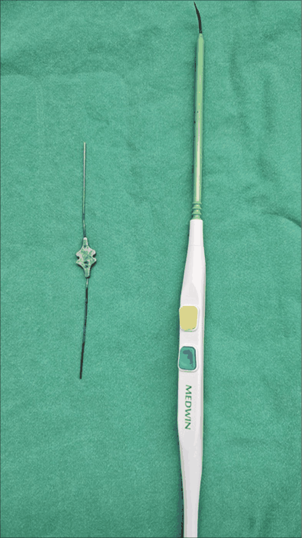
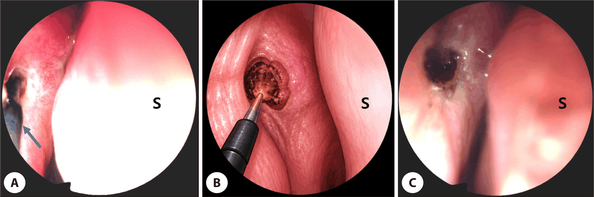
안과와 이비인후과의 협진하에 국소마취로 수술을 시행하였다. 30° 비강내시경을 이용하여 ostium과 ICO의 관계를 확인한 뒤, 안과 술자가 하부 눈물소관을 통해 size 00 lacrimal probe(누도 탐침)를 공통눈물소관을 따라 ICO 방향으로 전진시켰다. 탐침으로 ostium 내측의 병변을 비강 방향으로 부드럽게 밀어낸 후, 비강 내로 돌출된 병변을 직접 확인하였다. Needle-tip monopolar electrocautery의 전극 끝을 접근 각도에 맞게 구부리고, 출력은 coagulation mode 15 W로 설정하였다(Fig. 2). 소작 범위는 비강 내로 돌출된 병변으로 한정하였다. 열손상을 줄이기 위해 전극을 1–2초씩 짧고 간헐적으로 활성화하였다. 최종적으로 누도 탐침에 의해 비강내로 돌출되는 병변이 없음과 비강내시경을 통해 DCR ostium 및 ICO의 개방성을 확인한 후 수술을 종료하였다(Fig. 3). 추가적인 실리콘관은 삽입하지 않았다. 1년간 추적 관찰한 결과 누런 비루와 코막힘은 재발하지 않았고, 비강내시경검사에서 DCR ostium과 ICO가 잘 개방되어 있었으며 추정 육아종은 관찰되지 않았다(Fig. 4). Probe-Assisted Intranasal Exteriorization 수술 전과 술 후 1년의 Munk score는 각각 3점과 0점이었다. 술 후 눈물길 관류검사에서는 원활한 통과가 확인되었고 역류는 관찰되지 않았다. Fluorescein dye disappearance test는 시행하지 않았다. 유루증은 소실되었으며, 코막힘과 누런 코 등의 비강 증상도 호소하지 않았다.
jcohns-37-3-119-g2
Fig. 2. A lacrimal probe (left) and a needle-tip monopolar electrocautery (right).
Download Original Figure
jcohns-37-3-119-g3
Fig. 3. Intraoperative endoscopic view of the presumed granuloma exteriorized into the nasal cavity with a lacrimal probe and selectively cauterized with a needle-tip monopolar electrocautery (blue arrow) (A), a schematic illustration of the probe-assisted exteriorization and cauterization technique (B), and the appearance after cauterization (C). S: septum.
Download Original Figure
jcohns-37-3-119-g4
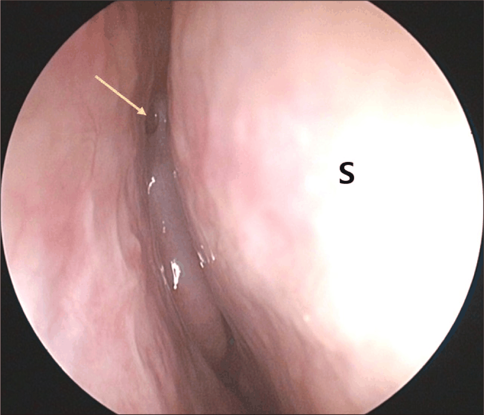
Fig. 4. Endoscopic view at the 1-year follow-up showing a patent internal common opening (yellow arrow) without recurrence of the presumed granuloma. S: septum.
Download Original Figure

고찰

DCR 후 ostium granuloma는 ICO와의 관계에 따라 임상적 의미가 달라진다. Jo 등은 병변을 inner, edge, extra type으로 분류하였고, Ali 등은 peri-ICO, bang-on-ICO, peritubal, bridge type 등을 포함하여 보다 세분하였다.5,6) 본 증례의 병변은 ostium 기저부에서 ICO를 직접 덮는 양상으로, inner 및 bang-on-ICO type에 해당하였다. 이러한 병변은 기능적 배액로를 직접 방해할 수 있고 비강 내에서 기저부가 충분히 노출되지 않아 반복적인 표면 절제 후에도 잔여 병변이 남을 수 있다.

모든 ostium granuloma가 수술적 치료를 필요로 하는 것은 아니다. Ma 등은 비강 내 스테로이드 치료와 경과 관찰 중 ostium granuloma가 시간 경과에 따라 퇴축할 수 있음을 보고하여, 초기 또는 비폐쇄성 병변에서는 보존적 치료가 우선되어야 함을 뒷받침하였다.8) 병소 내 triamcinolone 주사 역시 진행성 병변에 대한 비수술적 치료로 보고되었다.5) 따라서 본 술식은 일차 치료가 아니라, 충분한 보존적 치료에도 병변이 지속되거나 재발하고, ICO를 직접 침범하며, 비강 내 접근만으로 병변의 기저부를 안전하게 노출하기 어려운 경우에 한정하여 고려해야 한다. 본 증례는 3개월간 반복적인 forceps 제거, 국소 안연고 및 비강 스테로이드 치료에도 재발하여 이러한 조건에 해당하였다.

본 술식의 독창성은 monopolar electrocautery의 사용이 아니라 누도 탐침을 이용한 병변의 비강 내 돌출(exteriorization)에 있다. 탐침을 개방된 공통눈물소관을 따라 ICO까지 전진시켜 병변을 비강 쪽으로 이동시키면, 깊은 inner 또는 bang-on-ICO 병변을 내시경 시야에 노출하여 병변과 정상 ostium 점막을 구분할 수 있다. 이에 따라 비강 내에서 맹목적으로 깊은 조직을 절제하는 대신, 실제로 배액로를 막는 돌출 병변만을 선택적으로 처리할 수 있다. 적용 대상은 충분한 보존적 치료에도 병변이 지속되거나 재발하며, 탐침으로 부드럽게 이동 가능한 국소성 inner 또는 bang-on-ICO granuloma이다. 또한 공통눈물소관이 개방되어 있고, ostium을 통해 병변을 비강 내로 충분히 돌출시킬 수 있어야 한다.5,6)

ICO와 공통눈물소관에 인접하여 monopolar electrocautery를 사용하는 만큼 thermal injury, canalicular injury 및 cicatricial stenosis의 위험을 간과할 수 없다. 전기수술의 조직 손상은 출력, 전류 밀도, 접촉 면적 및 활성화 시간의 영향을 받으므로, 전극을 가장 낮은 유효 출력으로 짧고 간헐적으로 활성화하고 비강 내로 돌출된 병변에만 접촉하며 정상 ICO 점막과 전극 사이의 거리를 유지해야 한다.9) 금속 탐침과 활성 전극의 직접 접촉, ICO rim의 원주형 소작 및 심부 탄화를 유발하는 반복 소작을 피해야 한다. 병변의 창백화·수축과 지혈이 확인되고 ICO가 직접 보이면 즉시 종료하는 것이 바람직하다. 이러한 예방 조치가 위험을 완전히 제거하는 것은 아니므로, 병변을 안전하게 노출할 수 없거나 병변과 정상 조직의 경계가 불명확하면 소작을 중단하고 냉기구를 이용한 절제, bipolar 기구 또는 revision DCR 등 다른 치료를 고려해야 한다.

본 술식은 섬유화되어 움직이지 않는 병변, 광범위한 diffuse 또는 bridge-type 병변, 완전한 공통눈물소관 폐쇄, 심한 반흔성 ostium 폐쇄, 활동성 감염, 종양 또는 특이 감염이 의심되어 조직검사가 필요한 병변에는 적합하지 않다. 또한 안과와 이비인후과의 협진, 누도 탐침 조작과 비강내시경 술기에 대한 숙련이 필요하다.6,7) 본 증례 보고의 제한점은 다음과 같다. 첫째, 단일 증례이다. 둘째, 조직검사를 시행하지 않아 조직학적 확진이 없다. 셋째, 단일 증례에서 얻은 기능적 결과와 소작 조건을 일반화할 수 없다. 따라서 누도 탐침 보조 비강 내 돌출 및 선택적 소작술은 기존 치료에 반응하지 않는 이동성 inner 또는 bang-on-ICO 추정 육아종에서 고려할 수 있는 구제적 노출-소작술(salvage exposure-and-ablation technique)일 수 있으나, 안전성과 재현성을 확인하기 위해 더 많은 증례와 장기 추적이 필요하다.

Acknowledgements

Not applicable.

Funding Information

Not applicable.

Conflicts of Interest

No potential conflict of interest relevant to this article was reported.

Author Contribution

Conceptualization: Koo HB, Lee JH.

Data curation: Koo HB, Kim JB, Lee JH.

Formal analysis: Koo HB, Kim JB, Lee JH.

Methodology: Koo HB, Lee JH.

Supervision: Lee JH.

Writing-original draft: Koo HB, Lee JH.

Writing-review & editing: Koo HB, Kim JB, Lee JH.

Ethics Approval

This study was approved for an exemption by the Institutional Review Board (IRB) of our institution (IRB No. 2026-02-016).

References

1.

Locatello LG, De Zan ER, Caiazza N, Tarantini A, Lanzetta P, Miani C. A critical update on endoscopic dacryocystorhinostomy. Acta Otorhinolaryngol Ital 2024;44(6):351-60.

2.

Woog JJ, Kennedy RH, Custer PL, Kaltreider SA, Meyer DR, Camara JG. Endonasal dacryocystorhinostomy: a report by the American Academy of Ophthalmology. Ophthalmology 2001;108(12):2369-77.

3.

Eichhorn K, Harrison AR. External vs. endonasal dacryocystorhinostomy: six of one, a half dozen of the other? Curr Opin Ophthalmol. 2010;21(5):396-403.

4.

Yim M, Wormald PJ, Doucet M, Gill A, Kingdom T, Orlandi R, et al. Adjunctive techniques to dacryocystorhinostomy: an evidence-based review with recommendations. Int Forum Allergy Rhinol 2021;11(5):885-97.

5.

Jo A, Lee SH, Song WC, Shin HJ. Effects of ostium granulomas and intralesional steroid injections on the surgical outcome in endoscopic dacryocystorhinostomy. Graefes Arch Clin Exp Ophthalmol 2018;256:1993-2000.

6.

Ali MJ, Wormald PJ, Psaltis AJ. The dacryocystorhinostomy ostium granulomas: classification, indications for treatment, management modalities and outcomes. Orbit 2015;34(3):146-51.

7.

Park J, Kim H. Office-based endoscopic revision using a microdebrider for failed endoscopic dacryocystorhinostomy. Eur Arch Otorhinolaryngol 2016;273:4329-34.

8.

Ma ST, Shin S, Navale A, Woo KI. Regression course of ostium granulomas under topical intranasal steroids coverage after endoscopic dacryocystorhinostomy. Sci Rep 2024;14:17089.

9.

Massarweh NN, Cosgriff N, Slakey DP. Electrosurgery: history, principles, and current and future uses. J Am Coll Surg 2006;202(3):520-30.