

Introduction
Cemento-ossifying fibroma (COF) is a subtype of ossifying fibroma, which is benign. COF is composed of fibrous connective tissue that includes varying degrees of calcified material similar to bone and/or cementum.1) COF commonly has a thin radiopaque line and mixed-density internal appearance on computed tomography (CT) scans. In some cases, COF may appear entirely radiopaque with a ground-glass pattern. Under microscopic examination, COF is characterized by lamellar bone and cementum-like droplets, which are lined by osteoblastic rimming.2) COF most commonly develops between the third and fourth decades of life and shows marked female predominance, with a female-to-male ratio of approximately 5:1.2) COF typically exhibits slow growth and often remains asymptomatic until facial swelling becomes noticeable. COF can develop in any region of the facial skeleton. However, unlike other subtypes of ossifying fibroma, COF shows a strong predilection for the mandible (approximately 70% of cases).3) We present a rare case of COF in a 28-year-old female patient in the middle turbinate, which is an unusual site of occurrence.
Case Report
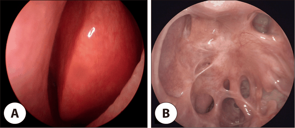
A 28-year-old female patient was referred by a plastic surgery clinic due to an abnormal finding of left nasal cavity on a CT scan. The patient reported no additional symptoms except for intermittent intranasal pain. No palpable head and neck mass were identified on physical examination. Neurological, respiratory examinations revealed no abnormalities. Endoscopic examination of the nasal cavity showed a hypertrophied left middle turbinate and a septal deviation toward the right; otherwise, the nasal cavity was unremarkable (Fig. 1).
A CT scan showed that, a focal sclerotic mass with an approximate diameter of 28 mm was confined to the left middle turbinate. The CT scan demonstrated a well-circumscribed sclerotic mass, clearly distinguishable from fibrous dysplasia (Fig. 2). The mass was extended to the left lateral lamella of cribriform plate superiorly and left lamina papyracea laterally. The border of the mass did not invade but was abutted to the lamina papyracea. No significant lymph nodes were visualized in the neck. The crista galli, bilateral nasal bones and cribriform plates were intact, and the bilateral orbits and its contents were normal.
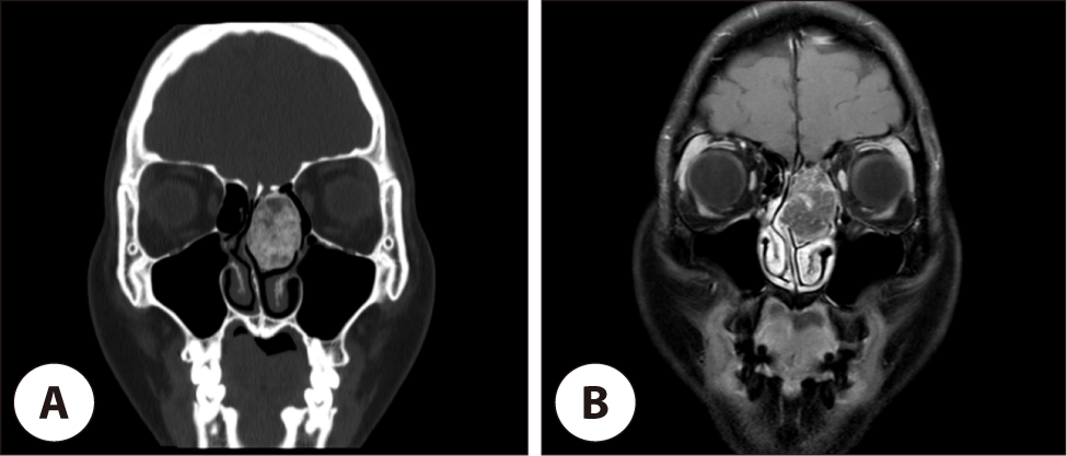
A magnetic resonance imaging (MRI) before the surgery revealed that a sclerotic mass, which was confined to the left middle turbinate, was adjacent to the vertical lamella of the left cribriform plate, left nasal septum and left lamina papyracea. It also showed that the mass did not invade the lateral lamella of cribriform plate and it was attached to the skull base (Fig. 2).
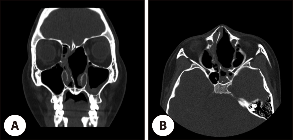
Based on the imaging studies, endoscopic piecemeal resection was planned due to the large size of the mass. Because the mass occupied nearly the entire left middle turbinate, a complete middle turbinate resection was also planned. Under general anesthesia, the left middle turbinate was resected with cut forceps and drill via navigation-assisted endoscopic approach. After resecting the left middle turbinate, the natural left frontal sinus ostium, which was obstructed by the mass, was visualized. The ostium was widened using a microdebrider. The left lateral lamella of cribriform plate, where the mass was attached, was also exposed. For the purpose of the complete resection of the mass, the remnant mass which was attached to the lateral lamella was gently trimmed with burr. During the trimming, no cerebrospinal fluid leakage was observed. The absorbable packing materials (Floseal, Algipack, and NEXPAK) were adequately applied to the area where the left middle turbinate was removed. The patient was discharged on postoperative day 1. Postoperative CT scan showed no defect of the skull base, lamina papyracea (Fig. 3). At approximately 6 months postoperatively, endoscopic examination revealed no remarkable findings (Fig. 1).
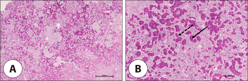
Histopathological examination revealed that the mass had well-defined margin, which consisted with the lamellar bone (Fig. 4). It also showed that cementum-like materials which contained acellular, round shape basophilic calcifications circumscribed by osteoblastic rimming. These histopathological features were distinguishable from the findings of psammomatoid ossifying fibroma (PsOF), which shows “water line” that corresponds to cystic changes and fluid accumulation. Additionally, PsOF is characterized by spherical concentric calcified ossicles (psammomatoid bodies) and its eosinophilic fringe.4) The histopathological examination supports a diagnosis of COF.
Discussion
Ossifying fibroma is a benign, bone-producing, fibrous tumor of the skeleton that is classified into two primary clinicopathologic entities: COF and juvenile ossifying fibroma. The latter is further subdivided into two distinct types, juvenile trabecular ossifying fibroma (JTOF) and PsOF.
The World Health Organization (WHO) defines COF as a well-circumscribed, sometimes encapsulated, neoplasm which consists of fibrous tissue containing variable amounts of mineralized material, such as bone and/or cementum. The WHO classifies COF as a fibro-osseous neoplasm arising from mesenchymal blast cells of the periodontal ligament with the potential to produce fibrous tissue, cementum, bone, or a combination of these components.1)
Two additional variants, JTOF and PsOF (formerly known as juvenile psammomatoid ossifying fibroma), were previously grouped with COF under fibro-osseous lesions in the WHO 2017 classification of head and neck tumors. However, in the 2022 edition, they were subsequently separated from odontogenic COF and are now classified individually as non-odontogenic benign fibro-osseous lesions.5)
The radiographic features of COF vary from well-defined radiolucent to mixed radiolucent and radiopaque lesions depending on the quantity of bone and cementum-like material. Histologically, COF is characterized by hypercellular fibroblastic stroma with variable amounts of calcified structures. The stromal fibroblastic cells show hyperchromatic nuclei and the calcified structures are composed of variable amounts of osteoid or bone and lobulated basophilic masses of cementum-like tissue resembling the cementicles that are normally found in the periodontal membrane.2) In contrast, JTOF is distinguished by long, slender strands of osteoid with hypercellular stroma composed of spindle cells with occasional mitosis.2) JTOF also shows multinucleated giant cells, pseudocystic stromal degeneration and hemorrhage cluster.6) PsOF is characterized by numerous basophilic psammoma-like bodies which shows concentric lamination with eosinophilic rimming.4)
COF arises from the periodontal ligament.7) The presence of COF in areas not associated with the periodontal ligament, such as the paranasal sinuses and orbit, remains unclear. One possibility is that primitive mesenchymal cells may differentiate similarly to the periodontal ligament, producing calcified material resembling bone and cementum. Another possibility is the presence of an ectopic periodontal membrane.8)
The most common method of managing OF is surgical excision, due to the good delimitation of the tumor.9) Small and well-defined lesions can be excised by enucleation and curettage; however, large expanding lesions require radical surgery within healthy margins and aesthetic recontouring.10)
Our case was unusual because the mass was confined to the middle turbinate, adjacent to the skull base and lateral lamella of the cribriform plate. While PsOF shows a predilection for the paranasal sinuses and nasal cavity, the occurrence of COF in such non-tooth-bearing sites is rare, reflecting its biological origin from the periodontal ligament. This can be explained either by the differentiation of primitive mesenchymal cells into cells resembling those of the periodontal ligament, which produce calcified material similar to bone and cementum, or by the presence of an ectopic periodontal membrane.8)
In our case, the mass was confined to the left middle turbinate, and no aesthetic concerns were anticipated following surgical intervention. Considering the characteristic expansile growth of COF, conservative observation posed a potential risk of continuous pressure on the skull base and orbital structures. Consequently, surgical intervention was planned and we performed left middle turbinectomy for complete surgical resection to minimize the risk of recurrence.
Conclusion
In the field of rhinology, COF specifically located within the nasal turbinate is rare. Due to its slow-growing nature, the frequency of clinical symptoms is low, and specific physical findings are often absent. Furthermore, since nasal endoscopy often fails to distinguish COF from a typical concha bullosa, diagnosis and treatment can be delayed. Such delays may inevitably necessitate a more extensive surgical resection. Therefore, performing imaging studies, such as a CT scan, when turbinate hypertrophy is observed would be beneficial for early diagnosis.


