Case Report
동측 경장근 및 두개내 농양과 반대측 외전신경 마비를 동반한 추체염 1예
Petrous Apicitis with Ipsilateral Longus Colli Abscess, Intracranial Abscesses, and Contralateral Abducens Nerve Palsy
Lee Hwangbo
1
, Sung-Won Choi
2, Se-Joon Oh
2, Seokhwan Lee
2,*
1부산대학교 의과대학 영상의학교실, 부산대학교병원 의생명연구원
2부산대학교 의과대학 이비인후과학교실, 부산대학교병원 의생명연구원
1Department of Diagnostic Radiology, Biomedical Research Institute, Pusan National University Hospital, Pusan National University School of Medicine, Busan, Korea
2Department of Otorhinolaryngology–Head and Neck Surgery, Biomedical Research Institute, Pusan National University Hospital, Pusan National University School of Medicine, Busan, Korea
*Corresponding author: Seokhwan Lee, Department of Otorhinolaryngology-Head and Neck Surgery, Biomedical Research Institute, Pusan National University Hospital, Busan 49241, Korea, Tel: +82-51-240-7335, Fax: +82-51-246-8668, E-mail:
entlsh85@gmail.com
© Copyright 2026. The Busan, Ulsan, Gyeoungnam Branch of Korean Society of Otolaryngology-Head and Neck Surgery. This is an Open-Access article distributed under the terms of the
Creative Commons Attribution Non-Commercial License (http://creativecommons.org/licenses/by-nc/4.0/) which permits
unrestricted non-commercial use, distribution, and reproduction in any
medium, provided the original work is properly cited.
Received: Jun 06, 2026; Revised: Jun 25, 2026; Accepted: Jul 05, 2026
Published Online: Sep 30, 2026
ABSTRACT
Petrous apicitis is a rare complication of acute otitis media and usually causes ipsilateral cranial neuropathy. We report a 26-year-old man with persistent left otorrhea, headache, vertigo, and fever despite prior antibiotics. Two days after admission, he developed left retro-orbital pain, horizontal diplopia, and right abducens nerve palsy. CT and MRI showed new bony erosion of the left petrous apex, abscesses in the left-dominant prepontine and left cerebellopontine cisterns, and a left longus colli abscess. Apparent diffusion coefficient (ADC) imaging showed a small diffusion-restricted focus near the right posterior cavernous sinus, without definite thrombosis on contrast-enhanced MRI. Conservative treatment with 7 weeks of broad-spectrum antibiotics and adjunctive hyperbaric oxygen therapy was selected because the primary otitis media improved rapidly and the deep lesions were difficult to drain. At 18 months, all lesions and abducens palsy had resolved. This case highlights the importance of repeat MRI when neurologic symptoms develop during otogenic infection.
Keywords: Petrous bone; Otitis media; Abducens nerve diseases; Abscess; Hyperbaric oxygenation
서론
추체염(petrous apicitis)은 급성 중이염 또는 유양돌기염의 염증이 추체첨부로 파급되어 발생하는 드문 이성 합병증이다.1,2) 추체첨부는 외전신경이 주행하는 Dorello관 및 삼차신경절이 위치한 Meckel굴과 인접하여, 병변이 진행하면 안와후방통, 외전신경 마비, 삼차신경통과 같은 두개신경 증상이 동반될 수 있다.2,3) 항생제 사용 이후 전형적인 Gradenigo 증후군은 흔하지 않지만, 진단이 지연될 경우 뇌수막염, 두개내 농양, 해면정맥동 혈전증과 같은 치명적인 합병증으로 진행할 수 있다.1,4)
추체염에서 외전신경 마비는 일반적으로 병변과 동측으로 나타나며, 양측 마비는 보고되었으나 반대측 단독 마비는 매우 드물다.3) 경장근(longus colli muscle) 농양 역시 드문 심부 경부 감염이다. 저자들은 좌측 급성 중이염 후 좌측 추체염, 동측 경장근 및 두개내 농양이 발생했으나 외전신경 마비는 반대측인 우측에 단독으로 나타났고, 보존적 치료로 호전된 1예를 보고한다.
증례
기저질환이 없는 26세 남자가 3주간 지속된 좌측 이루, 두통, 어지럼 및 38.2℃ 발열로 내원하였다. 환자는 좌측 급성 중이염으로 경구 ciprofloxacin 2주, 이후 타 병원에서 vancomycin과 ciprofloxacin 정주 치료를 1주간 받았으나 호전되지 않았다. 이전 치료 과정에서 균배양검사와 vancomycin 치료적 약물농도감시(therapeutic drug monitoring, TDM)는 시행되지 않았다.
이내시경 검사에서 좌측 고막의 소천공과 농성 이루가 관찰되어 즉시 이루 배양검사를 시행하고 1.52 mm 환기관을 삽입하였다. 순음청력검사(6분법)에서 우측 기도청력은 11 dB, 좌측은 기도 34 dB 및 골도 22 dB이었다. 내원 1주 전 측두골 전산화단층촬영(computed tomography, CT)에서는 좌측 중이강, 유양동 및 추체첨부의 연조직 음영이 있었으나 명확한 골 결손은 없었다. 내원 당시 백혈구 18,030/μL, C-반응단백 12.78 mg/dL로 상승되어 있었고, 세균성 미로염 가능성을 고려하여 vancomycin, ceftazidime 및 metronidazole 정주 치료를 시작하였다.
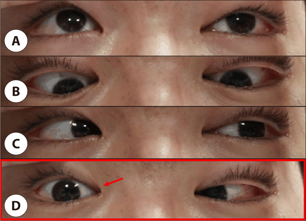
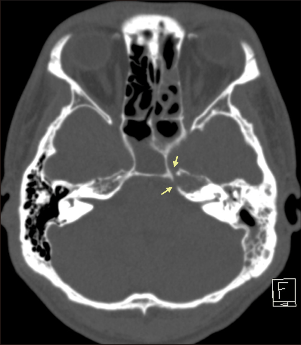
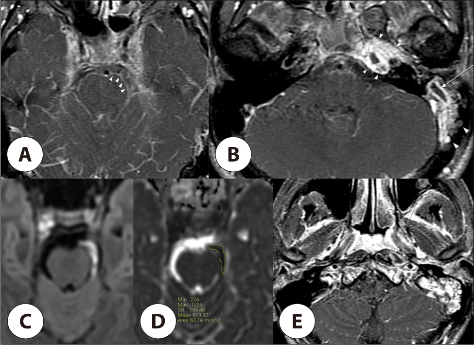
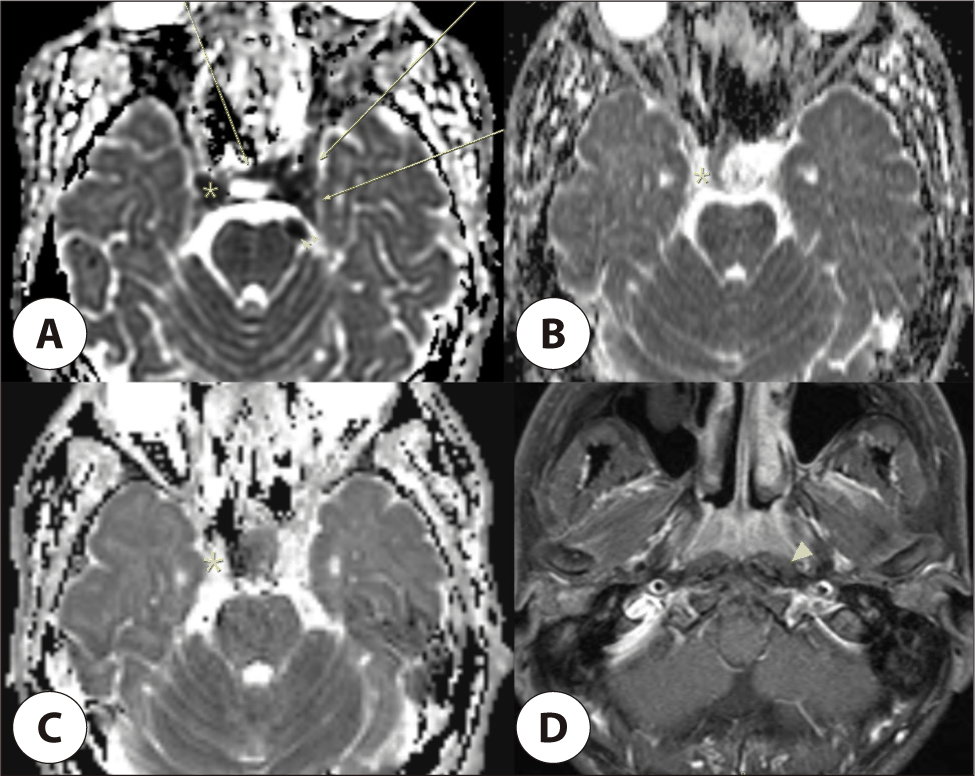
입원 2일째 좌측 안검 부종, 좌측 안와후방통 및 수평 복시가 새로 발생하였다. 안구운동검사에서 우측 주시 시 우측 안구 외전 제한이 확인되었고, 좌측 외전 제한은 없어 우측 제6뇌신경 마비로 진단하였다(Fig. 1). 이전 CT 촬영 1주일 뒤 추적 CT에서 좌측 추체첨부의 새 골 결손이 관찰되었다. 우측 접형동염도 보였으나 골벽 결손이나 안와첨부 침범은 없었다(Fig. 2). 조영증강 뇌 자기공명영상(magnetic resonance imaging, MRI)에서는 좌측 유양동 및 추체첨부 염증과 함께 좌측 우세의 전교조(prepontine cistern), 좌측 소뇌교각조(cerebellopontine cistern), 좌측 경장근 내 농양성 병변이 확인되었다(Fig. 3). 명확한 해면정맥동 혈전증은 없었으나, clinoid process 높이의 ADC(apparent diffusion coefficient) 영상에서 우측 후방 해면정맥동과 해면간정맥동 부위의 국소 제한확산이 관찰되었다(Fig. 4A). 이는 소량의 감염성 내용물 또는 초기 정맥동 침범 가능성을 시사하였지만, 다른 시퀀스에서 혈전증이나 환형 조영증강 병변으로 확인되지는 않았다.
Fig. 1.
Ocular motility findings demonstrating isolated right abducens nerve palsy. The patient was instructed to look upward (A), downward (B), leftward (C), and rightward (D). Left gaze showed preserved left-eye abduction. During right gaze, the right eye failed to abduct fully (arrow), consistent with right sixth cranial nerve palsy.
Download Original Figure
Fig. 2.
Axial temporal bone CT obtained after neurologic deterioration. Cortical dehiscence and bony erosion of the left petrous apex are noted (arrows), with persistent inflammatory opacification of the left otomastoid region.
Download Original Figure
Fig. 3.
Initial brain MRI. Axial fat-suppressed contrast-enhanced T1-weighted images show leptomeningeal enhancement in the prepontine and left cerebellopontine cisterns (A, arrowheads). Contiguous left mastoiditis and petrous apicitis are also seen, with focal nonenhancing components compatible with abscess formation (B, arrows; arrowheads indicate surrounding inflammatory change). The nonenhancing component demonstrates restricted diffusion on diffusion-weighted imaging (C) and a low apparent diffusion coefficient value of 614×10-6 mm2/s on apparent diffusion coefficient (ADC) mapping (D). Contrast-enhanced T1-weighted imaging also demonstrates ipsilateral longus colli muscle abscess formation (E, arrow).
Download Original Figure
Fig. 4.
Serial MRI findings from the initial study to 18-month follow-up after treatment. On the initial apparent diffusion coefficient (ADC) map at the level of the clinoid processes (A), focal low-ADC areas suggestive of restricted diffusion are seen in the right posterior cavernous sinus/paraclinoid region (asterisk) and along the left cavernous and intercavernous sinus regions (arrows). A portion of the cisternal abscess is also visible (arrowheads). These diffusion-restricted foci decreased on the 2-month follow-up ADC map (B) and were no longer evident on the 18-month follow-up ADC map (C). The 18-month fat-suppressed contrast-enhanced T1-weighted image shows complete resolution of the left longus colli muscle abscess (D, arrowhead).
Download Original Figure
뇌척수액 검사에서는 호중구 우세 세포증가(총 세포 250개/uL중 호중구 23.8%), 단백 상승(27.2mg/dL)이 확인되어 세균성 뇌수막염에 합당하다고 판단하였다. 내원 전부터 항생제가 사용된 영향으로 이루, 혈액 및 뇌척수액 배양검사에서 동정된 균은 없었다. 항생제는 cefepime과 vancomycin 중심으로 조정하였고, 치료 중 혈액검사 이상으로 meropenem 단독요법을 약 1주간 일시 사용한 뒤 cefepime과 vancomycin을 재개하여 총 7주간 유지하였다.
유양동삭개술 및 추체 병변 배농을 검토하였으나, 환기관 삽입과 항생제 투여 후 이루가 빠르게 감소하고 중이염이 조절되었으며 청력도 비교적 보존되어 있었다. 또한 주된 농양성 병변이 전교조, 소뇌교각조 및 경장근에 위치하여 유양동 수술만으로 직접 배농의 이득이 제한적이라고 판단하였다. 이에 수술적 배농 없이 항생제 치료를 지속하고 영상 소견을 면밀히 추적하였다.
항생제 치료 2주 후 중이염과 이루는 호전되었으나 두개내 및 경장근 농양의 영상학적 호전은 충분하지 않아, 보조적 고압산소치료를 2기압에서 120분씩 하루 1회, 총 14회 시행하였다. 치료 2개월째 MRI에서 우측 후방 해면정맥동 인접 병변과 좌측 해면정맥동 및 해면간정맥동 제한확산 병변은 감소하여 항생제 중단 후 퇴원하였다(Fig. 4B). 우측 외전신경 마비와 복시는 치료 3개월째에 완전 호전되었다. 치료 종료 5개월째 시행한 MRI는 퇴원당시(2개월) 촬영 MRI와 동일한 상태로 악화가 없음이 확인되었으며, 치료 종료 18개월 뒤 MRI에서는 두개저, 두개내 및 경장근 병변이 소실되었다(Fig. 4C and D).
고찰
본 증례의 가장 중요한 특징은 좌측 급성 중이염에 의한 좌측 추체염과 심부 농양이 확인되었음에도, 외전신경 마비는 반대측인 우측에 단독으로 발생했다는 점이다. 추체염에서 외전신경 마비는 대개 병변과 동측으로 나타나며, 양측 마비가 보고된 경우는 있으나 반대측 단독 마비는 흔하지 않다.2,3) 따라서 이성 감염 환자에서 외전신경 마비의 방향이 중이염 병변과 일치하지 않더라도 추체염 및 두개내 합병증을 배제해서는 안 된다.
반대측 외전신경 마비의 기전은 가설로만 해석할 수 있다. 조영증강 MRI에서 명확한 해면정맥동 혈전증이나 우측 추체첨부 병변은 없었고, 경미한 우측 접형동염도 골벽 결손이나 안와첨부 침범이 없어 주된 원인으로 보기 어려웠다. 다만 ADC 영상에서 우측 후방 해면정맥동 및 해면간정맥동 부위의 국소 제한확산이 관찰되었다. 제한확산은 농성 또는 감염성 내용물을 시사할 수 있지만, 단독 소견으로 농양이나 혈전증을 확정하거나 인과관계를 증명할 수는 없다.5) 따라서 좌측 추체염에 의한 기저수막염 또는 전교조 농양의 반대측 파급과 ADC에서만 보인 미세한 우측 해면정맥동 또는 해면간정맥동 침범이 함께 작용했을 가능성이 있다.
반복 영상검사도 중요하였다. 내원 1주 전 CT에서는 골파괴가 없었으나, 안와후방통과 외전신경 마비 후 시행한 CT와 MRI에서 좌측 추체첨부 골 결손, 두개내 농양, 경장근 농양 및 ADC에서만 뚜렷한 우측 해면정맥동 인접 제한확산 병변이 확인되었다. CT는 골파괴 평가에 유용하지만, 조영증강 MRI는 수막염, 두개내 파급, 두개저 및 상경부 연부조직 침범, 해면정맥동 또는 정맥동 합병증 평가에 더 적합하다.6) DWI(diffusion-weighted imaging)/ADC는 감염성 내용물을 확인하는 보조 시퀀스이나 조영증강 영상 및 임상 경과와 함께 해석해야 한다.5) 따라서 신경학적 증상이 새로 발생하거나 발열이 지속될 때는 조영증강 MRI와 DWI/ADC를 포함한 영상검사를 반복 시행해야 한다.
경장근 농양도 특이한 소견이다. 경장근은 경추 전방 추전공간의 심부 근육으로, 감염성 농양은 매우 드물다.7) 진행된 세균성 부비동염에서 양측 경장근 농양이 발생한 보고가 있으나, 본 증례의 우측 부비동염은 경미하며, 병변과 반대측인 좌측 경장근 농양은 유발하지 않았을 것으로 판단된다. 특히, 추체염에서 동측 경장근으로 국소 파급된 증례는 현재까지 보고된바가 없다.8) 본 증례에서는 좌측 추체첨부의 새 골 결손과 두개저 심부 공간의 연속성을 통해 염증이 하방으로 파급된 것으로 추정된다. 따라서 추체염이 의심될 때 병변을 측두골에 한정하지 말고 두개저와 상경부까지 포함하여 평가할 필요가 있다.
추체염 치료에는 아직 확립된 단일 기준이 없다. Gadre와 Chole은 경험적 항생제를 우선 투여하고, 48시간 이내 호전이 없거나 이후 악화되는 경우 수술을 고려하는 접근을 제시하였다.4) 농양의 진행, 골괴사, 조절되지 않는 감염, 청력 소실 또는 진행성 신경학적 악화가 있으면 배농을 적극적으로 검토해야 한다.4,9,10) 본 증례에서는 중이염이 빠르게 조절되었고 주요 병변이 이과적 접근만으로 충분히 배농되기 어려워 보존적 치료를 선택하였다. 고압산소치료는 항생제와 수술을 대체하는 치료가 아니라 보조요법으로 해석해야 하며, 두개내 농양 및 악성외이도염에서 보조적으로 사용된 보고가 있다.11-13) 본 증례에서는 두개저 및 두개내 농양이 남아 있고 수술적 접근의 이득이 제한적인 상황에서 선택적으로 병행하였다.
결론
급성 중이염 환자에서 반대측 외전신경 마비가 발생하더라도 추체염과 두개내 합병증 가능성을 고려해야 한다. 초기 CT에서 골파괴가 명확하지 않더라도 신경학적 증상, 안와후방통, 지속 발열이 새로 발생하면 조영증강 MRI와 DWI/ADC를 포함한 영상평가로 두개내, 두개저 및 상경부 파급 범위를 재평가해야 한다. 원발 감염이 빠르게 조절되고 심부 병변의 직접 배농 이득이 제한적인 경우에는 장기 항생제 치료, 면밀한 영상 추적 및 선택적 고압산소치료를 포함한 보존적 치료를 고려할 수 있다.