

Introduction
Infectious mononucleosis (IM), caused by the Epstein-Barr virus (EBV), typically presents with the classic triad of fever, lymphadenopathy, and exudative tonsillopharyngitis.1) While usually self-limiting, the intense systemic inflammatory response triggered by EBV can act as a profound physiological stressor, potentially precipitating life-threatening metabolic emergencies such as diabetic ketoacidosis (DKA).1,2) We report a case of acute EBV tonsillitis triggering severe DKA in a young adult with type 2 diabetes. This case highlights the diagnostic challenge of distinguishing EBV from bacterial tonsillitis; symptomatic overlap often leads to inappropriate aminopenicillin administration, which frequently induces characteristic maculopapular rashes in EBV patients.3,4) Through this report, we emphasize the necessity of early differential diagnosis in refractory tonsillitis and the importance of vigilant metabolic monitoring during acute viral infections.
Case Report
A 20-year-old female with a medical history of type 2 diabetes mellitus (T2DM) and asthma (both diagnosed at age 10) presented with a 5-day history of severe odynophagia, fever (>38.0℃), and intractable vomiting. Despite her early diagnosis, she had been non-compliant with insulin and inhaler therapy for three years. Family history was notable for T2DM in both parents. Before transfer, she was treated for acute tonsillitis with intravenous ampicillin/sulbactam at an outside facility. However, as her clinical condition deteriorated and her mental status shifted from alert to drowsy, she was emergently transferred to the tertiary medical center.
Upon arrival, vital signs were: blood pressure 120/80 mmHg, heart rate 88 bpm, respiratory rate 17/min, and temperature 36.0℃. Initial arterial blood gas analysis (ABGA) revealed life-threatening metabolic acidosis (pH 6.80, pCO2 7 mmHg, HCO3- 3.0 mEq/L) and hyperglycemia (357 mg/dL). With an anion gap of 28, urinalysis confirmed significant glucosuria (2+) and ketonuria (2+). Laboratory tests showed a white blood cell count of 23,200/µL, C-reactive protein 4.64 mg/dL, HbA1c 12.0%, and serum ketone bodies 9,654.1 µmol/L. Notably, markedly elevated pancreatic enzymes (amylase 937 U/L, lipase 2,530 U/L) and ammonia (295 µg/dL) initially raised suspicion of acute pancreatitis.
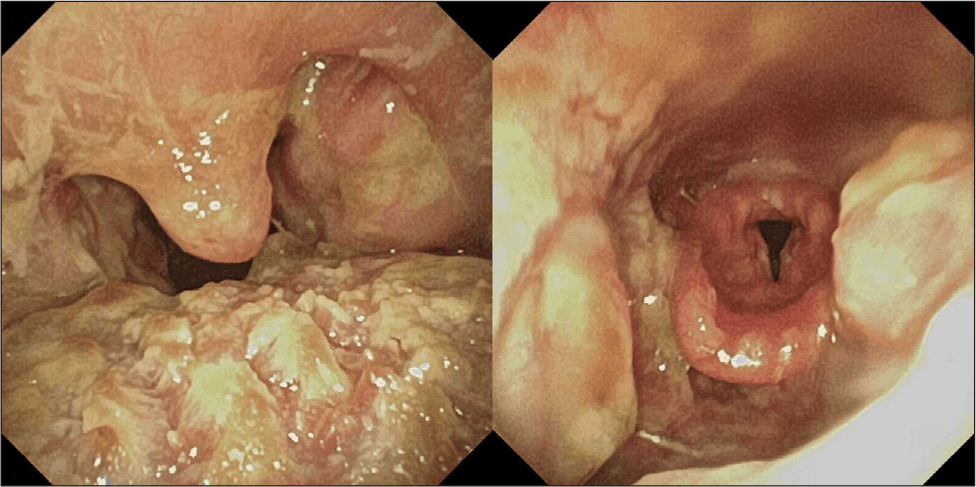
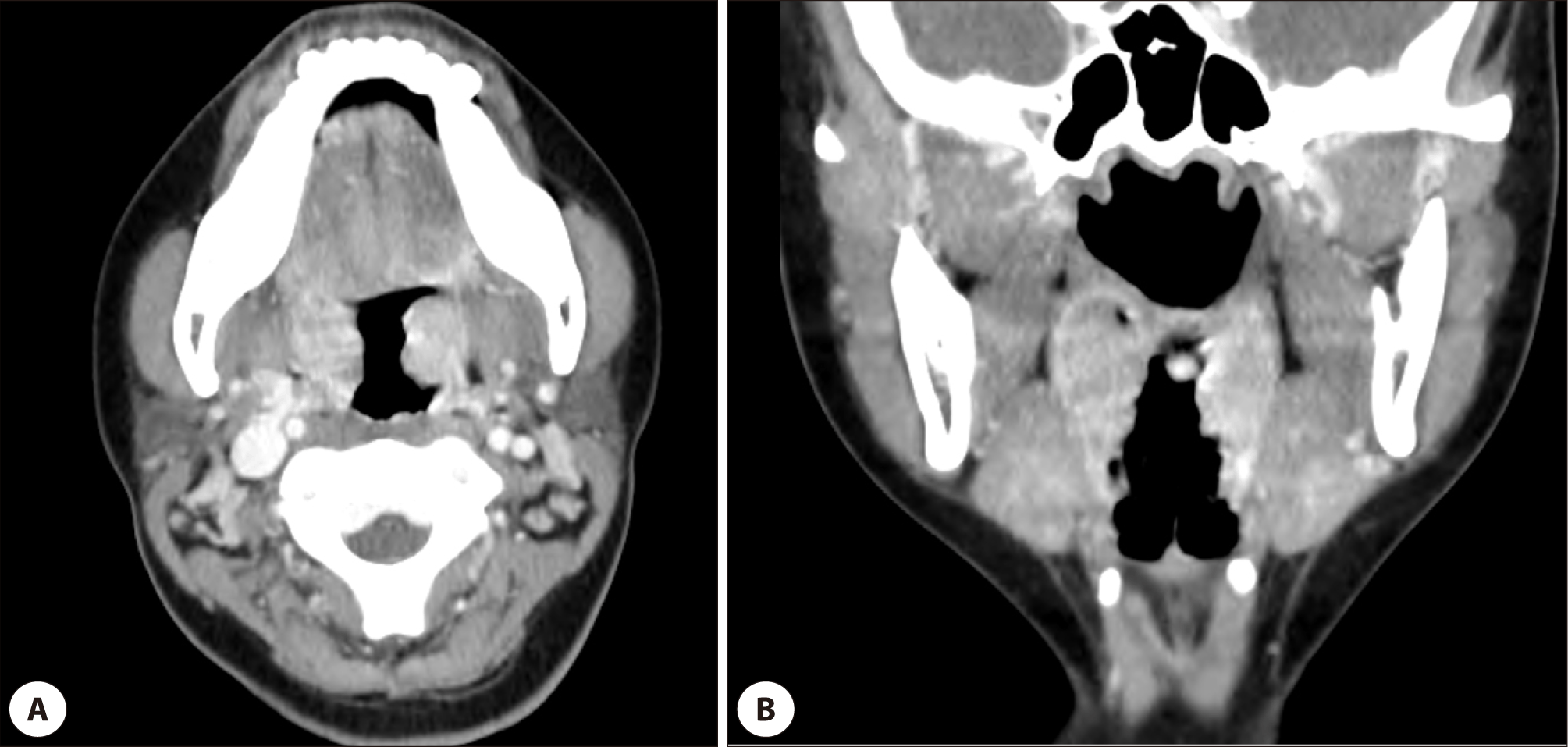
Physical examination of the neck revealed no palpable cervical lymphadenopathy or tenderness. Flexible laryngoscopy demonstrated significant bilateral tonsillar hypertrophy covered with thick, yellowish exudates (Fig. 1). To exclude deep neck space infection and evaluate the suspected pancreatitis, contrast-enhanced computed tomography (CT) of the neck and abdomen was performed. Neck CT revealed diffuse pharyngotonsillitis without abscess formation (Fig. 2), and abdominal CT showed a normal pancreas, with no evidence of hepatosplenomegaly. Given the absence of typical epigastric pain or tenderness, the gastroenterology department attributed the enzyme elevations to a non-specific biochemical manifestation of severe DKA rather than clinical acute pancreatitis.
The patient was admitted to the Department of Endocrinology for intensive DKA management. Further immunological and endocrinological evaluations confirmed that the patient’s underlying metabolic condition was T2DM rather than type 1 diabetes mellitus (T1DM). This was definitively established by negative results for both anti-insulin antibodies (2.8 U/mL) and anti-glutamic acid decarboxylase (GAD) antibodies, alongside preserved fasting and 2-hour postprandial serum C-peptide levels (1.0 ng/mL and 1.9 ng/mL, respectively). These findings collectively ruled out autoimmune-mediated pancreatic beta-cell destruction, confirming the diagnosis of pre-existing T2DM. While her metabolic status stabilized with aggressive fluid resuscitation and insulin infusion, the pharyngeal symptoms remained refractory to ampicillin/sulbactam. Blood cultures obtained on the day of admission showed no bacterial growth. Additionally, a swab culture of the tonsillar exudate isolated Streptococcus mitis/oralis and Candida albicans, which were interpreted as normal oral flora and superficial colonization rather than a true pyogenic bacterial co-infection.
Subsequent serological testing confirmed primary acute EBV infection (positive VCA IgM, VCA IgG, and EA-DR IgM). Although ampicillin/sulbactam had been administered at the outside facility and during the initial admission period, the patient did not develop any skin rash. Upon serological confirmation of acute EBV infection, the antibiotic was discontinued pre-emptively to avoid aminopenicillin-induced eruption. Under supportive care and glycemic control, the exudates and symptoms resolved. The patient was discharged on hospital day 15 with normalized inflammatory markers and stabilized glucose levels
Discussion
IM, primarily caused by EBV, is characterized by the classic triad of fever, pharyngitis, and lymphadenopathy.1,3) While typically self-limiting, it acted as a profound physiological stressor in the patient, precipitating DKA. A significant diagnostic challenge arises because EBV-induced tonsillitis often presents with thick, confluent exudates that are clinically indistinguishable from bacterial tonsillitis.1,4)Notably, EBV is identified as the primary pathogen in approximately 75% of severe exudative tonsillitis cases.5)
Therefore, clinicians must maintain a high index of suspicion for EBV in cases of refractory tonsillitis to avoid prolonged, presumptive antibiotic therapy.4) This is critical because aminopenicillins induce a characteristic maculopapular rash in 80%–100% of EBV patients.1,3) Although this reaction is not a true IgE-mediated allergy, it causes significant morbidity and complicates future drug allergy assessments.1) If a bacterial co-infection is suspected, non-beta-lactam alternatives such as macrolides or clindamycin should be prioritized over aminopenicillins or cephalosporins to minimize the risk of cutaneous reactions.1) In this case, prompt serological confirmation enabled discontinuation of the antibiotic, avoiding aminopenicillin-induced rash. The diagnosis of acute EBV infection was established by positive VCA IgM, VCA IgG, and EA-DR IgM results. While VCA IgM is a sensitive indicator of acute primary infection, the co-existence of EA-DR IgM reflects active viral replication, confirming that the acute phase of EBV infection acted as the physiological stressor triggering DKA.
Based on this case, we suggest immediate DKA screening (blood glucose, ketones, and blood gas analysis) for diabetic patients presenting with severe otorhinolaryngologic infections. This screening should be aggressively considered for: (1) exudative tonsillitis refractory to empirical antibiotics, (2) deep neck space infections, or (3) head and neck infections accompanied by systemic red flags like altered mental status or intractable vomiting. Early assessment is critical to prevent metabolic collapse.
EBV precipitates DKA through synergistic mechanisms: systemic inflammation-induced insulin resistance, direct pancreatic infiltration, and immunological beta-cell stunning via molecular mimicry.2,6–9) First, EBV triggers a systemic inflammatory response resembling a cytokine storm. Pro-inflammatory cytokines (e.g., IL-6, TNF-α) interfere with insulin signaling, while a simultaneous surge in counter-regulatory hormones—such as cortisol and catecholamines—accelerates hepatic gluconeogenesis and lipolysis.2,6,7) The marked leukocytosis and elevated CRP in the patient reflect this intense systemic stress.
Second, EBV can directly infiltrate pancreatic tissue, potentially impairing beta-cell function.2,7,8) Although radiographic evidence of pancreatitis was absent in the patient, the markedly elevated enzymes likely reflected micro-inflammation or extreme physiological stress within the pancreatic tissue. Third, molecular mimicry between EBV antigens and host proteins can trigger immunological cross-reactivity and transient beta-cell failure.2,6,9) The preservation of C-peptide levels following DKA resolution in the patient supports the hypothesis of transient functional suppression rather than permanent autoimmune destruction.6)
Differentiating this case from fulminant type 1 diabetes mellitus (FT1DM) was essential, given the rapid onset of DKA following viral infection. FT1DM is a distinct subtype characterized by the extremely rapid destruction of pancreatic beta-cells, often linked to viral triggers like EBV.6)
Several clinical indicators distinguished this case from FT1DM. First, the HbA1c level was decisive. According to the FT1DM diagnostic criteria, patients typically progress to DKA within one week, resulting in an HbA1c usually below 8.7%.8) In contrast, the patient’s HbA1c was 12.0%, suggesting an exacerbation of underlying T2DM rather than sudden de novo destruction. Second, preserved C-peptide levels further excluded FT1DM, where endogenous insulin secretion is typically exhausted (C-peptide <0.3 ng/mL).8) Finally, while 98% of FT1DM patients exhibit elevated pancreatic enzymes reflecting inflammatory damage,8) the patient’s imaging revealed no structural abnormalities. Combined with preserved C-peptide, these elevations likely reflected subclinical viral involvement or non-specific DKA manifestations. Ultimately, this case represents a severe metabolic collapse of pre-existing T2DM triggered by EBV-induced stress.
EBV infection necessitates vigilance for systemic complications, most notably splenic rupture. Although rare (0.1%–0.5%), over 50% of ruptures occur spontaneously.1) Clinicians must monitor for abdominal pain and advise patients to avoid strenuous activities or contact sports for at least three to four weeks to mitigate this life-threatening risk.1,10) While uncomplicated EBV is managed with supportive care, the routine use of antivirals or corticosteroids is not recommended unless specific complications, such as airway obstruction, occur.3,11)
However, as demonstrated here, the hierarchy of treatment shifts when EBV precipitates a metabolic crisis. DKA is a medical emergency; thus, aggressive fluid resuscitation and intensive insulin therapy must take precedence over supportive care for the viral infection.6,8) This case underscores that in patients with underlying comorbidities, prognosis depends not on treating the virus itself, but on the timely correction of the severe physiological imbalances it triggers.


