
Journal of Clinical Otolaryngology Head and Neck Surgery
The Busan, Ulsan, Gyeoungnam Branch of Korean Society of Otolaryngology-Head and Neck Surgery
Original Article

토끼 귀연골의 전기기계적 변형

나한슬1https://orcid.org/0000-0002-0118-3887, 김지아2https://orcid.org/0000-0001-9797-5135, 이정우3https://orcid.org/0000-0003-0568-2883, 이석환1https://orcid.org/0000-0002-5044-6735, 최성원1,4https://orcid.org/0000-0002-7463-7720, 오세준1,4,*https://orcid.org/0000-0001-8910-0064
Han-Seul Na1https://orcid.org/0000-0002-0118-3887, Ji-A Kim2https://orcid.org/0000-0001-9797-5135, Jung Woo Lee3https://orcid.org/0000-0003-0568-2883, Seokhwan Lee1https://orcid.org/0000-0002-5044-6735, Sung-Won Choi1,4https://orcid.org/0000-0002-7463-7720, Se-Joon Oh1,4,*https://orcid.org/0000-0001-8910-0064
1부산대학교병원 이비인후과 및 의생명연구원
2울산하나이비인후과병원
3창원탑이비인후과의원
4부산대학교 의과대학 이비인후과교실
1Department of Otorhinolaryngology and Biomedical Research Institute, Pusan National University Hospital, Busan, Korea
2Ulsan Hana ENT Hospital, Ulsan, Korea
3Changwon Top Otolaryngology Clinic, Changwon, Korea
4Department of Otorhinolaryngology, College of Medicine, Pusan National University, Busan, Korea
*Corresponding author: Se-Joon Oh, Department of Otorhinolaryngology, Pusan National University Hospital and Pusan National University School of Medicine, Busan 49241, Korea, Tel: +82-51-240-7824, Fax: +82-51-246-8668, E-mail: o3jdoc@hanmail.net

© Copyright 2026. The Busan, Ulsan, Gyeoungnam Branch of Korean Society of Otolaryngology-Head and Neck Surgery. This is an Open-Access article distributed under the terms of the Creative Commons Attribution Non-Commercial License (http://creativecommons.org/licenses/by-nc/4.0/) which permits unrestricted non-commercial use, distribution, and reproduction in any medium, provided the original work is properly cited.

Received: Dec 31, 2025; Revised: Feb 02, 2026; Accepted: Apr 15, 2026

Published Online: Jun 30, 2026

ABSTRACT

Background and Objectives: Electromechanical reshaping (EMR) is an emerging, non-thermal cartilage reshaping technique that induces permanent shape change by applying low-voltage direct current to mechanically deformed cartilage. This study aimed to evaluate the effects of applied voltage and application time on the degree of cartilage reshaping, temperature changes, and histologic alterations using an ex vivo rabbit auricular cartilage model. Materials and Methods: Auricular cartilage specimens (1×2 cm) were harvested ex vivo from 10-week-old female New Zealand White rabbits. Cartilage samples were mechanically bent to a standardized 90-degree configuration using a custom-made jig and subjected to EMR using a custom-built device. Direct current voltage (2, 4, 6, and 8 V) and application time (2, 4, 6, and 8 minutes) were systematically varied. The degree of reshaping was quantified by measuring the bend angle after EMR. Surface temperature changes were continuously monitored using infrared thermography, and histologic evaluation was performed to assess chondrocyte and matrix integrity. Results: No significant cartilage deformation was observed with a 2-minute application at any voltage. Time-dependent reshaping became significant from 4 minutes onward at 6 V, with bend angles increasing progressively with longer application times. Voltage-dependent reshaping was evident at a fixed duration of 4 minutes, with significant deformation observed at voltages ≥4 V. Temperature elevation during EMR increased in a voltage-dependent manner but remained below 2℃ at voltages ≤4 V. Histologic analysis demonstrated preserved cartilage architecture at 2–6 V, whereas focal chondrocyte injury and matrix disruption were observed at 8 V, particularly around electrode insertion sites. Conclusion: EMR effectively induces controlled reshaping of rabbit auricular cartilage in an ex vivo setting. Significant shape change occurs at application times ≥4 minutes and voltages ≥4 V, while higher voltages may increase the risk of cartilage injury. These findings provide fundamental data for defining safe and effective EMR parameters and support further in vivo and clinical investigations.

Keywords: Ear; Electric stimulation; Biomechanical phenomena; Cartilage

서론

이개 연골은 외이의 형태와 심미적 특성을 결정하는 핵심 구조물로, 선천성 기형, 외상 또는 종양 절제 후 재건 과정에서 연골의 형태를 조절하는 술식이 빈번히 요구된다. 기존의 이개 성형술은 절개, 봉합 또는 연골 절제를 기반으로 하여 효과적인 교정이 가능하나, 흉터, 출혈, 비대칭 및 재발 등의 한계를 가진다.13) 이에 따라 절개를 최소화하거나 비침습적으로 연골의 형태를 변형시키기 위한 다양한 시도가 이루어져 왔다.46) 전기기계적변형, EMR(electromechanical reshaping)은 연골을 원하는 형태로 변형시킨 상태에서 저전압 직류 전류를 인가하여, 국소적인 전기화학 반응을 통해 연골의 기계적 이완과 영구적인 형태 변화를 유도하는 기술이다.7) EMR은 열 손상을 유발하지 않는 비열성 방식으로 작용하며, 장비가 비교적 단순하고 비용이 낮다는 장점이 있어 기존의 레이저 기반 연골 변형 기법의 대안으로 주목받고 있다.7,8) 기존 연구에서는 토끼 이개를 이용한 in vivo 모델에서 바늘 전극 기반 EMR이 연골의 안정적인 형태 변형을 유도할 수 있음이 보고되었다. 특히 전압과 적용 시간이 증가함에 따라 연골 변형 각도가 증가하는 용량-반응 관계가 관찰되었으며, 장기 추적 관찰에서도 변형된 형태가 유지됨이 확인되었다. 이러한 결과들은 EMR의 임상 적용 가능성을 뒷받침한다. 그러나 in vivo 연구에서는 피부, 연부조직, 혈류 및 염증 반응과 같은 생체 변수가 개입되어, 전압과 적용 시간이라는 개별 인자가 연골 자체의 변형, 온도 변화 및 조직 손상에 미치는 영향을 정밀하게 평가하는 데 한계가 있다. 특히 EMR 적용 시 발생하는 온도 변화와 연골세포 손상 간의 관계는 충분히 규명되지 않았다.

이에 본 연구에서는 토끼 이개 연골을 ex vivo 환경에서 채취하여 EMR을 적용함으로써, 생체 변수의 영향을 최소화한 상태에서 전압과 적용 시간에 따른 연골 변형 정도, 온도 변화 및 조직학적 변화를 체계적으로 분석하고자 하였다. 본 연구는 EMR의 안전하고 효과적인 적용 조건을 규명하기 위한 기초 자료를 제공하는 것을 목적으로 한다.

대상 및 방법

본 연구는 병원 동물실험윤리위원회(Institutional Animal Care and Use Committee, IACUC)의 승인 하에 수행되었다(승인번호: PNUH-2019-141). 10주령 암컷 토끼(New Zealand White rabbit) 20마리를 CO2 흡입을 통해 희생하였으며, 각 개체의 양측 이개 중간 부위에서 1×2 cm 크기의 연골 조직을 각 2개씩 채취하여 총 80개의 시편을 확보하였다. 채취된 연골은 연부조직을 제거한 뒤 0.9% 생리식염수에 침지하여 4℃에서 보관하였으며, 채취 후 2시간 이내에 모든 실험을 완료하였다. EMR은 조직의 수분 함량에 민감한 전기화학 반응임을 고려하여, 보관 중 건조 및 대사 변화를 최소화하기 위해 위와 같은 조건을 유지하였다. EMR 적용 전 모든 연골 시편의 두께는 디지털 캘리퍼스를 이용하여 측정하였으며, 평균 두께는 1.9±0.47 mm이었다. 실험군 간 연골 두께에는 통계적으로 유의한 차이가 없었다(one-way analysis of variance, ANOVA; p>0.05).

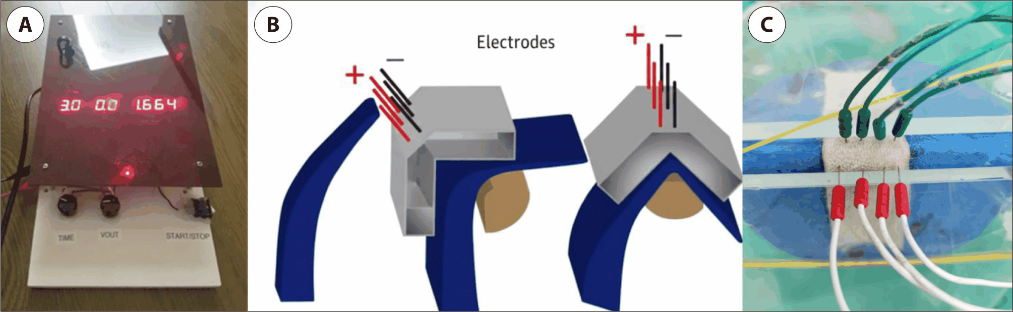
EMR은 연구진이 직접 제작한 EMR 기기를 이용하여 시행하였다(Fig. 1A). 해당 기기는 직류 전원을 안정적으로 출력할 수 있도록 설계되었으며, 0–8 V 범위의 직류 전압을 선택적으로 인가할 수 있도록 구성되었다. 전압은 실험 조건에 따라 2, 4, 6, 8 V로 설정하였고, 전류 인가 시간은 타이머를 통해 2, 4, 6, 8분으로 제어하였다. 연골은 EMR 적용 전, 연구진이 제작한 투명 아크릴 소재의 고정 지그(jig)에 삽입하여 90도의 일정한 각도로 굴곡된 상태로 고정하였다(Fig. 1B). 지그는 상·하부 플레이트 구조로 되어 있으며, 연골이 실험 중 이동하거나 초기 변형 각도가 달라지지 않도록 설계되었다. 모든 실험에서 동일한 지그를 사용하여 연골의 초기 변형 상태를 표준화하였다.
jcohns-37-2-51-g1
Fig. 1. Experimental setup for electromechanical reshaping (EMR) of rabbit auricular cartilage. (A) Custom-built EMR device developed by the authors, allowing precise control of direct current voltage and application time. (B) Schematic illustration of the experimental configuration showing auricular cartilage fixed in a standardized 90-degree bent position using a custom-made jig. Four anodes (+) and four cathodes (–) were inserted perpendicular to the cartilage surface with fixed inter-electrode spacing of 2 mm (same polarity) and 3 mm (opposite polarity). (C) Representative photograph of the experimental setup demonstrating platinum needle electrodes penetrating the cartilage and connected to the EMR device during ex vivo application.
Download Original Figure

전기 자극 전달을 위해 백금(platinum) 바늘 전극을 사용하였다. 전극은 연골 표면에 수직으로 삽입하여 연골을 관통하도록 배치하였으며, 양극(+) 4개와 음극(−) 4개, 총 8개의 전극을 동일한 배열로 삽입하였다(Fig. 1C). 동일 극성 전극 간 간격은 2 mm로 유지하였고, 서로 반대 극성 전극 간 간격은 3 mm로 설정하였다. 전극은 실험 중 위치 변화가 없도록 고정한 상태에서 EMR 기기에 연결하여 직류 전류를 인가하였다.

연골 시편은 전압(2, 4, 6, 8 V)과 적용 시간(2, 4, 6, 8분)에 따라 총 16개의 실험군으로 분류하였으며, 각 실험군당 5개의 연골 시편을 사용하였다. 대조군은 동일하게 지그에 고정하고 전극을 삽입하였으나 전압을 인가하지 않은 0 V 조건으로 정의하였으며, 이를 통해 전극 삽입 자체에 의한 기계적 영향을 통제하였다. EMR 적용 후 지그에서 연골 시편을 제거한 뒤 30분간 상온에서 안정화한 후 굴곡 각도(angle of bending)를 측정하여 변형 정도를 평가하였다. 조직학적 분석을 위한 시편은 각도 측정 직후 즉시 10% neutral buffered formalin에 고정하였다. 또한 EMR 적용 중 연골 표면의 온도 변화는 적외선 열화상 카메라(infrared thermal imaging camera)를 이용하여 실시간으로 측정하였다. 온도 변화(ΔT)는 EMR 적용 직전 연골 표면 온도를 기준값(baseline)으로 설정하여, EMR 적용 중 동일 부위에서 측정된 최대 온도와의 차이로 정의하였다. 모든 실험은 실험실 내 상온(22℃–23℃) 환경에서 수행되었다. 조직학적 분석은 실험 조건에 대해 맹검(blinded) 상태인 독립적인 평가자에 의해 수행되었으며, 연골세포의 형태 보존 여부와 기질 구조의 변화 여부를 중심으로 정성적으로 평가하였다.

통계 분석은 각 실험군의 변형 각도 및 온도 변화 값을 평균±표준편차로 제시하였다. 전압 및 적용 시간에 따른 굴곡 각도의 차이, 그리고 실험군 간 연골 두께의 동질성 검증은 ANOVA을 이용하여 비교하였으며, 사후 분석은 Tukey 검정을 시행하였다. 통계적 유의성은 p값이 0.05 미만인 경우로 정의하였다.

결과

EMR 적용에 따른 연골 형태 변화

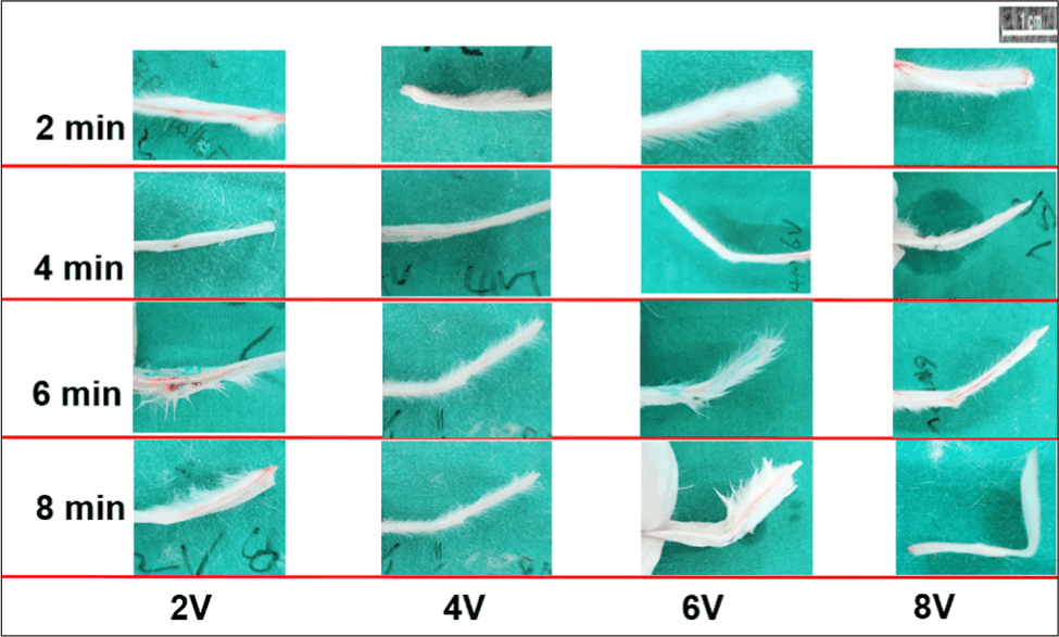
Ex vivo 이개 연골에서 EMR 적용 후 전압과 적용 시간에 따른 형태 변화를 육안적으로 관찰할 수 있었다(Fig. 2). 대조군(0 V, 전극 삽입 후 전압 미인가)에서는 지그 제거 후 연골의 굴곡 각도 변화가 관찰되지 않았다. 2분 적용군에서는 모든 전압 조건에서 의미 있는 연골 굴곡 변화가 관찰되지 않았다. 반면, 4분 이상 적용한 경우 전압 증가에 따라 연골의 굴곡 각도가 점진적으로 증가하는 양상이 관찰되었다.
jcohns-37-2-51-g2
Fig. 2. Time- and voltage-dependent reshaping of auricular cartilage following ex vivo electromechanical reshaping (EMR). Representative gross photographs of auricular cartilage specimens after EMR application under varying voltage and time conditions. Rows represent application time (2, 4, 6, and 8 min), and columns represent applied voltage (2, 4, 6, and 8 V). Minimal shape change was observed at 2 minutes across all voltage conditions. In contrast, increasing application time and voltage resulted in progressively greater cartilage bending, with marked reshaping observed at ≥4 min and ≥4 V.
Download Original Figure
적용 시간에 따른 연골 변형

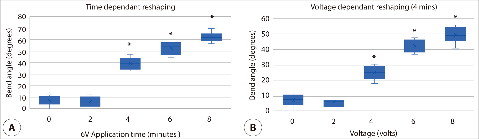
6 V 조건에서 적용 시간에 따른 연골 변형 각도를 비교한 결과, 4분부터 통계적으로 유의한 형태 변화가 관찰되었다(Fig. 3A). 평균 굴곡 각도는 4분에서 41.8°, 6분에서 55.6°, 8분에서 63.2°로, 적용 시간이 증가함에 따라 연골 변형 정도가 유의하게 증가하였다(p<0.05). 반면, 2분 적용 시에는 6 V 조건에서 6.6°로 대조군과 유의한 차이가 없었으며, 2 및 4 V 조건에서는 5개 시편 모두에서 육안적으로 측정 가능한 변형이 관찰되지 않아 굴곡 각도를 0°로 기록하였다(표준편차=0; Table 1).
jcohns-37-2-51-g3
Fig. 3. Quantitative analysis of time- and voltage-dependent electromechanical reshaping (EMR) of auricular cartilage. (A) Time-dependent changes in bend angle following EMR application at a fixed voltage of 6 V. Bend angle increased significantly with longer application time, with marked reshaping observed from 4 min onward. (B) Voltage-dependent changes in bend angle following EMR application for a fixed duration of 4 min. Increasing voltage resulted in progressively greater cartilage bending, with significant differences observed at ≥4 V compared with controls. Box plots represent median values with interquartile ranges; whiskers indicate minimum and maximum values. Asterisks denote statistically significant differences compared with the control condition (p<0.05).
Download Original Figure
Table 1. Angular deformation according to applied voltage and duration
Time (min) 2 V 4 V 6 V 8 V
2 0 0 6.6 ± 4.2 7.0 ± 3.6
4 4.5 ± 2.3 27.3 ± 3.7* 41.8 ± 6.7* 48.3 ± 7.3*
6 8.3 ± 4.5 35.0 ± 6.3* 55.6 ± 5.4* 64.3 ± 7.9*
8 10.1 ± 4.2 40.4 ± 6.4* 63.2 ± 4.6* 88.2 ± 7.4*

Values are presented as mean ± SD (degrees).

Control was defined as cartilage fixed in the jig with needle electrodes inserted but without voltage application (0 V).

p<0.05 compared with control.

Values of 0° with no standard deviation indicate that no measurable deformation was observed in any of the five specimens under the given condition.

Download Excel Table
전압에 따른 연골 변형

적용 시간을 4분으로 고정한 상태에서 전압에 따른 연골 변형을 분석한 결과, 4 V부터 유의한 연골 굴곡 변화가 관찰되었다(Fig. 3B). 평균 굴곡 각도는 4 V에서 27.3°, 6 V에서 41.8°, 8 V에서 48.3°로, 전압 증가에 따라 연골 변형 각도가 유의하게 증가하였다(p<0.05). 2 V 조건에서는 4.5°로 통계적으로 유의하지 않았다(Table 1).

EMR 적용 중 온도 변화

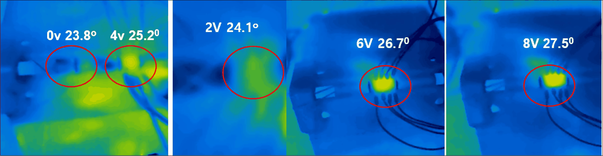
EMR 적용 중 연골 표면의 온도 변화는 적외선 열화상 카메라를 이용하여 측정하였다(Fig. 4). 4분 적용 기준에서, 0 V와 비교하였을 때 전압 증가에 따라 국소적인 온도 상승이 관찰되었다. 온도 변화 범위는 2 V에서 0.2±0.1℃, 4 V에서 1.6±0.3℃, 6 V에서 3.1±0.4℃, 8 V에서 4.4±0.4℃로 나타났으며, 전압 증가에 따라 유의하게 상승하였다(ANOVA, p<0.05). 사후 검정에서 4 V 이상의 모든 조건은 대조군(0 V)과 유의한 차이를 보였다.
jcohns-37-2-51-g4
Fig. 4. Infrared thermographic assessment of temperature changes during electromechanical reshaping (EMR). Representative infrared thermographic images acquired during ex vivo EMR application at different voltages (0, 2, 4, 6, and 8 V) for a fixed duration of 4 min. Localized temperature elevation was observed around the electrode insertion sites, with progressively greater temperature increases at higher applied voltages. Minimal temperature change was noted at 0 and 2 V, whereas distinct focal heating was evident at 6 and 8 V. Red circles indicate regions of interest used for temperature measurement, each centered on the electrode insertion site as a consistent anatomical reference point.
Download Original Figure
조직학적 분석

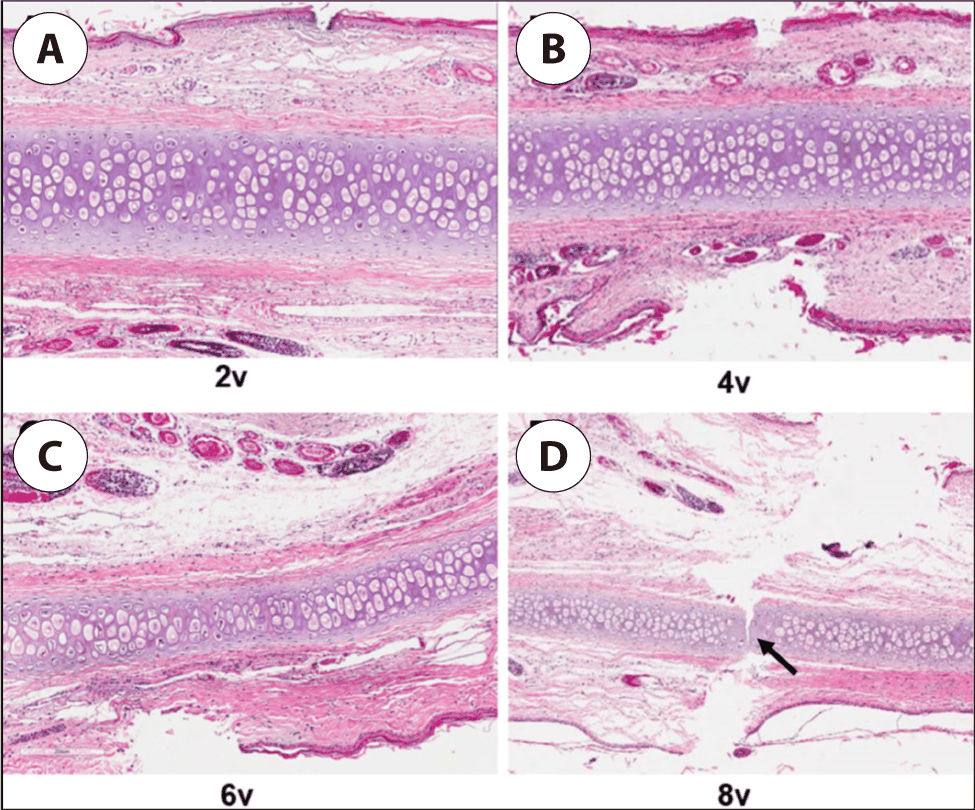
조직학적 분석 결과, 2 및 4 V 조건에서는 연골 구조가 비교적 잘 유지되었으며 뚜렷한 연골세포 손상은 관찰되지 않았다(Fig. 5). 6 V 조건에서는 일부 국소적인 변화가 관찰되었으나 연골 전반의 구조는 유지되었다. 반면, 8 V 조건에서는 전극 삽입 부위를 중심으로 연골세포 손상 및 조직 구조의 붕괴가 관찰되었다.
jcohns-37-2-51-g5
Fig. 5. Histologic findings of auricular cartilage following ex vivo electromechanical reshaping (EMR). Representative hematoxylin and eosin–stained sections of auricular cartilage after EMR at different voltages: (A) 2 V, (B) 4 V, (C) 6 V, and (D) 8 V. At 2, 4, and 6 V, the overall cartilage architecture and chondrocyte distribution were largely preserved, with histologic changes mainly attributable to mechanical penetration by the needle electrodes. In contrast, specimens treated at 8 V demonstrated focal structural disruption of the cartilage matrix, including partial fissuring of the cartilage, consistent with electrochemically induced tissue injury. Arrows indicate areas of chondrocyte injury and matrix disruption.
Download Original Figure

고찰

본 연구에서는 토끼 이개 연골을 ex vivo 환경에서 대상으로 하여 EMR의 전압과 적용 시간이 연골 변형, 온도 변화 및 조직학적 손상에 미치는 영향을 체계적으로 분석하였다. 본 연구의 주요 발견은 (1) EMR에 의한 연골 변형이 전압과 적용 시간에 모두 의존적이며, (2) 의미 있는 형태 변화가 4분 및 4 V 이상의 조건에서 관찰되었고, (3) 8 V 조건에서는 연골세포 손상이 동반될 수 있다는 점이다.

기존의 이개 연골 변형 술식은 절개, 봉합 또는 연골 절제를 기반으로 하여 효과적인 교정이 가능하나, 흉터, 출혈, 비대칭 및 재발 등의 한계를 가진다.13) 이러한 한계를 극복하기 위해 무절개 또는 최소 침습적 이개 성형술이 제안되어 왔으나, 변형의 예측 가능성과 장기 안정성 측면에서는 여전히 제한점이 존재한다.46) EMR은 연골을 원하는 형태로 기계적으로 변형시킨 상태에서 저전압 직류 전류를 인가하여 영구적인 형태 변화를 유도하는 기술로, 기존 술식의 대안으로 주목받고 있다.7,8)

Wong 등은 토끼 이개를 이용한 in vivo 연구를 통해 바늘 전극 기반 EMR이 실제 생체 환경에서도 연골의 안정적인 형태 변형을 유도할 수 있음을 처음으로 보고하였다.9) 이후 장기 추적 연구에서는 EMR로 변형된 이개의 형태가 수개월 이상 안정적으로 유지됨이 확인되었으며, 비교적 낮은 전압 범위에서도 임상적으로 의미 있는 변형이 가능하다고 보고되었다.10) 이러한 연구들은 EMR의 임상 적용 가능성을 뒷받침하는 중요한 근거를 제공한다. 그러나 기존 in vivo 연구들은 피부, 연부조직, 혈류, 염증 반응 및 부목 고정과 같은 생체 변수의 영향을 배제하기 어렵다는 한계를 가진다. 특히 전압과 적용 시간이라는 개별 인자가 연골 자체의 변형, 온도 변화 및 연골세포 손상에 미치는 영향을 독립적으로 평가하기에는 제약이 있다. 본 연구는 이러한 한계를 극복하기 위해 ex vivo 모델을 사용하였으며, 동일한 90도 굴곡 지그를 이용해 초기 변형 조건을 표준화함으로써 연골의 전기–기계적 반응을 보다 직접적으로 평가할 수 있었다. 특히 ex vivo 환경에서는 EMR 적용 전 모든 시편의 연골 두께를 디지털 캘리퍼스로 정밀하게 측정하고 실험군 간 동질성을 통계적으로 확인할 수 있다는 점에서, 연골 두께의 정확한 측정이 어려운 in vivo 연구에 비해 명확한 장점을 가진다.

본 연구 결과, 연골 변형은 적용 시간에 따라 점진적으로 증가하였으며, 2분 적용에서는 모든 전압 조건에서 의미 있는 형태 변화가 관찰되지 않았다. 반면 4분 이상 적용 시에는 시간 의존적인 연골 변형이 명확히 나타났으며, 이는 EMR에 의해 유도되는 전기화학 반응이 일정 시간 이상 누적되어야 연골의 기계적 이완과 영구적인 형태 변화가 발생함을 시사한다. 이러한 결과는 EMR이 시간 의존적인 전기화학적 과정임을 보고한 기존 연구들과도 일치한다.7,11) EMR의 기전을 보다 구체적으로 살펴보면, 직류 전류 인가 시 양극 주변에서는 산화 반응에 의한 국소적 산성화가, 음극 주변에서는 환원 반응에 의한 국소적 알칼리화가 발생한다.7,12) 이러한 국소적 pH 변화가 연골 기질의 기계적 이완에 기여하는 것으로 추정되나, 정확한 기전은 아직 완전히 규명되지 않은 상태이다.12) 또한 누적 전기량(total charge transfer)이 증가할수록 연골 변형 정도와 연골세포 손상이 함께 증가한다는 점이 보고되어 있으며,7,11,13) 본 연구에서 전압 및 적용 시간 증가에 따라 변형 각도와 조직 손상이 동반 증가한 결과는 이와 일치한다. 따라서 EMR 임상 적용 시에는 충분한 변형 효과를 얻으면서도 연골세포 손상을 최소화할 수 있는 누적 전기량의 최적 범위를 설정하는 것이 중요하다.

전압에 따른 분석에서도 4 V 이상에서 의미 있는 연골 변형이 관찰되었으며, 이는 기존 in vivo 연구에서 제시된 임계 전압 범위와 유사한 결과이다.9,10) 특히 6 V 이상에서는 변형 각도가 크게 증가하였으나, 8 V 조건에서는 조직 손상이 동반되었다는 점에서, 전압 증가가 항상 바람직한 변형 효과로 이어지지는 않음을 시사한다. 이는 EMR 적용 시 전압과 적용 시간의 균형이 중요함을 강조한다.

온도 분석 결과, EMR 적용 중 연골 표면의 온도는 전압 증가에 따라 점진적으로 상승하였으나, 4 V 이하에서는 온도 상승이 2℃ 미만으로 제한적이었다. 이는 EMR이 본질적으로 비열성(non-thermal) 메커니즘을 기반으로 작용함을 지지하는 결과이다.7,11) 다만 6 및 8 V 조건에서는 국소적인 온도 상승이 보다 뚜렷하게 관찰되었으며, 이는 전극 주변의 전기저항 증가 및 국소적인 전류 밀도 상승과 연관될 가능성이 있다.

조직학적 분석에서는 2 및 4 V 조건에서 연골 구조가 비교적 잘 유지되었으며, 뚜렷한 연골세포 손상은 관찰되지 않았다. 반면 8 V 조건에서는 전극 삽입 부위를 중심으로 연골세포 손상과 조직 구조의 붕괴가 관찰되었다. 이러한 결과는 전극 주변에서 발생하는 국소적인 전기화학 반응 및 pH 변화가 세포 손상에 기여할 수 있음을 시사하며, 기존의 세포 손상 패턴에 대한 보고들과도 일치한다.13)

임상적 관점에서 본 연구 결과는 EMR의 안전하고 효과적인 적용 조건을 제시한다는 점에서 의미가 있다. Ex vivo 모델에서 4–6 V 범위의 전압과 4–6분의 적용 시간은 연골 변형 효과와 조직 안전성 간의 균형 측면에서 가장 적절한 조건으로 판단된다. 이러한 조건은 향후 in vivo 연구 및 임상 적용 시 초기 설정값으로 활용될 수 있을 것이다.

본 연구의 한계로는 ex vivo 모델을 사용하였으므로 생체 내 조건을 완전히 재현하는 데 한계가 있다. 구체적으로, 혈관 공급 부재로 인한 산소 및 영양 차단, 연골주위막(perichondrium)의 기능적 기여 배제14), 그리고 면역 반응 및 조직 리모델링 평가의 불가능이라는 제한점이 존재한다. 또한 동일 개체의 양측 이개에서 복수의 시편을 채취함으로써 시편 간 생물학적 독립성이 완전히 보장되지 않으며, 단일 굴곡 각도(90도)만을 적용하여 다양한 변형 조건에서의 EMR 효과를 평가하지 못하였다. 조직학적 평가는 정성적 수준에 그쳤으며 표준화된 정량적 grading system을 적용하지 않았고, 적외선 열화상 이미지에서 ROI(region of Interest)의 픽셀 크기를 모든 패널에서 완전히 통일하지 못하였다는 점도 제한점으로 인정된다. 향후 연구에서는 본 연구에서 도출된 최적 조건을 바탕으로 in vivo 모델에서의 장기적 형태 유지 및 조직 반응을 검증하는 연구가 필요할 것이다.

Acknowledgements

Not applicable.

Funding Information

This study was supported by Biomedical Research Institute Grant (20220191), Pusan National University Hospital.

Conflicts of Interest

No potential conflict of interest relevant to this article was reported.

Author Contribution

Conceptualization: Oh SJ.

Data curation: Na HS, Lee S, Choi SW.

Formal analysis: Choi SW, Lee S, Oh SJ.

Methodology: Kim JA, Lee JW, Oh SJ.

Software: Choi SW, Lee S.

Validation: Kim JA, Lee JW.

Investigation: Na HS, Kim JA, Lee JW.

Writing - original draft: Na HS, Oh SJ.

Writing - review & editing: Na HS, Kim JA, Lee JW, Lee S, Choi SW, Oh SJ.

Ethics Approval

All animal experiments were conducted in accordance with relevant institutional and national guidelines for the care and use of laboratory animals. The study protocol was reviewed and approved by the Institutional Animal Care and Use Committee (IACUC) of Pusan National University Hospital, Korea (Approval No. PNUH-2019-141).

References

1.

Mustardé JC. The correction of prominent ears using simple mattress sutures. Br J Plast Surg 1963;32(4):486.

2.

Kelley P, Hollier L, Stal S. Otoplasty: evaluation, technique, and review. J Craniofac Surg 2003;14(5):643-53.

3.

Nuara MJ, Mobley SR. Nuances of otoplasty: a comprehensive review of the past 20 years. Facial Plast Surg Clin North Am 2006;14(2):89-102.

4.

Fritsch MH. Incisionless otoplasty. Facial Plast Surg 2004;20(4):267-70.

5.

Obadia D, Quilichini J, Hunsinger V, Leyder P. Cartilage splitting without stitches: technique and outcomes. JAMA Facial Plast Surg 2013;15(6):428-33.

6.

Mehta S, Gantous A. Incisionless otoplasty: a reliable and replicable technique for the correction of prominauris. JAMA Facial Plast Surg 2014;16(6):414-8.

7.

Manuel CT, Foulad A, Protsenko DE, Sepehr A, Wong BJ. Needle electrode-based electromechanical reshaping of cartilage. Ann Biomed Eng 2010;38(11):3389-97.

8.

Manuel CT, Foulad A, Protsenko DE, Hamamoto A, Wong BJF. Electromechanical reshaping of costal cartilage grafts: a new surgical treatment modality. Laryngoscope 2011;121(9):1839-42.

9.

Yau AY, Manuel CT, Hussain SF, Protsenko DE, Wong BJF. In vivo needle-based electromechanical reshaping of pinnae: New Zealand white rabbit model. JAMA Facial Plast Surg 2014;16(4):245-52.

10.

Manuel CT, Tjoa T, Nguyen T, Su E, Wong BJF. Optimal electromechanical reshaping of the auricular ear and long-term outcomes in an in vivo rabbit model. JAMA Facial Plast Surg 2016;18(4):277-84.

11.

Wu EC, Protsenko DE, Khan AZ, Dubin S, Karimi K, Wong BJF. Needle-electrode-based electromechanical reshaping of rabbit septal cartilage: a systematic evaluation. IEEE Trans Biomed Eng 2011;58(8):2378-83.

12.

Kuan EC, Hamamoto AA, Manuel CT, Protsenko DE, Wong BJF. In-depth analysis of pH-dependent mechanisms of electromechanical reshaping of rabbit nasal septal cartilage. Laryngoscope 2014;124(10):E405-10.

13.

Protsenko DE, Ho K, Wong BJF. Survival of chondrocytes in rabbit septal cartilage after electromechanical reshaping. Ann Biomed Eng 2011;39(1):66-74.

14.

Mo JH, Lee DJ, Chung PS, Chung YJ. Regenerative and proliferative activities of chondrocyte based on the degree of perichondrial injury in rabbit auricular cartilage. Eur Arch Otorhinolaryngol 2014;271:1573-80.