
Introduction
A perilymphatic fistula (PLF) is a condition that results in abnormal communication between the perilymph fluid-filled inner ear and middle ear. This condition causes leakage of perilymgh from the cochlea or vestibule, leading to hearing loss and vestibular symptoms. Hearing manifestations include sudden, progressive, or fluctuating sensorineural hearing loss.1) A descending pattern of hearing loss, with poorer hearing at higher frequencies, is common; however, the pattern of hearing loss can vary from almost normal to profound.1,2) Dizziness, which is typically aggravated by positional changes, may present as a vestibular symptom.3)
PLF primarily results from trauma or middle ear diseases, such as cholesteatoma. Trauma that can causes PLF includes direct labyrinthine and mild head trauma, as well as internal and external barotrauma.4,5) Additionally, we believe that sudden and strong acoustic trauma can also lead to PLF. Here, we describe two cases of suspected PLF that resulted from acoustic trauma and were successfully treated with surgical repair.
Case Report
A 12-year-old boy visited the emergency department due to sudden right-sided hearing loss that occurred immediately after exposure to a loud sound from a speaker while preparing for a school art festival. He first visited a local clinic and was referred for the diagnosis of sudden sensorineural hearing loss on pure tone audiometry (PTA). However, PTA performed in the emergency department showed only mild high-frequency disturbances (Fig. 1A). The patient was discharged from the emergency department with high-dose steroids prescribed by the local clinic. Unfortunately, the symptoms worsened when he visited the otolaryngology out-patient department three days later. The PTA revealed moderate-to-severe sensorineural hearing loss (Fig. 1B). Moreover, he experienced vertigo while moving his head. Despite no pathological nystagmus during the fistula test on the right ear or a positional test, we suspected PLF due to acoustic trauma and decided to perform exploratory tympanotomy and PLF repair.
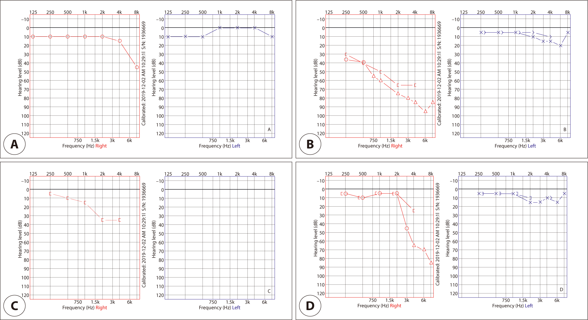
Exploratory tympanotomy was performed under general anesthesia using a microscopic endaural approach. The tympanomeatal flap was elevated, and the middle ear was explored. After careful examination of the middle ear, a clear fluid around the oval window was discovered. We harvested a small piece of soft tissue and used it cover the stapes foot plate and the round window to repair the fistula. The middle ear was then packed with fibrin glue and absorbable gelatin powder. Finally, the tympanomeatal flap was repositioned, and the external auditory canal (EAC) was packed with absorbable gelatin powder. Positional vertigo disappeared immediately after surgery, and bone conduction PTA also improved 2 days after surgery (Fig. 1C). EAC packing was removed 3 weeks after the surgery, and PTA performed a month after the surgery showed normal hearing at low- to mid-frequency, but there was disturbance at hearing high-frequency (Fig. 1D). The patient could hear without any discomfort.
A 13-year-old boy visited the emergency department with sudden right-sided hearing loss that occurred immediately after a friend shouted loudly in his right ear. He also experienced whirling-type vertigo. No abnormal findings were observed on otoscopic examination. There was no pathologic nystagmus, but the patient complained of dizziness while performing a fistula test and positional test. PTA revealed profound right-sided sensorineural hearing loss (Fig. 2A). PLF caused by acoustic trauma was suspected; therefore, we decided to perform exploratory tympanotomy and PLF repair.
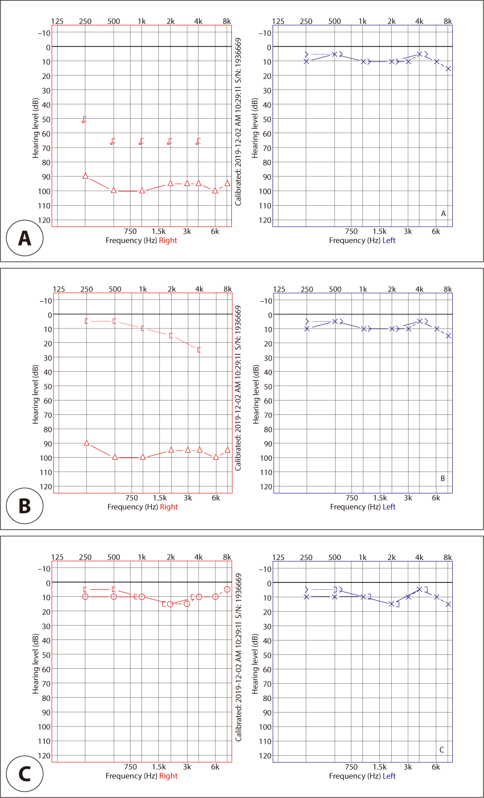
We performed surgery under general anesthesia using a microscopic endaural approach. The tympanomeatal flap was elevated, and middle ear exploration was performed. Due to the narrow bony annulus, we could not fully evaluate the stapes footplate. Therefore, we drilled the superoposterior bony annulus to expose this area sufficiently. Clear fluid was observed near the oval window. Therefore, we harvested a small amount of soft tissue and sealed the oval and round windows. The middle ear was packed with fibrin glue and absorbable gelatin powder. Finally, the tympanomeatal flap was repositioned, and the EAC was packed with absorbable gelatin powder.
Vertigo improved after surgery. Bone conduction PTA performed 4 days after surgery showed normalized bone conduction hearing (Fig. 2B). The EAC packing was removed 3 weeks after surgery, and PTA performed approximately a month after surgery revealed normal hearing at all frequencies (Fig. 2C). The patient can hear well without any discomfort after then.
Discussion
The concept of PLF was first proposed in the early 1900s. Initially, it was thought to be due to labyrinthine capsule dehiscence associated with stapes hypermobility or cholesteatoma, or fistula that develops between the inner ear and middle ear that occurs during stapes surgery. After Fee reported PLF caused by head trauma in 1968,6) several studies identified trauma as a common cause of PLF. Such traumas can be mild head injuries or iatrogenic trauma, as well as barotrauma. Although acoustic trauma has rarely been proposed as a cause of PLF, we believe that sudden and intense acoustic trauma can also instigate PLF, as observed in our cases.
Two hypotheses may be proposed regarding the mechanism by which acoustic trauma causes PLF. After exposure to high-intensity sound, the contraction of the stapedial muscle induces an acoustic reflex (AR) to reduce ossicular mobility. The AR serves to protect the inner ear from damage by mitigating the transmission of vibrational energy. However, there is a potential for injury to the anterior annulus of the stapes footplate, induced by a strong backward contraction of the stapedial muscle due to sudden and loud sound stimulation. Injury to the annulus of footplate may lead to an oval-window PLF, resulting in sensorineural hearing loss and dizziness.
The second hypothesis is that the round window membrane can be damaged by a strong wave traveling inside the cochlea. Acoustic stimulation transmits vibrational energy through the oval window to the inner ear, where it travels along the basilar membrane, passes through the helicotrema, and finally delivers pressure to the round window. Although AR can reduce the vibrational energy that reaches the inner ear, it is known to have a latency of tens to hundreds of milliseconds, depending on its frequency.7) If strong vibrational energy of sudden acoustic trauma cannot be migiated due to the latency of the AR, there exists a significant possibility of damaging the round window membrane, which is the most vulnerable part of the labyrinth, resulting in PLF.
Some previous reports note the occurrence of spontaneous PLF without any identiable trauma. These reports indicate a potential weakness in the otic capsule, particularly near the oval and round windows, in patients with PLF.8,9) Acoustic trauma is also likely to cause PLF in individuals who have this preexisting weakness.
Combining the above hypotheses, if PLF occurs due to acoustic trauma, it is likely to occur in the oval or round window membrane. Therefore, performing exploratory tympanotomy and repairing the oval and round windows in our two cases resulted in immediate recovery from both vertigo and hearing loss.
PLF should be differentiated from several conditions such as sudden deafness with vertigo, and Meniere’s disease or cerebellar infarction, which can cause sudden hearing loss and vertigo together. PLF is suspected when there is progressive or fluctuating sensorineural hearing loss and vertigo following clinically identifiable trauma.1) Traditionally, PLF has been diagnosed through the identification of perilymph leakage in the middle ear during exploratory surgery, with symptom improvement after surgical repair in clinically suspected patients. Recently, the suspicion of PLF has been strengthened when pneumolabyrinth is visible on high-resolution temporal bone computed tomography (CT) scans. Methods to measure biomarkers, such as Cochlin-tomoprotein in fluid in the middle ear, are currently under study; however, there is no definite diagnostic method yet available, making diagnosis is difficult.5) In our cases, CT was not conducted. Instead, we diagnosed PLF on traditional methods by checking for leakage and observing symptom improvement after surgical repair.
Due to the absence of a definitive diagnostic method, there is some controversy surrounding the treatment of PLF. Some reports have suggested conservative management,8,10) while a recent review recommended5) the blood patch procedure as first-line treatment. Conversely, other researchers advocate for surgical intervention, stating that prompt surgical repair can offer a better prognosis.3,6,11) In both of our cases, surgical treatment was performed within a few days following the initial trauma, resulting in favorable hearing outcomes.
Previously, sudden sensorineural hearing loss following acoustic trauma was primarily treated with high-dose steroids or intratympanic steroid injections. However, acute inner ear injury caused by explosive noise generally does not respond well to these treatments and often have poorer prognosis.12) If dizziness and hearing loss progress or fluctuate in these patients, acoustic trauma-induced PLF should be suspected, and appropriate treatment should be administered.