Journal of Clinical Otolaryngology Head and Neck Surgery
The Busan, Ulsan, Gyeoungnam Branch of Korean Society of Otolaryngology-Head and Neck Surgery
Case Report

전두동 중격봉소의 점액낭종: 드문 임상적 사례

박혜진1https://orcid.org/0000-0003-4237-6834, 박지환1https://orcid.org/0000-0001-9301-6957, 김성동1https://orcid.org/0000-0002-8436-5722, 조규섭1,*https://orcid.org/0000-0002-4381-6996
Hye-Jin Park1https://orcid.org/0000-0003-4237-6834, Ji-Hwan Park1https://orcid.org/0000-0001-9301-6957, Sung-Dong Kim1https://orcid.org/0000-0002-8436-5722, Kyu-Sup Cho1,*https://orcid.org/0000-0002-4381-6996
1부산대학교 의과대학 이비인후과학교실, 부산대학교병원 이비인후과
1Department of Otorhinolaryngology and Biomedical Research Institute, Pusan National University School of Medicine, Pusan National University Hospital, Busan, Korea
*Corresponding author: Kyu-Sup Cho, Department of Otorhinolaryngology and Biomedical Research Institute, Pusan National University School of Medicine, Pusan National University Hospital Busan 49241, Korea, Tel: +82-51-240-7824, Fax: +82-51-246-8668, E-mail: choks@pusan.ac.kr

© Copyright 2025 xxxxxxxxxxxxxxx. This is an Open-Access article distributed under the terms of the Creative Commons Attribution Non-Commercial License (http://creativecommons.org/licenses/by-nc/4.0/) which permits unrestricted non-commercial use, distribution, and reproduction in any medium, provided the original work is properly cited.

Received: Feb 15, 2025; Revised: Mar 06, 2025; Accepted: Mar 14, 2025

Published Online: Mar 31, 2025

ABSTRACT

A mucocele is a benign, slow-growing, epithelium-lined lesion filled with mucus that expands within the paranasal sinus. Interfrontal sinus septal cells refer to pneumatized structures located in the midline or paramedian bony labella (septum) between the frontal sinuses. While mucoceles commonly develop in the frontal or ethmoidal sinuses, those arising in interfrontal sinus septal cells are extremely rare. This report presents a case of interfrontal sinus septal cell mucocele causing severe headache, which was successfully managed through wide marsupialization using a transnasal endoscopic approach.

Keywords: Mucocele; Frontal sinus; Marsupialization; Headache

서론

부비동 점액낭종(mucocele)은 부비동의 개구부 폐쇄로 인해 점막으로 둘러싸인 공간 내에 점액이 저류되어 발생하는 낭성 병변이다. 이 중 감염으로 인해 화농성 변화를 동반한 경우를 점액농류(mucopyocele)라고 한다.1) 전두동간 중격봉소(interfrontal sinus septal cell, IFSSC)는 전두동 사이의 중격이 함기화된 형태를 의미하며, “frontal intersinus septum” 또는 “frontal wishbone”으로도 알려져 있다.2,3) 점액낭종은 주로 전두동이나 사골동에서 발생하는데, IFSSC에서 발생하는 경우는 매우 드물다. 본 연구에서는 전두부 통증을 주소로 내원한 환자에서 IFSSC에 발생한 점액낭종을 진단하고, 내시경적 조대술(marsupialization)을 통해 성공적으로 치료한 증례를 문헌 고찰과 함께 보고하고자 한다.

증례

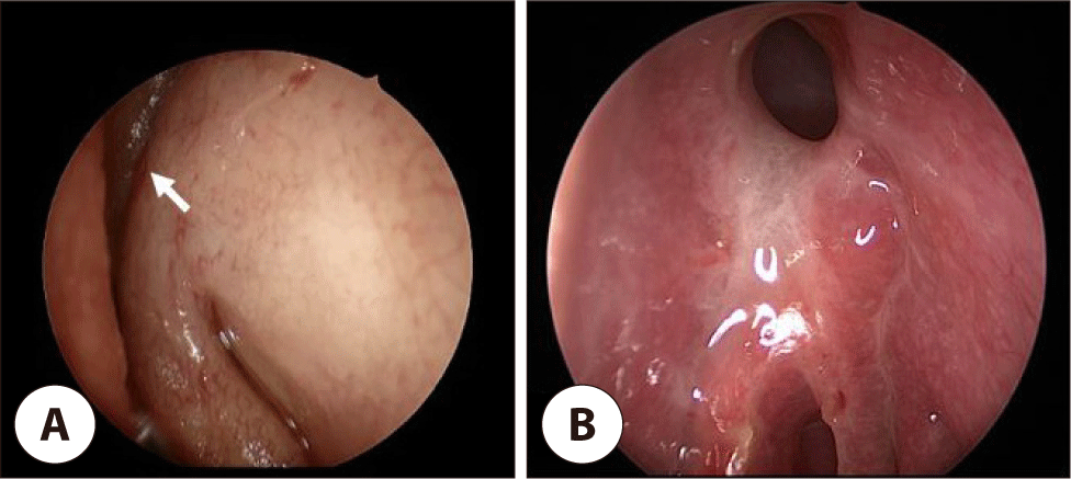
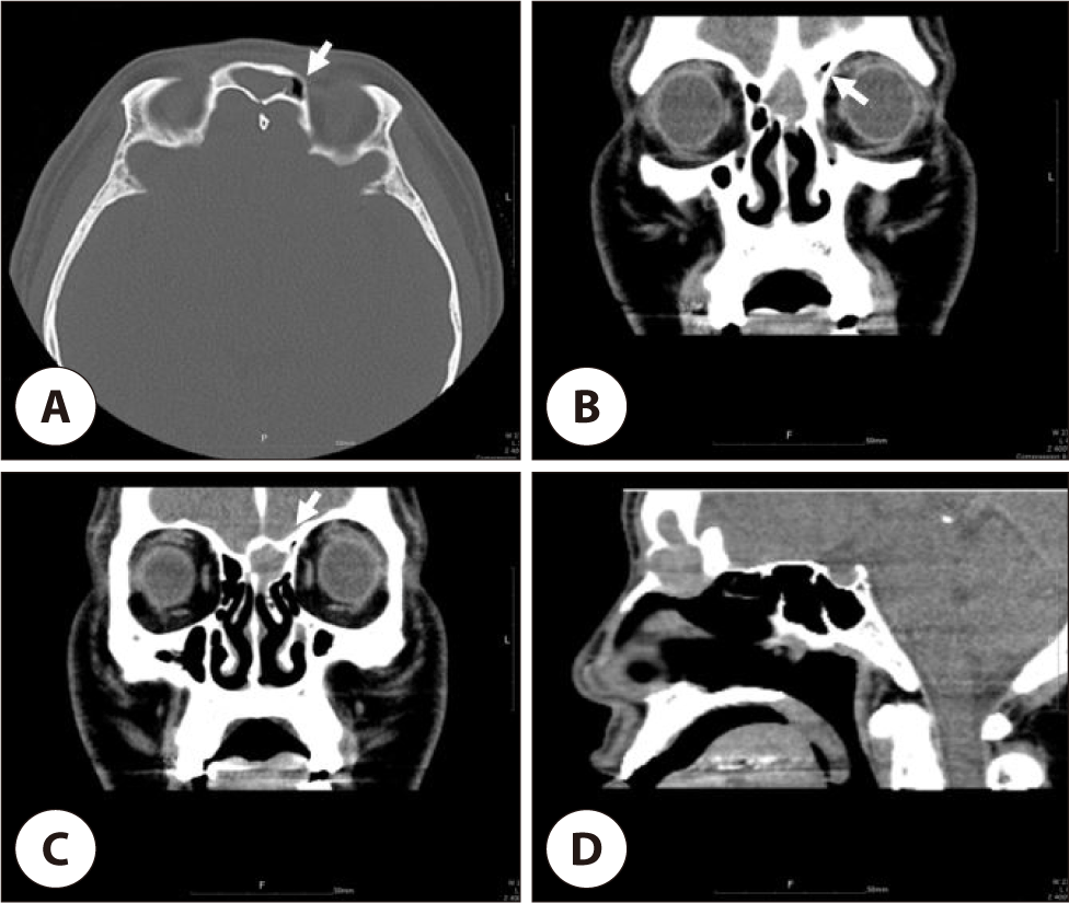
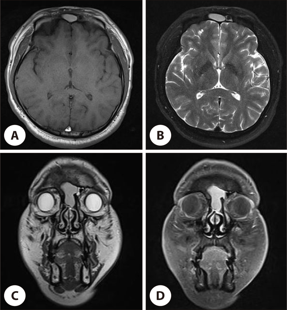
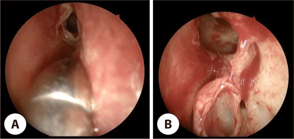
43세 남성 환자가 몇 달간 지속된 좌측 전두부 통증을 호소하며 내원하였다. 코막힘, 후각저하, 비루, 후비루 등의 증상은 동반되지 않았으며, 고혈압이나 당뇨 등의 기저 질환도 없었다. 비내시경 검사에서 특이 소견은 보이지 않았으나, 비중격 상부와 중비갑개 사이에서 낭성 종물이 관찰되었다(Fig. 1A). 부비동 전산화 단층촬영에서 IFSSC를 가득 채운 병변이 확인되었으며, 좌측 전두동을 압박하고 전두동의 앞뒤 골벽이 얇아져 있었다(Fig. 2A–C). 부비동 자기공명영상에서 병변은 T1 강조 영상에서 고 신호 강도(high signal intensity; Fig. 3A), T2 강조 영상에서도 고 신호 강도를 보였고(Fig. 3B, C), gadolinium 조영제 투여 후 가장자리가 조영 증강되어 경계가 명확하게 구분되는 양상이었다(Fig. 3D). 병변의 크기는 약 3.5×2.0 cm로, 좌측 전두엽을 압박하고 있었으나 뇌 실질의 신호 강도 변화는 관찰되지 않았다. IFSSC에 발생한 점액낭종으로 진단하고, 내시경적 조대술을 시행하였다. 네비게이션 유도 하에 IFSSC의 낭성 종물을 천자하여 다량의 농성 분비물을 배출한 후(Fig. 4A), 재발 방지를 위해 IFSSC 개구부를 확장하고(Fig. 4B), 항생제를 혼합한 생리식염수로 여러 차례 세척을 시행하였다. 수술 중 종양성 병변은 관찰되지 않았으며, 뇌척수액 누출이 없는 것을 확인한 후 수술을 종료하였다. 수술 후 환자의 두통 증상은 즉시 호전되었고, 수술 8주 후 시행한 비내시경 검사에서 IFSSC 개구부가 잘 유지되고 있음을 확인하였다(Fig. 1B).

jcohns-36-1-8-g1
Fig. 1. Preoperative and postoperative endoscopic findings. (A) Preoperative endoscopic image shows a cystic mass between nasal septum and middle turbinate (white arrow). (B) Postoperative endoscopic image shows that the interfrontal sinus septal cell opening is well maintained.
Download Original Figure
jcohns-36-1-8-g2
Fig. 2. Preoperative paranasal sinus computed tomography (CT). Contrast-enhanced CT images show a round well defined non-enhancing mass thinning the posterior bony wall of frontal table in the midline on axial (A), coronal (B, C) and sagittal view (D). The remnant of the left small frontal sinus is located in the left side of the cystic mass (white arrows).
Download Original Figure
jcohns-36-1-8-g3
Fig. 3. Preoperative paranasal sinus magnetic resonance (MR) imaging. MR images show expansile mass compressing left frontal lobe with high signal intensity on T1-weighted image (A) and high signal intensity on T2-weighted images (B, C). Post-contrast T1-weighted image (D) shows rim enhancement.
Download Original Figure
jcohns-36-1-8-g4
Fig. 4. Intraoperative endoscopic findings. (A) The mucocele cavity was punctured and mucopurulent discharge spilled out. (B) The cavity of mucocele was completely evacuated and outflow tract was widened.
Download Original Figure

고찰

IFSSC는 전두동 발생 과정에서 과도한 함기화로 인해 중격 내 공간이 확장되면서 형성된다.2) 일부 경우에서는 사골동 세포가 전두동 사이로 확장되면서 발생할 수도 있다.3) IFSSC의 발생 빈도는 연구마다 차이가 있지만, 약 10–20%사이로 드물게 보고되고 있다.2) IFSSC는 최소한 한 개의 전두동과 연결되는 1형과, 양측 전두동과 연결되지 않는 2형으로 분류되는데, 후자가 더 드문 것으로 알려져 있다.4) 본 증례에서 관찰된 점액낭종은 2형에 해당하며, 양측 전두동의 발달이 미약하여 선천적으로 형성된 것으로 판단된다.

부비동 점액낭종은 만성 염증, 외상, 수술 후 합병증 등에 의해 배출구가 막히면서 부비동 내부에 점액이 고여 발생하는 낭성 병변이다.1) 성장 속도가 느려 초기에는 증상이 거의 없지만, 점액이 축적으로 크기가 커지면 주변 조직을 압박하고 골벽을 얇게 만들며 다양한 증상을 유발할 수 있다.5) 또한 세균 감염이 동반될 경우, 농류를 형성하여 안와 또는 두개 내 감염의 원인이 될 수도 있다.5)

임상 증상은 병변의 위치, 크기, 주변 조직으로의 침범 여부에 따라 다양하게 나타난다.1) 전두동 및 사골동에 발생한 점액낭종은 주로 안구 돌출, 시력 저하, 안구 운동 장애, 두통 등을 유발하며, 상악동에 발생한 경우에는 비폐색, 협부 종창 및 통증이 흔하다. 후사골동이나 접형동에서 발생하면 시력 저하나 복시 등의 증상이 나타날 수 있다.6) 본 증례에서는 IFSCC의 점액낭종이 전두동과 연결되지 않아 전두동염을 동반하지 않았고, 좌측 전두엽을 압박하면서 두통을 유발하였으나, 안와 침범이 없어 안구 돌출이나 복시는 없었다.

부비동 점액낭종의 진단에서 병변의 위치, 크기, 침범 범위, 골 미란 여부를 평가하기 위해 영상검사가 필수적이다. 전산화 단층촬영에서는 병변이 부비동을 가득 채워 공기가 보이지 않고, 팽창과 함께 골 미란이 동반될 수 있다.7) 자기공명영상검사는 점액낭종과 다른 부비동 질환을 감별하는 데 유용하다. T1 강조 영상에서 점액 낭종은 단백질 함량에 따라 다양하게 보일 수 있으나, 액체 함량이 많기 때문에 T2 강조 영상에서 대부분 고 신호 강도를 나타내고 조영 증강이 되지 않는 특징이 있다.1) IFSCC에서 발생한 점액낭종의 영상학적 특징은 양측 전두동을 압박하는 전두골의 중앙에 위치한 낭종과 이로 인한 정중선의 전두 골벽의 앞쪽 또는 뒤쪽의 미란 소견으로 정의할 수 있다.2) 본 증례에서는 IFSCC에서 발생한 3.5×2.0 cm 크기의 점액낭종이 전두골 중앙에 위치하여 좌측 전두동을 압박하고 있었고, 전두골 전벽의 골 미란을 동반한 특징을 보였다. 또한, 자기공명영상에서 T1 및 T2 강조 영상에서 모두 고 신호 강도를 보였으며, 점도가 비교적 낮은 점액 및 농성 분비물이 다량 포함된 상태를 의미한다. 이는 수술 소견과 영상학적 소견이 일치함을 보여준다.

치료는 내벽을 포함하여 점액낭종을 완전히 제거하는 방법과, 내벽을 보존한 채 환기 및 배액 통로를 유지하는 조대술(marsupialization)이 있다. 최근에는 대부분 내시경적 접근을 통한 조대술이 시행된다.1,2) 본 증례에서도 내시경적 조대술을 시행하였으며, 수술 후 5년간 개구부가 잘 유지되고 있다. 이는 IFSSC에 발생한 점액낭종의 배액 통로가 중앙 하부에 위치하여 점액낭종 자체에 의해 자연적으로 확장되는 특성이 영향을 미친 것으로 생각된다.

IFSSC에서 발생한 점액낭종은 매우 드문 질환이지만, 만성 두통을 호소하는 환자의 감별 진단에서 고려해야 한다. 정확한 영상학적 평가와 적절한 내시경적 조대술을 통해 효과적으로 치료할 수 있다.

Acknowledgements

Not applicable.

Funding Information

This work was supported by the year 2024 clinical research grant from Pusan National University Hospital.

Conflicts of Interest

No potential conflict of interest relevant to this article was reported.

Author Contribution

Conceptualization: Cho KS.

Data curation: Park JH.

Methodology: Kim SD.

Writing - original draft: Park HJ.

Writing - review & editing: Park HJ, Park JH, Kim SD, Cho KS.

Ethics Approval

Not applicable.

References

1.

Kim HB, Lee DJ, Kim SD, Cho KS. A case of giant mucopyocele of the frontal sinus after endoscopic sinus surgery. J Clin Otolaryngol Head Neck Surg 2021; 32(3):276-80.

2.

Som PM, Lawson W. Interfrontal sinus septal cell: a cause of obstructing inflammation and mucoceles. Am J Neuroradiol 2008;29(7):1369-71.

3.

Milonski J, Pietkiewicz P, Urbaniak J, Kuśmierczyk K, Olszewski J. Inflammation of the frontal intersinus septal air cell as a cause of headaches. Int J Surg Case Rep 2014;5(12):1292-4.

4.

Joo YH. Two cases of isolated frontal septal cell inflammation treated with the Draf IIb procedure. J Clin Otolaryngol Head Neck Surg 2021;32(1):85-9.

5.

Suri A, Mahapatra AK, Gaikwad S, Sarkar C. Giant mucoceles of the frontal sinus: a series and review. J Clin Neurosci 2004;11(2):214-8.

6.

Kim YW, Yi KI, Kang MJ, Kim SD, Mun SJ, Cho KS. The clinical and radiologic features affecting the ocular symptoms in patients with paranasal sinus mucoceles involving the orbit. J Craniofac Surg 2021;32(3):e247-51.

7.

Aggarwal SK, Bhavana K, Keshri A, Kumar R, Srivastava A. Frontal sinus mucocele with orbital complications: management by varied surgical approaches. Asian J Neurosurg 2012;7(3):135-40.