Journal of Clinical Otolaryngology Head and Neck Surgery
The Busan, Ulsan, Gyeoungnam Branch of Korean Society of Otolaryngology-Head and Neck Surgery
Original Article

산화아연이 Aspergillus fumigatus 에 의한 진균성 부비동염 토끼 모델 제작에 미치는 영향

김태균1https://orcid.org/0000-0003-0811-2026, 황보창호1http://orcid.org/0000-0002-9156-6696, 이승환1http://orcid.org/0009-0008-6022-4045, 예미경1https://orcid.org/0000-0003-3732-9670, 신승헌1,*https://orcid.org/0000-0002-9118-0590
Tae Gyun Kim1https://orcid.org/0000-0003-0811-2026, Chang Ho Whangbo1http://orcid.org/0000-0002-9156-6696, Seung Hwan Lee1http://orcid.org/0009-0008-6022-4045, Mi Kyung Ye1https://orcid.org/0000-0003-3732-9670, Seung Heon Shin1,*https://orcid.org/0000-0002-9118-0590
1대구가톨릭대학교 의과대학 이비인후과 교실
1Department of Otolaryngology-Head and Neck Surgery, School of Medicine, Daegu Catholic University, Daegu, Korea
*Corresponding author: Seung Heon Shin, Department of Otolaryngology-Head and Neck Surgery, School of Medicine, Catholic University of Daegu, Daegu 42472, Korea Tel: +82-53-650-4530, Fax: +82-53-650-4533, E-mail: hsseung@cu.ac.kr

© Copyright 2022 The Busan, Ulsan, Gyeoungnam Branch of Korean Society of Otolaryngology-Head and Neck Surgery. This is an Open-Access article distributed under the terms of the Creative Commons Attribution Non-Commercial License (http://creativecommons.org/licenses/by-nc/4.0/) which permits unrestricted non-commercial use, distribution, and reproduction in any medium, provided the original work is properly cited.

Received: Nov 03, 2023; Revised: Dec 29, 2023; Accepted: Jan 05, 2024

Published Online: Mar 31, 2024

ABSTRACT

Background and Objectives:

Aspergillus fumigatus is commonly associated with airway inflammatory diseases. Zinc oxide (ZO) is known to be an essential microelement that facilitates fungal survival, growth, and proliferation. This study aimed to investigate the impact of ZO on A. fumigatus-induced fungal sinusitis in rabbit.

Materials and Methods:

Sixteen New Zealand white rabbits divided into five groups for this study. Group 1 (5 sides) served as the negative control, Group 2 (6 sides) received phosphate buffer saline (PBS) without ZO, Group 3 (7 sides) received PBS with ZO, Group 4 (7 sides) was exposed to A. fumigatus without ZO, and Group 5 (7 sides) was exposed to A. fumigatus with ZO. After 28 days, histological and mycologic examinations, cytokine analysis, and transcription factor evaluation were conducted.

Results:

The intramaxillary instillation of A. fumigatus conidia led to increase protein and mRNA expression of interleukin (IL)-1β and IL-8 in the maxillary sinus mucosa. These changed in chemical mediator production were associated with the activation of nuclear factor-κB and p38. Furthermore, intramaxillary instillation of fungal conidia resulted in significant enhancement of inflammatory cell infiltration and epithelial thickening. Notably, fungal biofilm formation was significantly increased in group 4 and 5. However, sub-mucoperiosteal application of ZO did not have a significant impact on A. fumigatus-induced inflammation of maxillary sinus mucosa.

Conclusion:

While intramaxillary instillation of A. fumigatus increased mucosal inflammation, cytokine production, and biofilm formation, the sub-mucoperiosteal application of ZO did not have a significant influence on inflammation in the maxillary sinus mucosa.

Keywords: Aspergillus fumigatus; Zinc oxide; Sinusitis; Rabbits; Inflammation

서론

만성부비동염은 바이러스와 세균 감염, 외부 환경 요인뿐 아니라 다양한 병인에 대한 선천성 혹은 후천성 비정상적인 국소 면역반응에 의해 발생한 비부동점막의 염증성 질환으로서 다양한 항원성 물질, 세균과 진균 등이 상호영향을 미치고 있다. 2000년 초 분자생물학적 기법의 발전과 함께 진균이 만성부비동염의 유발에 중요한 역할을 담당할 것이라는 제안이 있었으나, 항진균제의 사용이 만성부비동염의 예후에 영향을 미치지 못한다는 보고들과 함께 만성부비동염 병인으로 진균의 역할에 대한 논쟁이 지속되고 있다.1,2) 하지만 진균이 호흡기 점막의 다양한 염증반응을 유발하는 등의 진균과 점막 면역반응의 연관성에 대한 연구 결과들이 지속적으로 보고되고 있다.2,3) 다앙한 진균 중 약 20여 종이 전신감염을 유발할 수 있으며, 그 중 Alternaria, Aspergillus, Caldosporium, Penicillium 등이 호흡기 질환과 연관이 있는 것으로 알려져 있다.4,5)Aspergillus는 상기도와 하기도의 진균성 질환을 유발하는 가장 흔한 원인균으로 알려져 있다.6)

진균성 부비동염은 만성 질환자의 증가, 장기이식 후 면역억제제의 사용, 인구의 노령화, 지구 온난화와 환경 오염 등에 의해 지속해서 증가하고 있다.7) 진균구는 진균성 부비동염 중 가장 흔한 질환으로 중년 이상의 여성에서 호발하며, 대부분의 경우 상악동에서 발생한다. 진균구의 정확한 발병기전과 위험 인자에 대해서는 다양한 가설들이 제시되고 있으나 근관치료나 치과 임플란트를 위한 상악동 이식(maxillary augmentation)이 상악동 진균구의 발생에 영향을 미칠 수 있음이 보고되었다.7,8) 상악 대구치의 경우 상악동과 근접하게 위치하고 있어 근관치료 과정에서 천공이 발생하거나 근관 충전물이 상악동으로 유입될 수도 있다. 또한 치근관치료에 사용되는 충전물의 주요성분은 산화아연(zinc oxide, ZO)으로 아연, 마그네슘, 칼슘과 같은 다양한 금속 이온물질들이 진균의 성장을 촉진할 수 있다.9) 또한 진균구 환자와 만성부비동염 환자의 부비동 점막에서 진균성 생물막과 세균성 생물막이 공존하고 있으며, 이러한 생물막은 상악동 점막의 염증성 변화, 예후에 중요한 영향을 미친다.10,11) 산화아연은 비강상피세포와 함께 존재하는 경우, 진균 생물막 형성을 촉진시키지만, 산화아연과 진균만으로는 진균의 생물막 형성에 영향을 미치지 못하였다.12)

Aspergillus fumigatus 포자들은 호흡을 통해 기도 내로 들어오게 되고 인체의 선천성 면역방어기전을 통해 제거된다. 하지만 호흡기 점막의 물리적 장벽의 장애나 점액섬모수송 능력의 장애, 상피세포에서 만들어지는 방어 단백물질이나 효소의 변화 등으로 진균과 상피세포의 접촉 시간이 길어지거나 진균에 대한 식균작용이 충분히 이루어지지 못하는 경우, 진균성 호흡기 질환을 유발하게 된다. 이에 저자들은 산화아연이 진균성 부비동염 발병에 중요한 원인 될 수 있다는 가설을 세웠으며, 이를 토대로 부비동 점막과 산화아연의 상호작용이 부비동 내 흡입된 진균의 생물막 형성과 부비동 점막의 염증반응에 미치는 영향을 알아보기 위하여 사람의 부비동 구조와 흡사한 해부학적 구조를 가지고 있는 토끼를 이용하여 연구를 진행하였다.

대상 및 방법

Aspergillus fumigatus 포자 채취

A. fumigatus는 American Type Culture Collection (46645, Rockville, MD, USA)에서 구매하여 사용하였다. 진균 포자는 potato dextrose/corn meal agar 배지에 접종하여 5–7일간 배양 후 0.05% tween-20을 함유한 인산완충식염수(phosphate buffer saline, PBS)로 세척하여 획득하였다. 획득한 포자 중 2×10⁷ /mL개씩 나누어 포자를 45℃에서 건조시킨 후 사용할 때까지 –80℃에 보관하였다.

진균성 부비동염 토끼 모델의 제작

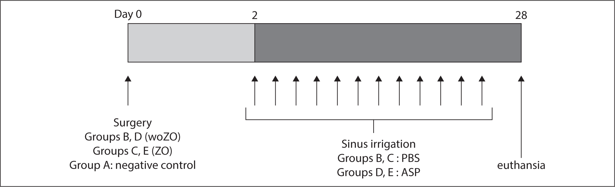
본 연구는 대구가톨릭대학교 동물윤리위원회의 심사와 승인 후 진행되었다. 토끼 모델은 뉴질랜드 수컷 흰토끼를 구매(Hyosung Science, Daegu, Korea) 하여 실험 전 3일 이상 최적의 환경에서 사육 후 실험을 진행하였다. 실험은 16마리의 토끼를 대상으로 하여 5군으로 나누어 진행되었다. 1군; 음성대조군(5측), 2군; 산화아연 없이 PBS를 상악동에 투여한 군(6측), 3군; 산화아연을 점막성골막하에 넣고 PBS를 상악동에 투여한 군(7측), 4군; 산화아연 없이 Aspergillus 포자를 상악동에 투여한 군(7측), 5군; 산화아연을 점막성골막하에 넣고 Aspergillus 포자를 투여한 군(7측)으로 하였다. 진균성 부비동염 모델의 제작은 기존의 만성부비동염 모델 제작 방법을 일부 변형하여 진행하였다.13) 산화아연 투여 방법은 0.35 mg/kg의 졸레틸과 0.15 mL/kg 럼푼을 근육주사하여 마취 후 콧등을 1:200,000 에피네프린이 섞인 2% 리도카인으로 국소 마취 후 절개를 가하여 피부와 피하조직을 박리하고 상악동 전벽을 노출하였다. 6.0 mm 드릴을 이용하여 골부를 제거하고 점막성골막을 노출시켰다. 점막성골막 부위를 박리 후 5 mg의 산화아연을 투여 후 4-0 나일론으로 피부를 봉합하였다. 술 후 2일째부터 일주일에 3회씩 상악동 전벽의 골 결손 부위를 통해 PBS 혹은 106개의 A. fumigatus 포자를 100 μL의 PBS에 섞어 상악동 내에 투여하였다. 실험 28일차에 4% 이소플루란과 CO2 가스를 이용하여 안락사를 유도 후 부비동 점막을 채취하였으며, 상악동 표면을 면봉으로 닦아 potato dextrose/corn meal agar 배지와 MacConkey agar 배지를 이용하여 각각 진균과 세균을 배양하였다(Fig. 1).

jcohns-35-1-3-g1
Fig. 1. Timetable of experiments among the different five groups. woZO: without zinc oxide, ZO: zinc oxide, PBS: phosphate buffer saline, ASP: Aspergillus fumigatus.
Download Original Figure
상악동 점막의 사이토카인 발현 측정

상악동 점막 조직의 interleukin(IL)-1β, IL-8, tumor necrosis factor(TNF)-α 단백과 mRNA 발현을 측정하였다. 단백 측정은 ELISA법(LSbio, Seattle, WA, USA)을 이용하여 측정하였으며 민감도는 IL-1β 7.8 pg/mL, IL-8 6 pg/mL, TNF-α 1.2 pg/mL이다. mRNA 측정은 정량적 역전사 중합효소연쇄반응을 이용하여 측정하였다. 상악동 점막을 1 mL의 trizole에 넣고, 총 RNA를 RNA isolation kit(Roche Diagnostics, Mannheim, Germany)를 이용하여 추출하였다. 역전사 반응은 1 μL의 RNA를 Perkin Elmer thermal cycle(PerkinElmer, Norwalk, CT, USA)을 이용하여 60℃에서 40 사이클로 반응시켰다. 사용한 시발체의 염기서열은 다음과 같다. IL-1β(sense, GTCTTCCTAAAGCAAGCCTTAC; antisense, GGGGTGTCACAATCTGTTTC, 92 bp), IL-8 (sense, GCATAAAGACACACTCCACAC; antisense, GTCCAGGCAGAGTTCTCTTC, 131 bp), TNF-α(sense, CCTGTGCCTCCCTTCACTTAT; antisense, TTTCTCGCCACTGACCAGTAG, 157 bp), GAPDH(sense, TAACTCTGGCAAAGTGGATGT; antisense, CGTGGGTGGAATCATACTG, 92 bp).

상악동 점막의 전사인자 발현 측정

상악동 점막의 nuclear factor(NF)-κB, c-Jun, p38, ERK, JNK 등의 전사인자 발현은 웨스턴 블롯을 이용하여 측정하였다. 점막을 용해 완충액(Thermo Fisher Scientific, Rockford, IL, USA)에 넣고 처리한 후 용해물을 전기영동 시킨 후 나이트로셀룰로스막에 옮겼다. 이후 각 전사인자의 항체로 일차 처리 후 2차 항체로 실온에서 2시간 처리하였다. 전사인자의 발현은 화학발광시스템(Pierece, Rockford, IL, USA)로 확인하였으며, 각각의 발현 정도는 multi Gauge v.2.02 소프트웨어(Fujifilm, Tokyo, Japan)을 이용하여 측정하였다.

상악동 점막의 진균성 생물막과 조직학적 검사

진균성 생물막은 상악동 점막을 생리식염수로 세척 후 0.1 mL의 LIVE/DEAD BacLight staining kit(Invitrogen, Carlsbad, CA, USA) 용액으로 빛이 차단된 상태에서 15분간 처리 후 니콘 A1 공초점 레이저 현미경(Nikon, Tokyo, Japan)으로 확인하였으며, 발현되는 형광 강도는 NIS-Elements AR 프로그램(Nikon)으로 확인하였다.

상악동 점막의 조직학적 검사는 상피층과 지층(stromal layer)의 두께, 및 호산구와 호중구의 침윤 정도는 hematoxylin-eosin 염색으로 확인하였다. 상피층과 지층의 두께는 400 μm 간격으로 5부분을 측정하여 평균하였으며, 염증세포의 침윤은 ×400 고배율로 3군데 측정하여 평균하였다. 점액분비세포의 측정은 periodic acid Schiff(PAS) 염색 시행 후 고배율로 3군데 측정하여 평균 수로 결정하였다.

결과

실험 28일째 희생시킨 토끼의 상악동 점막 채취 과정에서 상악동 내 농성 혹은 점액성 분비물이 확인되는 경우는 없었다. 상악동 점막의 진균과 세균 배양에서 A. fumigatus를 투여한 경우 14측 중 12측(85.7%)에서 진균이 배양되었고 A. fumigatus를 투여하지 않은 경우 1측(5.6%)에서 진균이 배양되었다. 세균의 경우 대부분 Bordetella bronchiseptica가 배양되었으며, A. fumigatus를 투여한 경우 2측(14.3%)에서 세균이 배양되었고 투여하지 않은 경우 8측(44.4%)에서 세균이 배양되었다(Table 1).

Table 1. Bacteriologic and mycological findings in maxillary sinus after 4 weeks experiments
NC (5) sZO+PBS (6) wZO+PBS (7) sZO+Asp (7) wZO+Asp (7)
Bacterial
 No growth 1 3 6 5 5
 Bordetella bronchiseptica 3 3 1 2 0
 Others 1 0 0 0 0
Fungal
 No growth 5 6 6 1 1
 Aspergillus 0 0 1 6 6

NC: negative control, sZO: without zinc oxide, wZO: with zinc oxide, PBS: phosphate buffer saline, Asp: Aspergillus fumigatus.

Download Excel Table

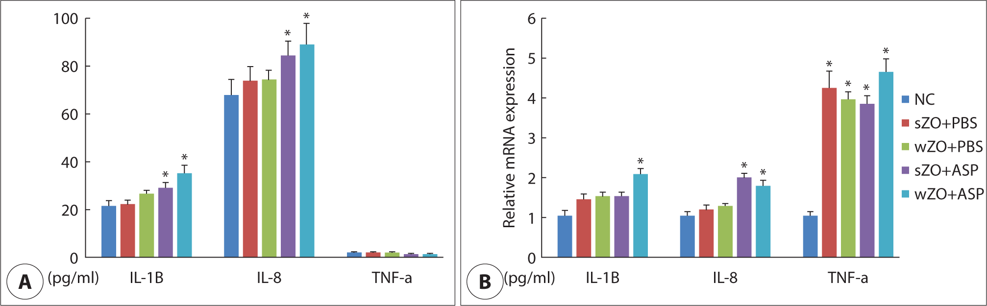
상악동 점막의 사이토카인 발현은 IL-1β의 경우 대조군 21.5±13.2 pg/mL에 비해 상악동 내 A. fumigatus를 투여한 경우 유의하게 증가하였으나, 상악동 골막하에 산화아연의 투여가 IL-1β의 생성에는 영향을 미치지 못하였다(wZO[with zinc oxide]: 35.2±16.9 pg/mL, sZO[without zinc oxide]: 29.1±17.5 pg/mL). IL-8의 경우 대조군 67.8±24.7 pg/mL에 비해 상악동 내 A. fumigatus를 투여한 경우 유의한 증가를 보였으나, 상악동 골막하 산화아연의 투여가 IL-8 생성에 영향을 미치지 못하였다(wZO: 89.1±32.3 pg/mL, sZO: 84.5±35.8 pg/mL). TNF-α 단백의 경우 실험의 조건에 관계없이 3.0 pg/mL 이하의 낮은 농도로 측정되어 비교할 수 없었다. mRNA 발현은 IL-1β와 IL-8의 경우 상악동 내 A. fumigatus를 투여한 경우 단백 발현과 유사하게 발현이 증가하였으며, TNF-α의 경우 상악동 내 PBS 혹은 A. fumigatus를 투여한 경우 대조군과 비교하여 실험군 모두에서 mRNA 발현이 증가하였다(Fig. 2).

jcohns-35-1-3-g2
Fig. 2. Maxillary sinus mucosal cytokine protein and mRNA expression. Intramaxillary inoculation of Aspergillus fumigatus (ASP) enhanced interleukin (IL)-1β, and IL-8 protein (A) and mRNA (B) expression. TNF-α mRNA was significantly increased in experimental groups in compare with negative control (NC) group. Zinc oxide (ZO) did not influence cytokine production by A. fumigatus. IL-1β: interlukin-1β, IL-8: interluikin-8, TNF-α: tumor necrosis factor-α, NC: negative control, PBS; phosphated buffer saline, wZO; with ZO, sZO: without ZO ASP: Aspergillus fumigatus. * p>0.05 compared with NC.
Download Original Figure

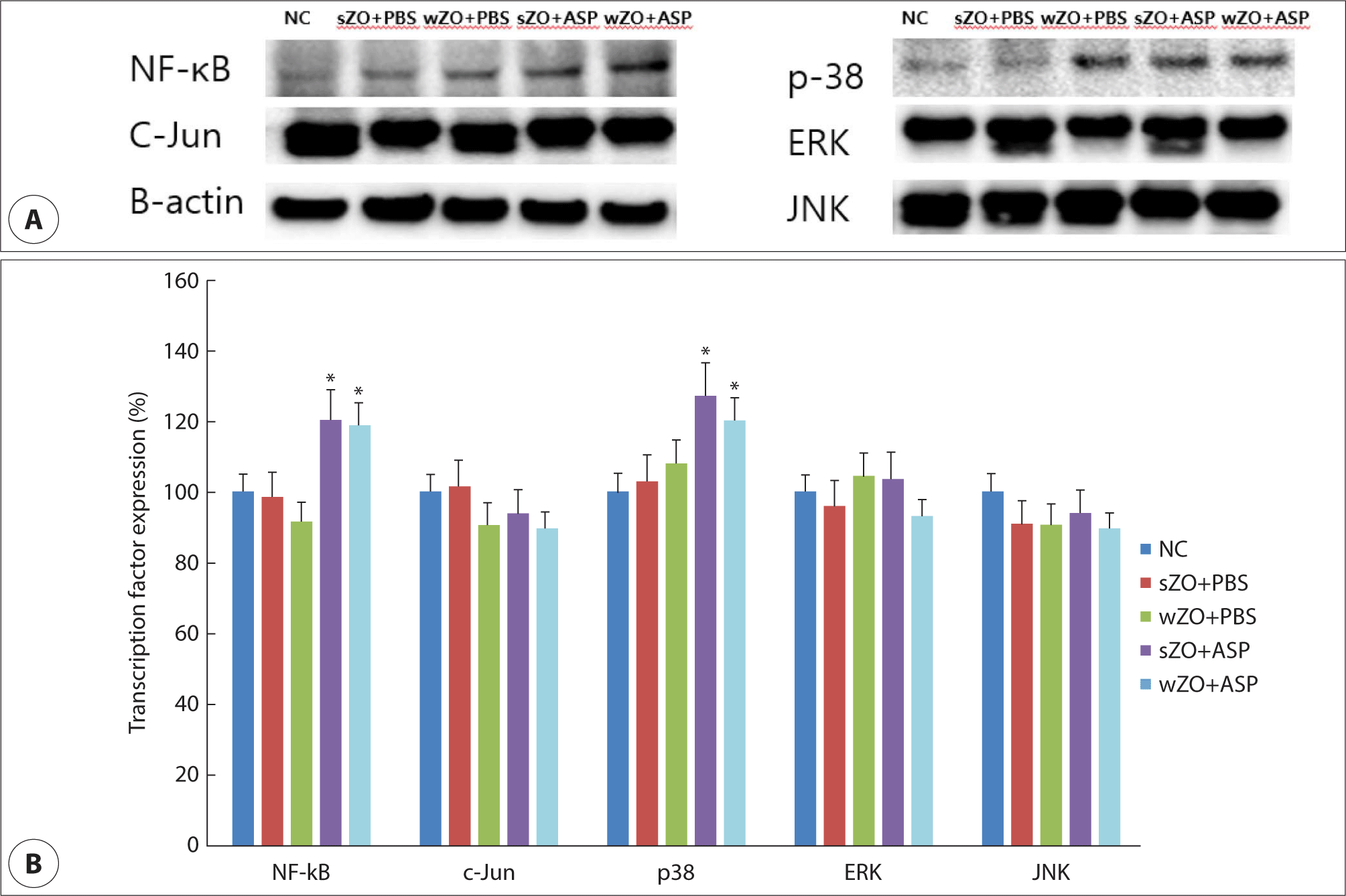
상악동 점막의 염증반응에 관여하는 대표적인 전사인자인 NF-κB, activated protein-1(c-Jun)과 mitogen-activated protein kinase(MAPK)를 분석하였다. 상악동 내 A. fumigatus를 투여한 경우 NF-κB와 p38의 발현이 유의하게 증가하였으며, 나머지 전사인자들은 그룹 간에 차이를 보이지 않았다. 또한 상악동 골막하 산화아연 투여는 A. fumigatus에 의한 전사인자 발현에 영향을 미치지 못하였다(Fig. 3).

jcohns-35-1-3-g3
Fig. 3. Transcription factor expression in the maxillary sinus mucosa. (A) shows representative finding of transcription factor expression. Transcription factor expression in maxillary sinus mucosa. Intramaxillary instillation of Aspergillus fumigatus (ASP) significantly upregulated NF-κB and p38 expression (B). NF-κB: nuclear factor-κB, C-Jun: activated protein-1 transcription factor transcription factor, mitogen-activated protein kinase: ß-actin: Actin beta, ERK: extracellular signal-regulated kinase, JNK: c-JUN NH2-terminal kinase: negative control, wZO: with zinc oxide, sZO: without zinc oxide, PBS: phosphate buffer saline. * p>0.05 compared with NC.
Download Original Figure

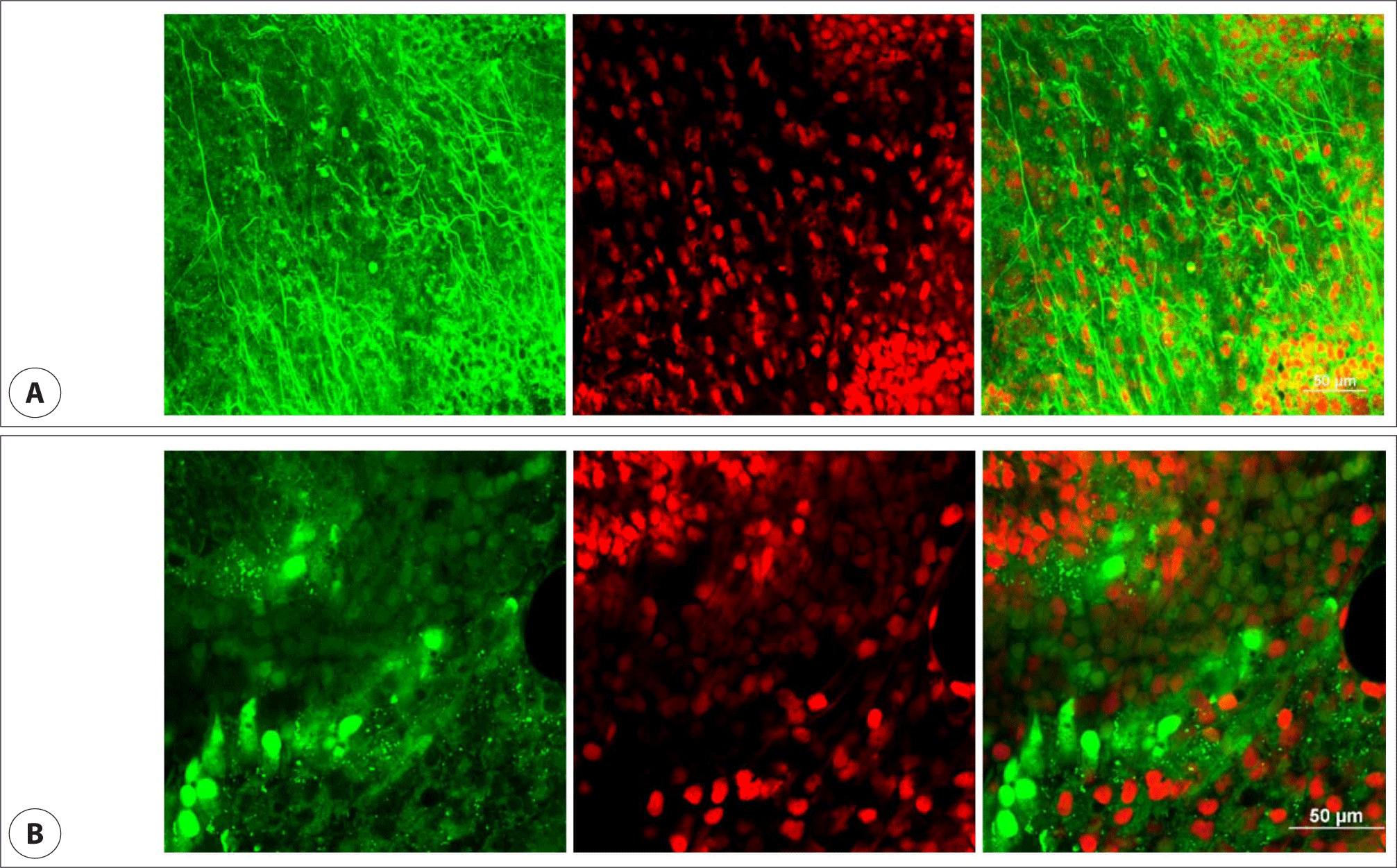
진균성 생물막의 경우 상악동 내 A. fumigatus 포자를 투여한 경우에만 공초점 레이저 현미경으로 생물막을 확인할 수 있었으며, 골막하 산화아연 투여는 생물막 형성에 영향을 미치지 못하였다(Fig. 4).

jcohns-35-1-3-g4
Fig. 4. Confocal scanning laser microscopic findings displaying the sinus mucosal cells and biofilms. A shows representative finding of fungal biofilms after intramaxillary instillation of Aspergillus fumigatus conidia. Fungal biofilm is fluorescing areas displayed as a highly coordinated network of hyphae, spreading in all directions and cross-lining each other. B shows no fungal biofilms without fungal instillation. Green: live cells, Red: damaged or dead cells (A & B: 40× magnification, scale bars = 50 μm).
Download Original Figure

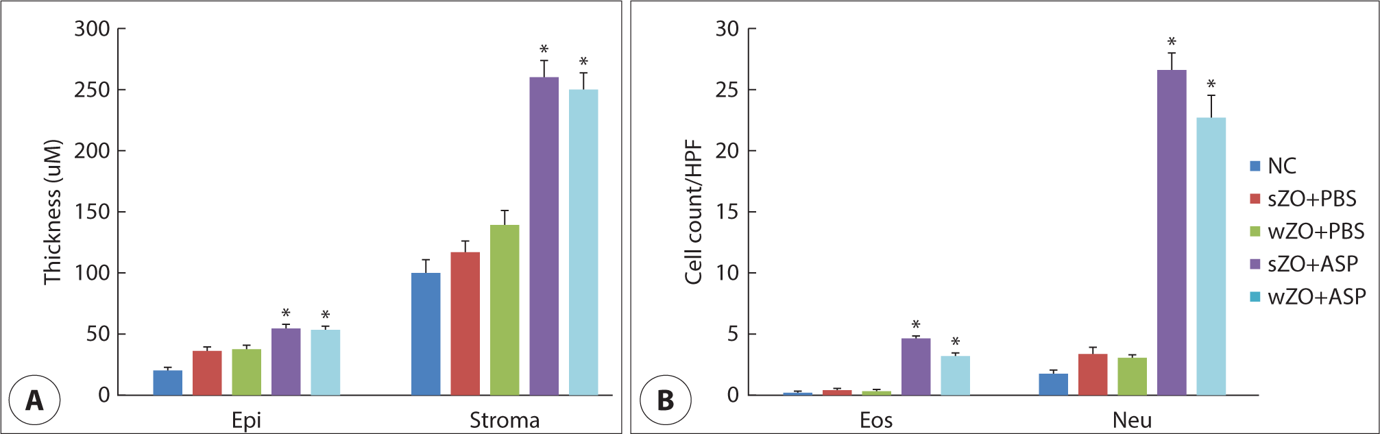
상악동 점막을 염증성 변화를 확인하기 위해 상피 두께의 변화, 염증세포의 침윤과 분비세포의 수를 확인하였다. 상악동 내 A. fumigatus 포자를 투여한 경우 상피층(wZO: 49.5±32.6 μm, sWO: 50.0±18.5 μm)과 지층(wZO: 214.9±56.5 μm, sWO: 261.7±49.5 μm)의 두께가 대조군에 비해 유의하게 증가하였다. 염증세포 또한 상악동 내 A. fumigatus 포자를 투여한 경우 호산구(wZO: 3.8±1.2 /HPF, sWO: 5.3±1.6 /HPF)와 호중구(wZO: 23.513.4 /HPF, sWO: 27.6±9.8 /HPF) 모두 대조군에 비해 유의하게 증가하였다(Fig. 5). 점액분비 세포의 경우에도 상악동 내 A. fumigatus 포자를 투여한 경우 유의하게 증가하였다(NC: 2±0.8 /HPF, sZO+PBS: 7.3±2.4 /HPF, wZO+PBS: 7.6±3.7 /HPF, sZO+ASP[Aspergillus fumigatus]: 17.6±8.7 /HPF, wZO+ASP: 19.5±5.6 /HPF). 하지만, 골막하 산화아연 투여가 상피세포의 두께, 염증세포의 침윤과 점액분비 세포 수에 영향을 미치지 못하였다.

jcohns-35-1-3-g5
Fig. 5. Histologic characteristics of the rabbit maxillary sinus mucosa. Epithelial (Epi) and Stromal (Stroma) thickness (A) and inflammatory cells infiltration (B) were significantly increased after intra-maxillary instillation of Aspergillus fumigatus (ASP) conidia. NC: negative control, wZO: with zinc oxide, sZO: without zinc oxide, PBS: phosphate buffer saline, Eos: eosinophils, Neu: neutrophils. * p>0.05 compared with NC.
Download Original Figure

고찰

Aspergillus는 상하기도에서 발생하는 진균성 호흡기 질환의 가장 흔한 원인균으로 기관지폐 아스페르길루스증, 진균구, 진균성 만성부비동염에서 흔히 검출되고 있다.2,14) 산화아연은 치근관 치료에 사용되는 충전제의 주요 구성물로 이용되고 있으나 진균의 생존, 성장과 증식을 촉진시키는 주요 원소로 진균성 상악동염을 유발하는 위험인자로 알려져 있다.8) 본 연구에서는 산화아연의 상악동 점막성골막하 존재가 상악동 내로 들어온 A. fumigatus 포자의 성장과 생물막 형성, 상악동 점막의 염증성 변화에 미치는 영향을 확인하고자 하였다. 상악동 내 진균 포자는 상악동 점막의 염증성 변화와 진균성 생물막의 형성을 유도하였지만, 골막하 산화아연이 진균 포자에 의한 염증 반응을 악화시키거나 생물막 형성을 촉진시키지는 못하였다.

토끼는 부비동의 해부학적 구조가 인간과 유사하고 상악동이 크기 때문에 부비동염의 동물 실험, 특히 상악동염 연구에 많이 활용되어왔다.15) 토끼 상악동의 평균온도는 35.2±0.7℃로 A. fumigatus의 성장에 유리한 조건이다. Dufour 등은 토끼모델을 이용하여 상악동 점막에 손상을 주거나 자연개구부를 폐쇄시킨 후 A. fumigatus 포자를 상악동 내에 투여하여 부비동염 모델을 제작할 수 있었다고 보고하였다. 특히 자연개구부 폐쇄가 진균성 상악동염을 유발하는 가장 중요한 요소로 고려하였으나, 연구 결과상, 진균은 배양되지 않았으며, 세균이 배양되거나, 농성 분비물이 확인되었다. 또한 본 연구와 달리 108 /mL의 포자를 상악동 내 한번 투여하여 염증을 유발시키고자 하였다.16) 저자들의 경우 상악동 점막 손상이나 자연개구부 폐쇄 없이 107 /mL의 포자를 일주일에 3번 4주간 투여하여 염증 반응을 유도하였으며, 육안적으로 농성 분비물이 확인되는 경우는 없었으나, 골막하 산화아연과는 무관하게 4군과 5군 모두에서 7측 중 6측에서 A. fumigatus가 배양되었다. 이는 상악동 점막의 손상이나 자연개구부가 폐쇄되는 경우, 상악동 내 존재하는 세균이 점막섬모운동 등을 통한 제거가 어려워짐에 따라 세균의 증식을 통해 발생하는 세균성 상악동염이 발생하는 것과 달리 본 연구에서는 상악동이 진균에 지속적으로 노출되는 경우 상악동 내 진균의 성장과 증식이 이루어짐을 확인할 수 있었고, 이로 인해 상악동 점막의 염증성 변화가 발생함을 확인할 수 있었다. 정상적인 호흡기 점막의 선천성 면역반응이 작동하는 경우 흡인된 진균이나 진균 포자는 자연배출되지만 점막섬모운동 능력의 손상이나 진균의 성장을 촉진하는 환경이 조성되는 경우 진균의 성장과 생물막이 형성될 수 있다. 이러한 현상은 상악동 점막의 A. fumigatus 생물막 형성이 증가된다는 사실로도 확인이 되었다. 하지만 골막하 산화아연이 진균의 성장이나 증식에는 영향을 미치지 못하였다.

일반적으로 진균이 인체에 침범했을 시 Th1 림프구와 호중구에 의한 선천성 세포매개면역의 탐식작용을 통한 국소면역방어 기전을 보이지만, Th2 림프구의 역할이 강화되는 경우 진균성 감염이 유발될 수도 있다.17) 본 연구에서도 상악동 내 A. fumigatus를 투여하는 경우 Th1과 Th2 염증세포인 호중구와 호산구 모두 증가하였다. 이는 상악동 점막에서 진균에 대한 방어기전과 염증반응이 동시에 이루어지고 있음을 의미할 것이다. 진균의 다양한 독소와 효소들은 호흡기 상피세포를 직접 손상시키거나 상피세포의 염증 반응을 유발한다. 특히 진균의 단백질분해효소는 상피세포의 패턴인식수용체와 반응하여 IL-1β, IL-8, TNF-α와 같은 다양한 염증유발 사이토카인(proinflammatory cytokine)의 생성을 유도한다.3)A. fumigatus 역시 상악동 점막의 IL-1β, IL-8, TNF-α mRNA의 발현과 IL-1β, IL-8 단백 생성을 촉진시켰다. IL-1β, IL-8은 염증반응을 조절하고 호중구의 이주를 통해 염증을 유지 악화시킬 뿐 아니라 병원성 물질의 제거에도 관여한다. TNF-α 또한 염증반응을 촉진하는 대표적인 화학매개물질이나 본 연구에서는 상악동 골막하 산화아연이 존재하는 경우 TNF-α mRNA만 증가하였다. 이는 상악동 내로 투여된 진균이 국소 염증반응을 충분히 유발하지 못하였지만, 골막하 산화아연이 상악동 점막의 염증반응을 유도할 수 있음을 유추해 볼 수 있었다. A. fumigatus는 NF-κB의 활성화를 통해 각종 염증유발 사이토카인의 생성을 유도한다.18) 저자들의 경우 상악동 내 A. fumigatus 포자를 투여하는 경우 NF-κB뿐 아니라 MAPK 중 하나인 p38의 발현도 증가시켰다. 하지만 골막하 산화아연이 진균에 의한 염증유발 사이토카인의 생성이나 전사인자 발현에 영향을 미치지는 못하였다.

A. fumigatus는 다세포 성분과 세포외 기질에 의한 생물막 형성을 통해 항진균제에 대한 감수성을 감소시킨다고 알려져 있다.19) 만성부비동염과 진균구에서 진균성 생물막을 확인할 수 있으며, 비강 상피세포와 A. fumigatus의 합동배양으로 진균성 생물막이 형성될 수 있으며, 황사와 같은 환경 오염물질에 의해 진균성 생물막 형성이 촉진된다.13,20) 또한 토양 성분의 하나인 아연은 A. fumigatus의 성장, 핵산 합성과 독소 생성을 유도할 수 있다.9) 근관 충전물의 주요 성분인 산화아연은 진균구 형성의 중요한 원인 인자로 알려져 있지만 저자들의 경우 점막성골막하에 존재하는 산화아연이 상악동 진균에 의한 염증 반응과 생물막 형성에는 영향을 미치지 못하는 것을 확인할 수 있었다. 이는 치근관 충전물과 같은 산화아연이 상악동염이나 진균구 형성에 영향을 미치기 위해서는 충전물이 점막성골막하에 위치하는 것으로는 충분하지 않고 골막을 통과하여 상악동 내로 이동한 후 상악동으로 흡입된 진균 포자와의 상호 작용을 통해 상악동염이나 진균구 형성에 영향을 미칠 수 있을 것으로 추정된다. 또한 본 연구에서 실험 기간이 4주로 산화아연과 진균 포자의 상호작용 시간이 충분하지 못함에 기인할 수 있을 것이다. 이러한 의문점을 해결하고 산화아연이 상악동 진균성 부비동염에 미치는 영향을 확인하기 위해서는 상악동 내 산화아연을 투여하거나, 산화아연과 진균 포자가 충분히 상호작용할 수 있는 실험 기간을 조건으로 하는 추가적인 연구가 필요할 것이다.

Acknowledgements

This work was supported by the National Research Foundation of Korea (NRF) grant funded by the Korea government (Ministry of Science and ICT) (2019R1F1A1047757).

Funding Information

Not applicable.

Conflicts of Interest

No potential conflict of interest relevant to this article was reported.

Author Contribution

Conceptualization: Shin SH.

Data curation: Kim TG, Shin SH.

Formal analysis: Whangbo CH.

Methodology: Lee SH.

Software: Kim TG, Shin SH.

Validation: Ye MK.

Investigation: Shin SH.

Writing - original draft: Ye MK, Shin SH.

Writing - review & editing: Kim TG, Whangbo CH, Lee SH, Ye MK, Shin SH.

Ethics Approval

Research was conducted according to all ethical standards, and written informed consent was obtained from all patients. This study was approved by the Institutional Review Board (IRB) of the Daegu Catholic University, Korea (Approval No. DCIAFCR-221129-36-Y)

References

1.

Fokkens WJ, van Drunen C, Georgalas C, Ebbens F. Role of fungi in pathogenesis of chronic rhinosinusitis: the hypothesis rejected. Curr Opin Otolaryngol Head Neck Surg 2012;20(1):19-23.

2.

Shin SH, Ye MK, Lee DW, Geum SY. Immunopathologic role of fungi in chronic rhinosinusitis. Int J Mol Sci 2023;24(3):2366.

3.

Rivas CM, Schiff HV, Moutal A, Khanna R, Kiela PR, Dussor G, et al. Alternaria alternata-induced airway epithelial signaling and inflammatory responses via protease-activated receptor-2 expression. Biochem Biophys Res Commun 2022;591:13-9.

4.

Zhao YC, Bassiouni A, Tanjararak K, Vreugde S, Wormald PJ, Psaltis AJ. Role of fungi in chronic rhinosinusitis through ITS sequencing. Laryngoscope 2018;128(1):16-22.

5.

Park GY, Kim HY, Min JY, Dhong HJ, Chung SK. Endodontic treatment: a significant risk factor for the development of maxillary fungal ball. Clin Exp Otorhinolaryngol 2010;3(3):136-40.

6.

Croft CA, Culibrk L, Moore MM, Tebbutt SJ. Interactions of Aspergillus fumigatus Conidia with airway epithelial cells: a critical review. Front Microbiol 2016;7:472.

7.

Mostafa BE, El Sharnoubi MMK, El-Sersy HAA, Mahmoud MSM. Environmental risk factors in patients with noninvasive fungal sinusitis. Scientifica 2016;2016:5491694.

8.

Nicolai P, Mensi M, Marsili F, Piccioni M, Salgarello S, Gilberti E, et al. Maxillary fungus ball: zinc-oxide endodontic materials as a risk factor. Acta Otorhinolaryngol Ital 2015;35(2):93-6.

9.

Cuero R, Ouellet T, Yu J, Mogongwa N. Metal ion enhancement of fungal growth, gene expression and aflatoxin synthesis in Aspergillus flavus: RT‐PCR characterization. J Appl Microbiol 2003;94(6):953-61.

10.

Wang X, Dong D, Cheng J, Fan X, Zhao Y. Relationship between biofilms and clinical features in patients with sinus fungal ball. Eur Arch Otorhinolaryngol 2015;272(9):2363-9.

11.

Foreman A, Boase S, Psaltis A, Wormald PJ. Role of bacterial and fungal biofilms in chronic rhinosinusitis. Curr Allergy Asthma Rep 2012;12(2):127-35.

12.

Geum SY, Park JW, Park HJ, Ye MK, Shin SH. In vitro studies on the role of zinc oxide in the development of Aspergillus fumigatus biofilm on nasal epithelial cells. Korean J Otorhinolaryngol Head Neck Surg 2022; 65(11):684-91.

13.

Braga AA, Valera FCP, Faria FM, Rossato M, Murashima AAB, Fantucci MZ, et al. An experimental model of eosinophilic chronic rhinosinusitis induced by bacterial toxins in rabbit. Am J Rhinol Allergy 2019;33(6):737-50.

14.

Singhal D, Baker L, Wormald PJ, Tan L. Aspergillus fumigatus biofilm on primary human sinonasal epithelial culture. Am J Rhinol Allergy 2011;25(4):219-25.

15.

Murat Ozcan K, Ozcan I, Selcuk A, Akdogan O, Gurgen SG, Deren T, et al. Comparison of histopathological and CT findings in experimental rabbit sinusitis. Indian J Otolaryngol Head Neck Surg 2011;63(1):56-9.

16.

Dufour X, Goujon JM, Rodier MH, Kauffmann-Lacroix C, Grollier G, Klossek JM. Experimental model of fungal sinusitis: a pilot study in rabbits. Ann Otol Rhinol Laryngol 2005;114(2):167-72.

17.

Bartemes KR, Kita H. Innate and adaptive immune responses to fungi in the airway. J Allergy Clin Immunol 2018;142(2):353-63.

18.

Du J, Chi Y, Song Z, Di Q, Mai Z, Shi J, et al. Crocin reduces Aspergillus fumigatus-induced airway inflammation and NF-κB signal activation. J Cell Biochem 2018;119(2):1746-54.

19.

Seidler MJ, Salvenmoser S, Müller FMC. Aspergillus fumigatus forms biofilms with reduced antifungal drug susceptibility on bronchial epithelial cells. Antimicrob Agents Chemother 2008;52(11):4130-6.

20.

Shin SH, Ye MK, Lee DW, Chae MH. Asian sand dust particles enhance the development of Aspergillus fumigatus biofilm on nasal epithelial cells. Int J Mol Sci 2022;23(6):3030.