Case Report
반대측 신경초종이 동반된 내시경 귀수술로 제거한 고실 사구종
A Case of Middle Ear Glomus Tympanicum Tumor Removed by Transcanal Endoscopic Ear Surgery with Contralateral Vestibular Schwannoma
Sang Hyo Lee
1
, Seok Hyun Kim
2, Hyun Min Lee
2, Il Woo Lee
2,*
2부산대학교 의과대학 이비인후과학교실, 양산부산대학교병원
1Department of Otorhinolaryngology-Head and Neck Surgery, Maryknoll Hospital, Busan, Korea
2Department of Otorhinolaryngology-Head and Neck Surgery, Pusan National University School of Medicine, Pusan National University Yangsan Hospital, Yangsan, Korea
*Corresponding author: Il Woo Lee, Department of Otorhinolaryngology-Head and Neck Surgery, Pusan National University School of Medicine, Pusan National University Yangsan Hospital, Yangsan 50612, Korea, Tel: +82-55-360-2651, Fax: +82-55-360-2162, E-mail:
entgate@pusan.ac.kr
© Copyright 2023 The Busan, Ulsan, Gyeoungnam Branch of Korean Society of Otolaryngology-Head and Neck Surgery. This is an Open-Access article distributed under the terms of the
Creative Commons Attribution Non-Commercial License (http://creativecommons.org/licenses/by-nc/4.0/) which permits
unrestricted non-commercial use, distribution, and reproduction in any
medium, provided the original work is properly cited.
Received: Jun 14, 2023; Revised: Jul 17, 2023; Accepted: Sep 04, 2023
Published Online: Sep 30, 2023
ABSTRACT
Glomus tympanicum tumors are a rare type of middle ear tumor that often present with symptoms such as pulsatile tinnitus, hearing loss, and ear fullness. Vestibular schwannomas are benign tumors of the cerebellopontine angle that can cause hearing loss, tinnitus, and vertigo. Although cases of both tumors occurring on the same side have been reported, only a few cases of contralateral glomus tympanicum tumors associated with vestibular schwannomas have been documented. We present a case of a 69-year-old female who was diagnosed with a vestibular schwannoma on the left side and a glomus tympanicum tumor on the right side. The patient underwent transcanal endoscopic ear surgery (TEES) for the removal of the glomus tympanicum tumor. This case emphasizes the importance of careful examination of both ears. And we suggest that TEES may be a viable option for treating early-stage glomus tympanicum tumors.
Keywords: Glomus tympanicum tumor; Neuroma, acoustic; Transcanal endoscopic ear surgery
서론
사구종양은 중이강 내에 발생하는 가장 흔하며 동시에 측두골에서는 두 번째로 가장 흔하게 발생하는 일차성 종양이다.1) 환자들은 박동성의 이명, 난청, 이충만감 등을 흔하게 호소한다.2),3) 전정신경초종은 연간 10만 명당 한 명꼴로 보고되는 소뇌교각의 가장 흔한 양성종양으로서, 진행성 혹은 돌발성의 난청과 이명을 흔히 동반하며, 크기가 큰 경우 안면신경 마비를 비롯한 다양한 신경증상을 동반하기도 한다.4),5) 이 두 종양이 동측에서 발생한 경우는 세 증례가 보고되어 있으나,6–8) 서로 다른 귀를 침범한 사례는 이제까지 단 2예만 보고되어 있다.9) 저자들은 좌측 난청으로 내원하여 확인된 좌측의 전정신경초종에 동반된 우측 고실사구종(glomus tympanicum tumor)을 발견하고 고실사구종에 대하여 내시경적 절제술을 통하여 치료하였기에 증례를 보고하고 연관된 문헌을 고찰해 보고자 한다.
증례
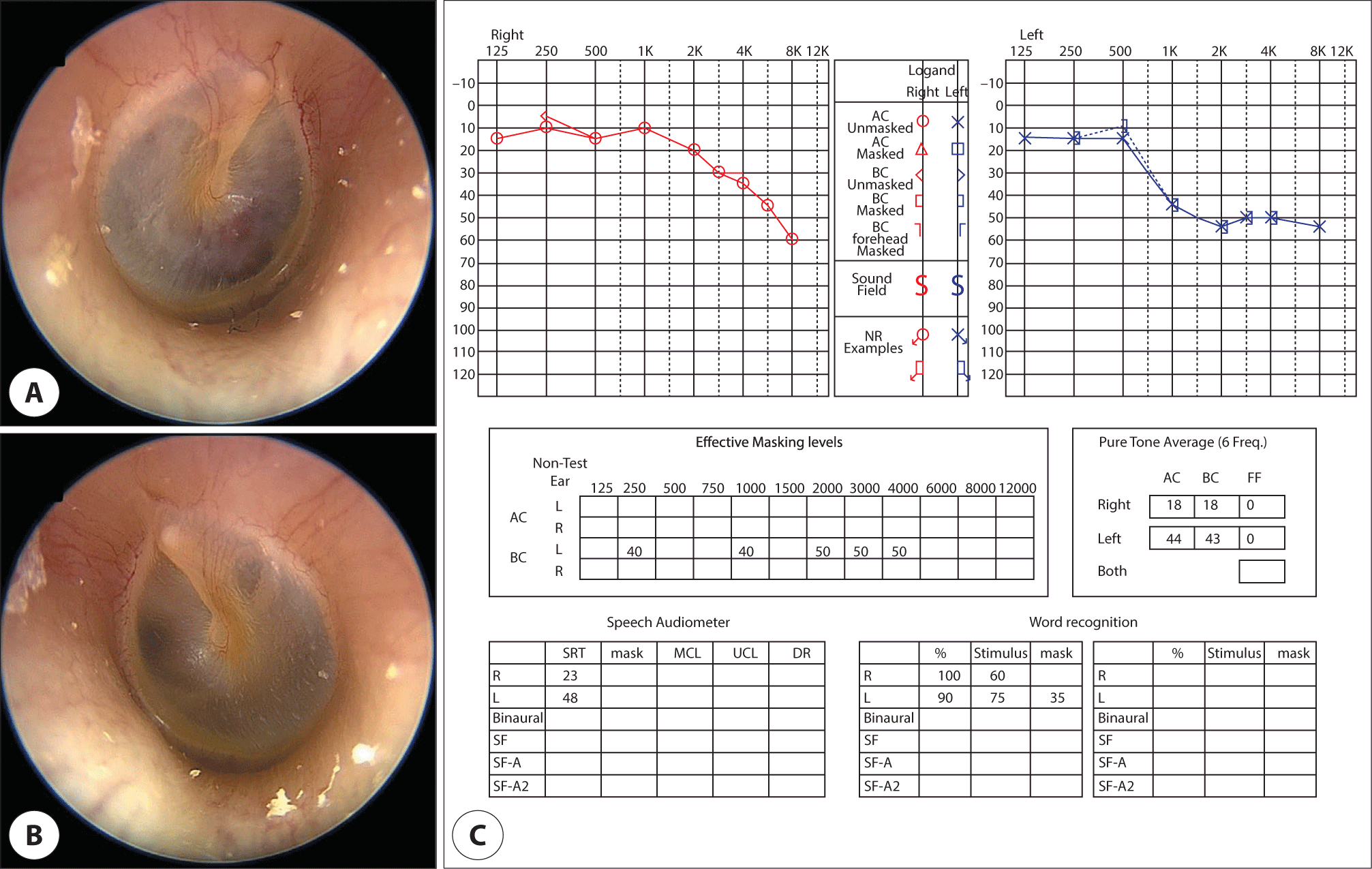
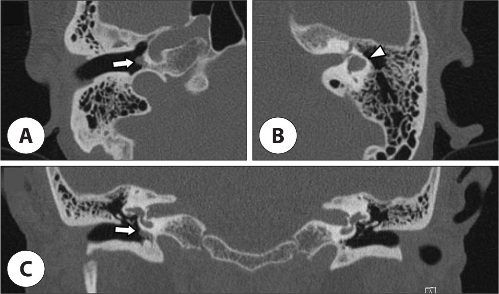
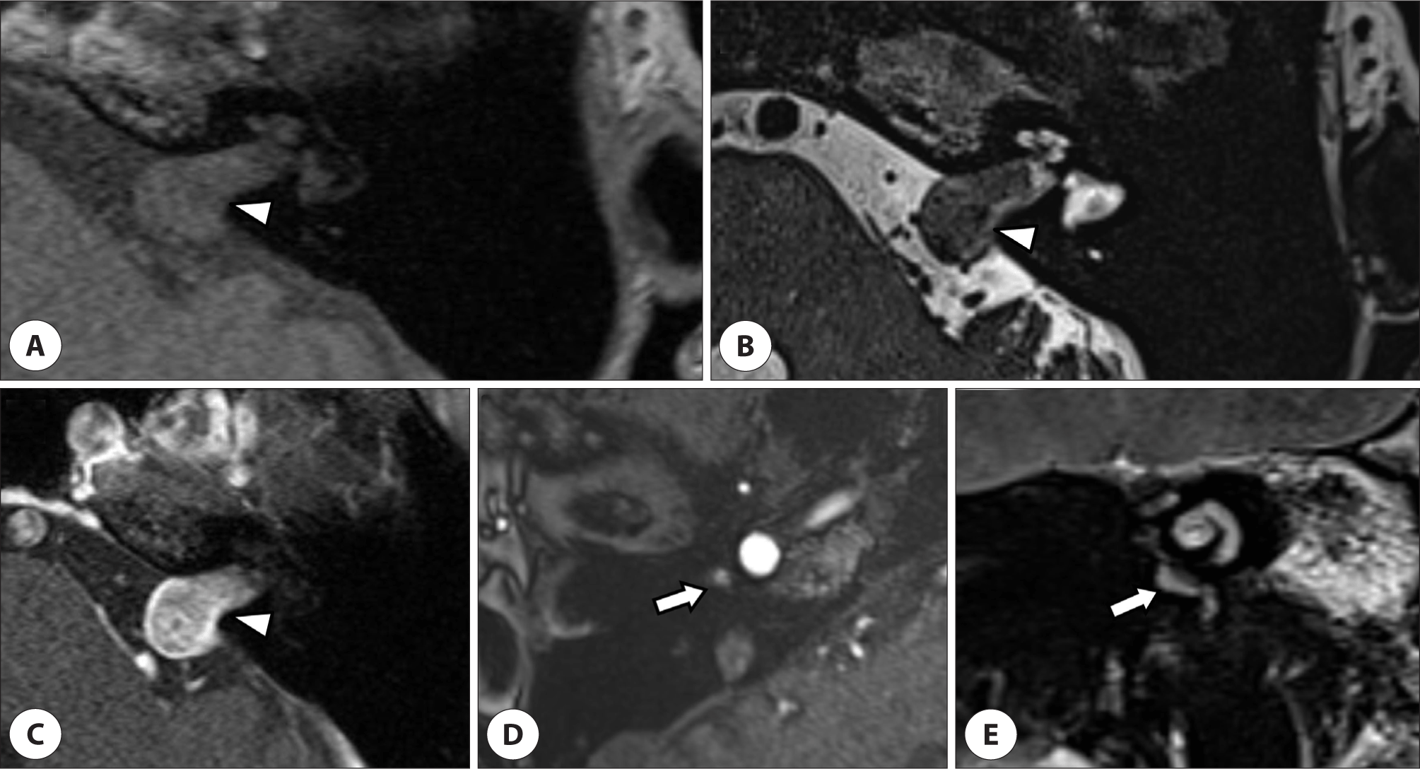
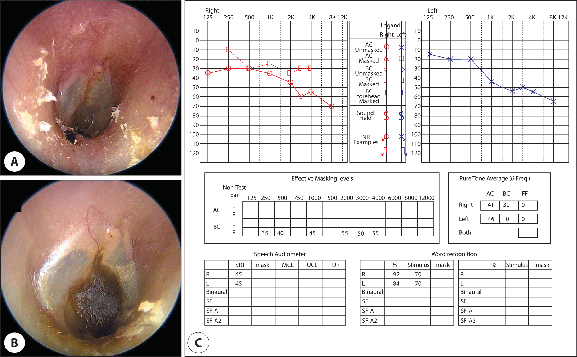
69세 여자 환자로 서서히 진행된 좌측 청력 저하 및 이명 주소로 개인의원에 내원하여 외이도 내시경 확인결과 양측 고막은 파열 등의 이상 소견이 없으나, 고막을 통해 관찰되는 우측 중이의 적색 종물이 확인되어 본원에 의뢰되었다. 청력검사 결과는 순음청력검사상 6분법으로 우측 기도 18 dB HL, 골도 18 dB HL, 좌측 기도 44 dB HL, 골도 43 dB HL의 역치를 보였으며, 어음청취역치는 우측 23 dB HL, 좌측 48 dB HL, 어음명료도는 우측 60 dB HL에서 100%, 좌측 75 dB HL에서 90%로 측정되었다(Fig. 1). 측두골전산화단층촬영 및 자기공명영상촬영을 시행하였으며 측두골전산화단층촬영에서는 우측 와우갑각의 연조직 음영(Fig. 2A, C)에 더하여 좌측 측반고리관의 이형성과 고위 경정맥구(high jugular bulb) 소견이 확인되었다(Fig. 2B, C). 측두골 자기공명영상에서는 좌측 내이도와 소뇌교각에 위치한 1.6×1 cm의 균질 조영증강되면서 T1, T2에서 등신호강도(isosignal intensity) 보이는 종양(Fig. 3A–C)이 발견되었고, 전정신경초종에 합당한 소견이었다. 우측의 중이강 내에서 와우갑각(promontory)에 밀착한 T1에서 저신호, T2에서 고신호강도를 보이며 조영증강이 잘 되는 4 mm 크기의 사구종양 가능성이 있는 병변이 확인되었으며(Fig. 3D, E), 이는 Fisch type A 및 Glasscock-Jackson type I에 해당된다. 전산화단층촬영상 좌측 측반고리관의 이형성이 관찰되었으나 환자 주관적인 전정증상을 호소하지 않고 추가적인 검사를 원하지 않아서 전정기능검사는 시행하지 않았다. 전정신경초종은 종양이 안정적인 경우, 경과를 관찰하는 것이 종합적으로 적절할 것이라는 판단하에 난청이 있는 상태로 경과관찰하기로 하였으며, 고실 사구종은 수술적으로 제거하기로 하였다. 술 중 출혈 경감을 위해 혈관조영 및 색전술 시행하기로 하여 수술 전일 우측 신경뇌막하고실동맥(inferior tympanic artery)으로 추정되는 종양의 공급 혈관(tumor feeder)을 선택하여 색전술을 시행하였다.
Fig. 1.
Initial findings of the tympanic membrane and audiometry results. A: Pinkish mass on the anterior-inferior quadrant of the right tympanic membrane. B: Normal tympanic membrane on the left side. C: The patient’s initial pure tone audiometry and speech audiometry results showed mild hearing loss in the left ear.
Download Original Figure
Fig. 2.
Preoperative temporal bone CT image. A: Axial image of CT scan of the right temporal bone. A protruding soft tissue (white arrow) was observed within the right middle ear. B: On the left side, the presence of a high jugular bulb and the aberrant lateral semicircular canal (white triangle) was confirmed. C: On the coronal image of the CT scan, protruding mass was observedinthe right middle ear cavity (white arrow). CT: computed tomography.
Download Original Figure
Fig. 3.
Preoperative temporal bone MRI findings. A: T1-weighted axial image. B: T2-weighted axial image. C: Gadolinium-enhanced T1-weighted images of left vestibular schwannoma (white triangle). On the MRI finding, heterogeneous iso/hyperintensity, strongly enhanced tumor with widening of the left internal acoustic canal was observed. D: Enhanced T1-weighted axial images. E: T2-weighted coronal image of the right glomus tympanicum tumor (white arrow). The tumor appears as a hyperintense mass on T2-weighted imaging, with enhancement on T1-weighted image. MRI: magnetic resonance image.
Download Original Figure
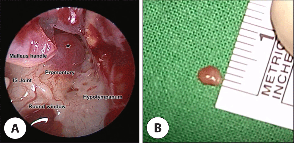
수술은 내시경만을 사용하여 실시하였으며, 외이고막 피판을 거상할 때에 추골병을 기준으로 10시–3시 방향까지 전방 및 하방의 외이도 절개를 시행하였다. 절개선을 따라 골막하 박리를 통해 섬유 고실륜을 포함하여 외이고막피판을 거상하였다. 이를 통해 충분한 시야 확보가 가능하여 일괴제거(en-block resection)를 시행할 수 있었다. 와우갑각에 줄기(stalk)가 확인되어 전기소작을 시행하였다. 중이 조작을 종료하고 외이고막피판을 원위치시키는 도중고막손상을 발견하여 인공진피(Megaderm®, L&C Bio, Seoul, Korea) 거치 후 피판을 원위치하고 mupirocin 연고를 도포한 거즈를 외이도에 충진한 후 수술을 종료하였다(Fig. 4).
Fig. 4.
Intraoperative findings of right glomus tympanicum tumor. A: Reddish mass (asterisk mark) was observed on promontory during transcanal endoscopic ear surgery. B: Image of a glomus tympanicum tumor removed en bloc. IS: incudostapedial.
Download Original Figure
술 후 2일째까지 특별한 합병증이 없어 퇴원하였으며, 술 후 2주차에 패킹 제거 후 특이소견은 없었으나 술 후 10주째 고막의 부분 천공이 확인되어 패치 적용을 하였으며, 순음청력검사상 우측 기도 42 dB HL, 골도 27 dB HL로 역치가 증가해 있었으며, 좌측은 기도 및 골도 48 dB HL로 큰 차이를 보이지 않고 있다. 패치사용에도 환자의 고막천공이 지속되어 수술 1년째에 인공진피를 이용한 sandwich graft 고막성형술을 시행하였다. 수술 이후 3개월까지 graft가 유지되면서 우측은 다른 증상 없이, 좌측은 난청 호전 없이 경과관찰 중이다(Fig. 5).
Fig. 5.
Postoperative tympanic membrane findings and audiometry results. A: Remaining perforation after endoscopic surgery of the right ear. B: Patch graft surgerywith cadaveric dermiswas performed. C: Puretone audiometry and speech audiometry results of patients confirmed 10 weeks afterthe initial surgery.
Download Original Figure
고찰
사구종양은 부신경절종(paraganglioma)으로도 불리며, 신경절 어디서든 생길 수 있으나 두경부에 발생하는 경우 고실신경(tympanic nerve; Jacobson’s nerve)과 미주신경의 귓바퀴가지(auricular branch of the vagus nerve; Arnold’s nerve) 또는 이들이 이루는 고실신경총(tympanic plexus)에서 기원하는 고실사구종(glomus tympanicum), 경정맥구의 신경절에서 발생하는 경정맥구사구종(glomus jugulare), 미주신경 주위에서 발생하는 미주신경 내 사구종(glomus intravagale) 등으로 나눌 수 있으며, 중이의 가장 흔한, 그리고 측두골에서 두 번째로 흔한 종양이다.1,2,10) 흔히 박동성 이명(pulsatile tinnitus), 이충만감, 청력저하 등을 유발하며 혈관이 풍부하여 내시경상 선홍색 혹은 분홍색을 띄는 종괴로 관찰된다. 중이 사구종의 크기 및 위치에 따른 분류는 Fisch의 분류법과 Glasscock-Jackson의 분류법이 있다(Table 1).
Table 1.
The Fisch-Mattox and Glasscock-Jackson classification system of glomus tympanicum tumor
|
The Fisch-Mattox classification system |
|
Grade |
Definition |
|
A |
Tumor entirely within the middle ear space |
|
B |
Tumor only within the middle ear or mastoid portion of the temporal bone |
|
C |
Tumor within the infralabyrinthine temporal bone or petrous apex |
|
D1 |
Tumors with <2 cm of intracranial extension |
|
D2 |
Tumors with ≥2 cm of intracranial extension |
|
Glasscock-Jackson classification system |
|
Type |
Description |
|
I |
Small mass limited to the promontory of the middle ear |
|
II |
Tumor completely filling the middle ear cleft |
|
III |
Tumor within the middle ear and mastoid extension |
|
IV |
Tumor within the middle ear and mastoid with extension into the external auditory canal |
Download Excel Table
방사선치료를 통해 효과를 본 사례들이 보고되고 있지만, 일반적으로는 수술적 제거를 최적의 치료로 간주하며,1–3,11) 최근 보고에서는 혈관 분포가 높은 종양임에도 내시경을 통한 성공적인 절제가 보고되고 있다.12) 본 증례에서는 Fisch type A 및 Glasscock-Jackson type I에 해당하는 중이 사구종의 수술적 제거에 있어 내시경을 통한 수술이 용이하리라 판단하였다. 종양의 공급혈관에 대한 술전 색전술에 관해서는 안면마비 등의 합병증이 드물게 보고되나 일시적이며,13) 술 중 출혈의 위험과 수술 소요시간을 감소시키는 것으로 보고되어 있어 유용함이 입증되어 있다.14)
청신경종양으로도 불리는 전정신경초종은 2005년 영국에서 보고된 바에 따르면 연간 10만 명당 1명(백만 명당 산발성 10.4명, NF-2[neurofibromatosis type 2] 연관환자군 포함 시 11.8명)꼴로 확인되며, 진단검사기법의 발달에 따른 무증상진단 비율이 높아짐에 따라 증가하는 것으로 추정된다.5) 진행성 청력저하와 편측 이명을 동반하며, 종양의 크기에 따라 현훈과 같은 전정신경이나 안면, 삼차신경의 장애나 수두증까지 동반하기도 한다.4) 치료는 크기와 증상에 따라 경과관찰 혹은 정위 방사선 수술(stereostatic radio-surgery)을 시행하는데, 종양이 안정적인 경우 경과관찰한 경우가 청력 및 안면신경의 경과가 보다 양호함이 보고되어 있으므로 본 증례에서는 경과관찰을 시행하기로 하였다.15)
동측에서 전정신경초종과 같이 발견된 사구종양은 2005년에서 2019년 사이에 국외에서 3건 보고된 바 있으나 모두 경정맥구사구종(glomus jugulare)으로, 본 증례는 Fisch type A, Glasscock-Jackson type I 및 와우갑각에 줄기가 존재하는 고실 사구종으로, 반대측의 전정신경초종과 함께 발견되어 이전에 국내에서 보고된 바 없는 사례로서 문헌고찰과 함께 보고하는 바이다(Table 2). 본 증례에서의 교훈은 다음과 같다. 첫째로, 환자 초진 시 증상이 없는 다른 이비인후과적 영역에도 국소 소견을 잘 관찰하는 것이 중요하다. 또한 환자의 주소와 국소 소견이 일치하지 않는 경우, 적극적인 검사를 통하여 환자의 증상에 맞는 원인을 찾는 노력을 해야 한다. 마지막으로 제한된 병기의 고실 사구종은 외이도절개를 통한 내시경수술만으로도 충분히 안전하게 제거할 수 있음을 확인하였다.
Table 2.
Features of 6 cases with vestibular schwannoma and glomus tumor
|
Author |
Age/sex |
Schwannoma |
Glomus tumor |
Treatment method |
|
Weber et al.6)
|
65/F |
Left |
20 mm |
Left |
Fisch type A |
Schwannoma and glomus tumor removed in one operation via the translabyrinthine approach |
|
Falcioni et al.7)
|
72/F |
Left |
5 mm |
Left |
Fisch type A |
Close observarion |
|
Schallawitz et al.8)
|
79/F |
Right |
12 mm |
Right |
Fisch type C |
Close observarion |
|
Moumoulidis et al.9)
|
66/F |
Right |
15 mm |
Left |
Fisch type D |
Schwannoma: observation Glomus tumor: embolization and radiotherapy |
|
Sass et al.11)
|
69/F |
Right |
8 mm |
Left |
Fisch type C |
Close observarion |
|
Lee et al.*
|
69/F |
Left |
16 mm |
Right |
Fisch type A |
Schwannoma: observation Glomus tumor: embolization and removed by transcanal endoscopic ear surgery |
Download Excel Table