Journal of Clinical Otolaryngology Head and Neck Surgery
The Busan, Ulsan, Gyeoungnam Branch of Korean Society of Otolaryngology-Head and Neck Surgery
Case Report

재발성 후각신경아세포종 환자에서의 감마나이프수술 1예

이근익1https://orcid.org/0000-0002-7924-5646, 박다희2https://orcid.org/0000-0001-7466-2793, 성순기3,4https://orcid.org/0000-0001-7138-9621, 노환중2,5,*https://orcid.org/0000-0002-3055-8713
Keun-Ik Yi1https://orcid.org/0000-0002-7924-5646, Da-Hee Park2https://orcid.org/0000-0001-7466-2793, Soon-Ki Sung3,4https://orcid.org/0000-0001-7138-9621, Hwan-Jung Roh2,5,*https://orcid.org/0000-0002-3055-8713
1인제대학교 의과대학 해운대백병원 이비인후과학교실
2양산부산대학교병원 이비인후과학교실
3부산대학교 의과대학 신경외과학교실
4양산부산대학교병원 신경외과학교실
5부산대학교 의과대학 이비인후과학교실
1Department of Otolaryngology-Head and Neck Surgery, Inje University College of Medicine, Busan, Korea
2Department of Otorhinolaryngology-Head and Neck Surgery, Pusan National University Yangsan Hospital, Yangsan, Korea
3Department of Neurosurgery, School of Medicine, Pusan National University, Busan, Korea
4Department of Neurosurgery, Pusan National University Yangsan Hospital, Yangsan, Korea
5Department of Otorhinolaryngology-Head and Neck Surgery, School of Medicine, Pusan National University, Busan, Korea
*Corresponding author: Hwan-Jung Roh, Department of Otorhinolaryngology-Head and Neck Surgery, Pusan National University Yangsan Hospital, Yangsan 50612, Korea, Tel: +82-55-360-1440, Fax: +82-55-360-2930, E-mail: rohhj@pusan.ac.kr

© Copyright 2023 The Busan, Ulsan, Gyeoungnam Branch of Korean Society of Otolaryngology-Head and Neck Surgery. This is an Open-Access article distributed under the terms of the Creative Commons Attribution Non-Commercial License (http://creativecommons.org/licenses/by-nc/4.0/) which permits unrestricted non-commercial use, distribution, and reproduction in any medium, provided the original work is properly cited.

Received: Apr 04, 2023; Revised: Apr 27, 2023; Accepted: Jun 07, 2023

Published Online: Jun 30, 2023

ABSTRACT

The olfactory neuroblastoma (ONB) is a rare malignant tumor that arises in the nasal cavity and paranasal sinuses. The most common treatment is the craniofacial resection, but recently endoscopic resection has been used as an initial treatment to reduce complications. In addition, depending on the stage of the disease, radiation therapy or chemotherapy can be performed before or after surgery. ONB can also recur, showing two patterns of recurrence: local recurrence and distant metastasis to cervical lymph nodes and lungs. Local recurrence is usually treated with craniotomy, but this is an invasive treatment method that can cause various complications. Therefore, non-invasive treatment methods such as stereotactic radiation therapy are now being considered. The authors report a recurred ONB case successfully treated with Gamma knife radiosurgery without craniofacial resection, as the patient showed delayed bilateral cervical lymph node metastasis and suspected recurrent intracranial invasion after treatment.

Keywords: Radiosurgery; Esthesioneuroblastoma, olfactory; Skull base; Endoscopic surgical procedure

서론

후각신경아세포종(olfactory neuroblastoma)은 미분화된 신경 외배엽에서 기원하는 악성 종양으로 비부비동 종양 중 3%–5% 정도 발생하는 드문 질환으로, 사골동 천정이나 비중격 등에서 주로 발생하여 부비동, 안와 및 두개 내로 침범한다.1) 두개안면절제술(craniofacial resection)이 원발암에 대한 보편적인 치료방법이며, 최근에는 내시경적 절제술이 두개안면절제술의 잠재적 합병증을 피하기 위한 초기 치료 전략으로 각광받고 있다.2) Kadish 병기, Hyams 등급, 림프절 전이 여부에 따라 수술 전 유도화학요법을 시행하거나 수술 후 방사선치료 및 보조 항암요법을 시행할 수 있다.

후각신경아세포종의 재발은 중추 신경계 전파 가능성이 있는 국소 재발과 경부 림프절 및 폐 전이의 두 가지 패턴을 보인다. 경부 림프절 전이 시 경부 림프절 절제술 및 방사선 치료를 주로 시행한다. 국소 재발의 경우에는 내시경적 두개안면절제술 등이 우선되나, 정위방사선수술(stereotactic radiosurgery) 개발로 인해 비침습적 종양 치료가 다른 치료의 합병증을 감소시킬 수 있는 치료 옵션이 될 수 있다. 저자들은 후각신경아세포종 수술 및 방사선 치료 이후, 두개 내 재발에 대해 감마나이프수술(Gamma knife radiosurgery, GKRS)로 치료한 증례를 경험하였기에 이를 보고하는 바이다.

증례

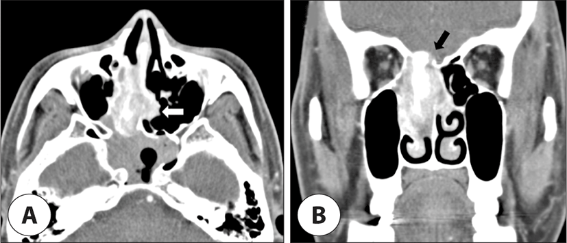
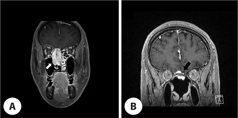
54세 남자 환자가 1년간 지속된 비루, 우측 비폐색 및 후각저하를 주소로 타 병원에서 시행한 비강 내시경상 우측 비강 내에 종물이 발견되어 내원하였다. 조직검사상 후각신경아세포종으로 진단되었으며 치료를 위해 의뢰되었다. 비강 내시경상 우측 비강 후열부에 쉽게 출혈되는 양상의 다발성 종물이 관찰되었고, 부비동 전산화단층촬영(paranasal sinus computed tomography, PNS CT)에서 우측 사골동을 가득 채우는 2.2×3.7×5.0 cm 크기의 연부조직음영이 우측 상·중비도 및 사상판을 지나 전두개와까지 확장되었고 반대측 비중격 후방까지 침범된 소견을 보였다(Fig. 1). 부비동 자기공명영상촬영(PNS magnetic resonance imaging, PNS MRI)에서 종물은 T1, T2 영상에서 중등도 신호, gadolinium으로 조영한 영상에서는 조영 증강이 되는 양상을 보였으며, 종물과 인접한 뇌막의 조영 증가도 관찰되어 두개 내 침범이 있는 것으로 판단되었다. 그 외 종물로 인한 폐쇄에 의해 전두동, 사골동, 접형동에 걸친 부비동염이 동반되어 있었다(Fig. 2A).

jcohns-34-2-52-g1
Fig. 1. Pre-operative contrast enhanced paranasal CT. A: Axial view. Right nasal cavity enhancing mass up to 5 cm shows septal bone erosion (white arrow). Right sphenoid sinus shows soft tissue density suggesting sinusitis caused by the mass effect. B: Coronal view. Enhancing mass shows cribriform plate destruction and sclerotic bony change in the anterior skull base to middle turbinate. There is suspicious intracranial extension at the level above cribriform plate (black arrow). CT: computed tomography.
Download Original Figure
jcohns-34-2-52-g2
Fig. 2. Gadolinium enhanced paranasal coronal MR images. A: Pre-operative T1 image shows heterogeneous signal intensity lesion (white arrow) at right superior and middle nasal cavity extending to anterior cranial fossa. B: Six-year post-operative T1 image shows high signal lesion (black arrow) centered on the planum sphenoidale with bony thickening. MR: magnetic resonance.
Download Original Figure

우선 유도화학요법(induction chemotherapy)을 VIP(Etoposide, Ifosfamide, and Cisplatin)를 이용하여 2회 시행하였으며, 4등급의 호중구감소증 및 3등급의 혈소판감소증이 발생하였고 병변의 치료효과는 불변(stable disease)으로 이후 수술적 치료를 계획하였다. 수술은 신경외과와 협의하여 내시경적 두개저수술을 시행하였다. 종양은 우측 비강 및 사골동을 침범하고 있었으며, 우측 안와 측벽을 침범한 소견으로 안와 지판(lamina papyracea) 일부를 제거하여 안와 골막을 노출시켰다. 좌측을 침범한 종양도 비중격, 중비갑개를 포함하여 제거하였다. Modified Lothrop 접근법을 시행하여 양측 전두동을 노출한 후 양측에서 두개저 골을 드릴링하여 종양이 부착된 두개저를 제거하였으며, 종양은 뇌경막(dura mater)을 침범하는 양상으로 경막을 포함하여 종물을 일괴로 제거하였다. 이후 결손된 두개저를 비중격피판(nasoseptal flap)으로 재건 후 수술을 마쳤다. 술 후 방사선 치료를 총 60–70 Gy를 계획하였으나 38 Gy 시행 후 비강의 심한 가피 및 잦은 출혈로 인한 합병증으로 잠정 중단하고 외래에서 경과 관찰하였다.

수술 시행 4년 후 시행한 MRI에서 우측 경부 level Ib에 림프절 크기가 증가한 소견이 관찰되었으며, 중심부침생검(core needle biopsy)상 전이가 확인되어 우측 선택적경부절제술(selective neck dissection; level I, II, III)을 시행하였다. 이후 경부 방사선 치료를 총 50 Gy를 계획하였으나 환자가 합병증 발생에 대한 두려움으로 거부하여 추가 치료 없이 경과 관찰하였다. 다시 1년 5개월 후 좌측 경부 level Ib에 크기가 증가한 림프절 소견이 확인되어 중심부침생검을 시행하였으며, 전이가 확인되어 좌측 변형 근치적 경부절제술(modified radical neck dissection)을 시행하였다.

9개월 후 시행한 영상검사상 좌측 접형골면을 따라 조영 증강된 소견으로 종양 재발이 의심되어(Fig. 2B), 신경외과와 협의하에 수술적 종물 제거 시 발생할 수 있는 합병증 및 환자의 치료 옵션 선호도 등을 감안하여 GKRS를 시행하기로 결정하였다. 비강 및 두개 내 종물의 용적은 1.96 cm3으로 50% isodose line에 20 Gy, 시신경은 평균 3.7±0.6 Gy, 수정체는 평균 1.5±0.6 Gy 방사선 조사를 시행하였다.

GKRS 시행 후 6개월마다 MRI 촬영을 하였고 GKRS 후 1년째 촬영한 MRI에서 좌측 후인두부 림프절 전이가 관찰되어 구강 내 접근을 통한 림프절 절제술을 시행하였고, 경부에 70 Gy의 방사선 치료를 시행하였다.

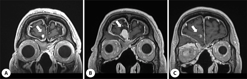
또한 첫 GKRS 이후 MRI 추적 결과 우측 전두엽 연수막의 전이가 관찰되었고, 6개월째에는 크기가 0.3 cm(Fig. 3A)이었으나 2년째에는 1.2 cm(Fig. 3B)까지 증가하였다. 따라서 GKRS 재수술을 계획하였고 연수막 종물의 용적은 2.62 cm3이며 50% isodose line에 20 Gy 방사선 조사를 시행하였다. GKRS 재수술을 시행한 후 2년간 6개월 간격으로 MRI 촬영 및 국소 소견을 확인 중이며 재발 소견 없이 경과 관찰 중이다(Fig. 3C).

jcohns-34-2-52-g3
Fig. 3. Gadolinium enhanced paranasal coronal MR T1 images. A: Six-month post-GKRS image shows 0.3 cm sized enhancing mass (white arrow) at right frontal convexity. B: Two-year post-GKRS image shows increased size up to 1.2 cm of enhancing mass (white arrow) at right frontal convexity. C: Two-year post-2nd GKRS image shows decrease in the size of enhancing mass (white arrow) at right frontal convexity. The difference in the shape of the right eye seen in the MRI image was due to the difference in the MRI cut, and the patient did not show ocular symptoms. GKRS: Gamma knife radiosurgery, MRI: magnetic resonance imaging.
Download Original Figure

고찰

후각신경아세포종은 매우 드문 암으로, 이는 수술, 고식적 방사선 치료 및 항암치료를 포함한 다양한 치료 옵션의 사용 및 시기에 관한 합의가 아직까지 없는 주요 이유가 될 수 있다. 대체로 치료는 수술과 방사선 치료가 가장 이상적인 것으로 알려져 있다. 경부절제술을 포함한 경부치료를 받지 않은 N0인 후각신경아세포종 환자를 대상으로 한 연구에서 수술 단독으로 치료한 군과 수술 및 술 후 방사선치료를 시행한 군으로 나누어 비교해보았을 때, 수술 단독을 시행한 군은 71%로 높은 국소 재발률을 보인 반면 수술 및 술 후 방사선치료를 받은 환자군은 17%에서 국소 재발이 발생하였다.3) 경부 림프절 전이 발생률도 수술만 단독으로 시행한 환자군에서 높았다. 진행된 병기이거나 수술로 완전 절제가 불가능한 경우는 항암치료를 할 수 있으며, 최근에는 내시경 수술만으로 종양을 제거하고 술 후 방사선 치료를 병행하여 좋은 성과를 보이고 있다.4)

후각신경아세포종의 예후는 현재까지 Kadish 병기 분류와 Hyams의 조직학상 분류가 중요한 것으로 알려져 있다.5) 본 증례의 경우는 두개 내 침범 소견을 보이고 있어 Kadish 병기 분류상 stage C로, Hyams 조직 분류상 grade II로 분류되었다. 예후를 예측하는 데 있어서는 Hyams의 조직학상 분류가 Kadish 분류보다 통계적으로 유의한 것으로 알려져 있으며,6) Hyams의 조직 분류에 따라 저도 악성(grade I, II)의 5년 평균 생존률은 56% 정도이고 고도 악성(grade III, IV)의 경우 25% 정도로 보고되고 있다.

후각신경아세포종의 재발은 두개 내 침범 가능성이 있는 국소 재발과 경부 림프절 전이 등의 양상을 주로 보인다. 경부 림프절 전이는 초기 진단 시 환자의 5%–8%에서, 재발로는 20%–25%에서 발생하는 것으로 보고되었고,7) 전이는 비교적 예측 가능한 패턴을 보이며 대부분은 Level I–III에서 발생한다.8,9) 경부 림프절 전이 시에는 경부 림프절 절제술 및 방사선 치료를 주로 시행하지만, 예방적 경부 림프절 치료에 관하여는 아직 의견이 분분하다. 메타분석에 따르면 진단 당시 임상적으로 N0 환자 중 14.1%에서 경부 림프절 전이가 이후 나타났으며, 경부 림프절 전이 환자의 사망률(60%)은 전이가 없는 환자(26%)에 비해 유의하게 높은 것으로 알려졌다.10) 다른 연구에서는 10년 지연성 경부 림프절 전이률은 33%로 나타났으며, 이 중 양측 경부 림프절 전이는 전체 경부 림프절 전이 환자 중 29%였다.11) 이는 예방적 경부 림프절 치료의 필요성을 주장하는 근거가 되며, N0 환자에서 예방적 경부 림프절 절제술 혹은 방사선 치료를 시행한 군이 그렇지 않은 군보다 현저히 낮은 재발률을 보였다.8) 또 다른 연구에서는 후각신경아세포종이 중앙선을 넘어선 병변인 경우나 경막의 침범이 있는 경우에는 양측 경부 림프절로 전이될 가능성이 높기 때문에 예방적 양측 경부 림프절 치료가 필요하다고 주장한다.10,12) 본 증례의 경우 처음 발생한 N0의 원발암이며 예방적 경부절제술의 시행에 대한 논란이 아직 존재하는 점, National Comprehensive Cancer Network(NCCN) 가이드라인에서 예방적 경부절제술에 대한 필수적인 권고가 없는 점 등으로 원발암에 대한 치료만을 먼저 시행하였으나, 수술 이후 술후 방사선 치료를 도중에 중단한 점, 진단 시 종물의 두개 내 침범이 의심되었던 점으로 경부 림프절 전이 발생률이 높아졌을 것으로 생각된다.

후각신경아세포종은 약 12%에서 두개 내 침범을 동반하는 것으로 알려져 있다.13) 두개 내 재발 소견이 있을 경우, 접근 가능한 병변이라면 외과적 제거를 우선적으로 고려할 수 있지만 구제 목적의 정위방사선수술이 국소 병변 억제(local disease control)에 있어 동등한 효과를 보일 수 있다. 처음 진단된 14명의 후각신경아세포종 환자에서 방사선치료를 시행한 연구에서 9명의 환자(64%)에서 재발 없이 치료하였고, 재발한 환자 5명(36%) 중 4명에게 다시 방사선치료를 시행하여 성공적으로 치료한 것을 보고하였다.14) 또한 두개 내 국소 재발성 후각신경아세포종 환자를 정위방사선수술을 이용하여 치료한 연구에서, 총 29케이스 중 26 케이스(89%)에서 억제력을 보여 좋은 치료 성적을 보고하였다.2) 본 증례의 경우는 이전에 수술, 방사선 치료 및 항암치료를 모두 시행한 환자로 추가적인 수술을 원치 않았던 경우이다. 수술을 시행을 원하지 않거나 시행할 수 없는 경우 방사선치료가 좋은 치료 방법이 될 수 있음을 보여주는 증례라고 할 수 있다. 마지막으로 시행 방사선량은 해당 병변에 두 번 모두 20 Gy를 조사하였는데, 여러 연구마다 조사 선량에 대한 수치는 일치하지는 않지만 대체로 한계 선량을 15 Gy 초과하여 조사했을 경우 치료 성공률이 높았다.2,14)

결론

후각신경아세포종의 국소 재발에 대해서, 수술 및 술후 방사선 치료는 합병증 발생의 위험이 높을 뿐 아니라, 여전히 재발의 가능성도 가지고 있다. 정위방사선수술은 고령, 개두술이 불가능한 의학적 상태, 환자가 수술을 거부하거나 수술이 허용할 수 없는 합병증을 유발할 수 있는 상황에서 후각신경아세포종의 국소 재발에 대한 비침습적인 치료 옵션으로 고려할 수 있을 것으로 보인다.

Acknowledgements

Not applicable.

Funding Information

This work was supported by a 2019 Research Grant of School of Medicine, Pusan National University.

Conflicts of Interest

No potential conflict of interest relevant to this article was reported.

Author Contribution

Conceptualization: Roh HJ.

Data curation: Sung SK.

Formal analysis: Park DH.

Methodology: Yi KI, Roh HJ.

Validation: Park DH.

Investigation: Sung SK.

Writing - original draft: Yi KI, Roh HJ.

Writing - review & editing: Yi KI, Park DH, Sung SK, Roh HJ.

Ethics Approval

Exemption from review was obtained from the Institutional Review Board of Pusan NationaI University Yangsan Hospital (Approval No. 05-2023-060).

References

1.

Bradley PJ, Jones NS, Robertson I. Diagnosis and management of esthesioneuroblastoma. Curr Opin Otolaryngol Head Neck Surg 2003;11(2):112-8.

2.

Van Gompel JJ, Link MJ, Sheehan JP, Xu Z, Mathieu D, Kano H, et al. Radiosurgery is an effective treatment for recurrent esthesioneuroblastoma: a multicenter study. J Neurol Surg B Skull Base 2014;75(6):409-14.

3.

Demiroz C, Gutfeld O, Aboziada M, Brown D, Marentette LJ, Eisbruch A. Esthesioneuroblastoma: is there a need for elective neck treatment? Int J Radiat Oncol Biol Phys 2011;81(4):e255-61.

4.

Castelnuovo PG, Delù G, Sberze F, Pistochini A, Cambria C, Battaglia P, et al. Esthesioneuroblastoma: endonasal endoscopic treatment. Skull Base 2006;16(1):25-30.

5.

Jethanamest D, Morris LG, Sikora AG, Kutler DI. Esthesioneuroblastoma: a population-based analysis of survival and prognostic factors. Arch Otolaryngol Head Neck Surg 2007;133(3):276-80.

6.

Oskouian RJ Jr, Jane JA Sr, Dumont AS, Sheehan JM, Laurent JJ, Levine PA. Esthesioneuroblastoma: clinical presentation, radiological, and pathological features, treatment, review of the literature, and the University of Virginia experience. J Neurosurg 2002;12(5):4.

7.

Zanation AM, Ferlito A, Rinaldo A, Gore MR, Lund VJ, McKinney KA, et al. When, how and why to treat the neck in patients with esthesioneuroblastoma: a review. Eur Arch Otorhinolaryngol 2010;267:1667-71.

8.

Yin ZZ, Luo JW, Gao L, Yi JL, Huang XD, Qu Y, et al. Spread patterns of lymph nodes and the value of elective neck irradiation for esthesioneuroblastoma. Radiother Oncol 2015;117(2):328-32.

9.

Banuchi VE, Dooley L, Lee NY, Pfister DG, McBride S, Riaz N, et al. Patterns of regional and distant metastasis in esthesioneuroblastoma. Laryngoscope 2016;126(7): 1556-61.

10.

Naples JG, Spiro J, Tessema B, Kuwada C, Kuo CL, Brown SM. Neck recurrence and mortality in esthesioneuroblastoma: implications for management of the N0 neck. Laryngoscope 2016;126(6):1373-9.

11.

Peacock JG, Harmsen WS, Link MJ, Van Gompel JJ, Giannini C, Olsen KD, et al. Risk of delayed lymph node metastasis in clinically N0 esthesioneuroblastoma. J Neurol Surg B Skull Base 2017;78(1):068-74.

12.

Marinelli JP, Janus JR, Van Gompel JJ, Link MJ, Moore EJ, Van Abel KM, et al. Dural invasion predicts the laterality and development of neck metastases in esthesioneuroblastoma. J Neurol Surg B Skull Base 2018;79(5):495-500.

13.

Morris L, Govindaraj S, Genden EM. Primary sphenoid sinus esthesioneuroblastoma. Am J Otolaryngol 2004; 25(5):350-3.

14.

Unger F, Haselsberger K, Walch C, Stammberger H, Papaefthymiou G. Combined endoscopic surgery and radiosurgery as treatment modality for olfactory neuroblastoma (esthesioneuroblastoma). Acta Neurochir (Wien) 2005;147:595-602.