Journal of Clinical Otolaryngology Head and Neck Surgery
The Busan, Ulsan, Gyeoungnam Branch of Korean Society of Otolaryngology-Head and Neck Surgery
Case Report

경장근 석회성 건염과 동반된 후인두 농양 1예

문성규1https://orcid.org/0000-0001-6690-2769, 백문승1https://orcid.org/0000-0003-1008-8168, 고소영1https://orcid.org/0000-0002-0638-3319, 김승우1,*https://orcid.org/0000-0002-7931-6977
Seong Kyu Moon1https://orcid.org/0000-0001-6690-2769, Moon Seung Beag1https://orcid.org/0000-0003-1008-8168, So Young Ko1https://orcid.org/0000-0002-0638-3319, Seung Woo Kim1,*https://orcid.org/0000-0002-7931-6977
1중앙보훈병원 이비인후-두경부외과
1Department of Otorhinolaryngology-Head and Neck Surgery, Veterans Health Service Medical Center, Seoul, Korea
*Corresponding author: Seung Woo Kim, Department of Otorhinolaryngology-Head and Neck Surgery, Veterans Health Service Medical Center, Seoul 05368, Korea, Tel: +82-2-2225-1384, Fax: +82-2-2225-1385, E-mail: entzzang1020@daum.net

© Copyright 2022 xxxxxxxxxxxxxxx. This is an Open-Access article distributed under the terms of the Creative Commons Attribution Non-Commercial License (http://creativecommons.org/licenses/by-nc/4.0/) which permits unrestricted non-commercial use, distribution, and reproduction in any medium, provided the original work is properly cited.

Received: Nov 01, 2022; Revised: Dec 11, 2022; Accepted: Dec 14, 2022

Published Online: Dec 31, 2022

ABSTRACT

Calcific tendinitis of the longus colli muscle (CTLC) is an inflammatory disease caused by the deposition of calcium hydroxyapatite crystal in longus colli muscle and its tendon. The incidence of it is rare, only a few cases have been reported so far, and its common symptoms and sings were neck pain, limitation of neck motion, dysphagia and odynophagia and so on. Clinically, it can be masqueraded as retropharyngeal abscess on account of its non-specific clinical presentations and rare incidence. We encountered a 39-year-old woman with five days duration of progressive bilateral neck pain, limitation of neck motion that was finally diagnosed as retropharyngeal abscess accompanied by the CTLC. We report this rare and didactic case with a brief review of relevant literature.

Keywords: Retropharyngeal abscess; Calcinosis; Tendinopathy

서론

경장근 석회성 건염은 1–3번 경추 사이에 있는 경장근(longus colli muscle)에 무정형성 석회화 침착이 되어 발생하는 드문 염증성 질환이다. 1,2) 흔한 증상은 경부 경직 및 움직임 제한, 연하통, 연하곤란 등이다. 3) 후인두 농양, 외상성 통증 또는 감염성 척추염 등으로 오인될 수 있으며, 대증치료로 증상이 호전되므로, 조기 진단이 매우 중요하다.3,4) 이제까지 경장근 석회성 건염은 후인두 농양으로 오인되어 보고된 경우는 국내에 몇 예가 있으나,1,5) 농양과 동반된 경우는 보고가 없었던 것으로 사료된다.

저자들은 경부 경직과 경부통을 호소하는 39세 여자 환자에서 경장근의 석회성 건염과 동반된 후인두 농양으로 진단된 드물고 교훈적인 증례를 경험하여, 참고 문헌과 함께 보고한다.

증례

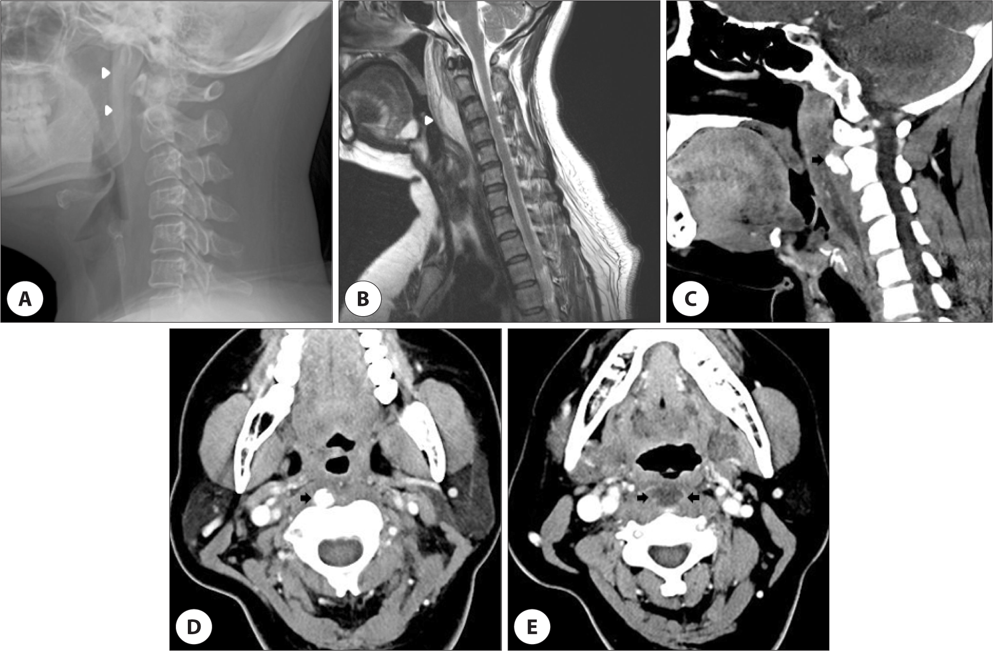
39세 여자 환자가 내원 5일 전부터 시작된 진행성 경부통, 경부강직 및 연하통 등을 주소로 내원하였다. 환자는 본원 정형외과에서 경추 자기공명영상을 촬영하였고, 후인두 공간의 체액 저류 소견과 악화된 연하통 등으로 본과로 진료 의뢰되었다. 이전 병력과 가족력 등에서 특이 소견은 없었고, 최근 경부 외상의 이력도 없었다. 신체 검사에서 경부의 신전 및 굴곡 등에 제한을 보였지만, 상지로의 방사통이나 신경학적 이상 소견 등은 없었고, 비정상적으로 촉지되는 경부 종물도 없었다. 경성 후두내시경 검사에서 후인두벽의 미만성 종창 소견이 관찰되었다(Fig. 1A, B). 일반혈액검사에서 백혈구는 15,700/μL(참고값: 3,000–9,300), 적혈구 침강속도는 92 mm/hr(참고값: 0–30), C-반응단백은 145 mg/L(참고값: 0–3) 등으로 상승된 소견이었다. 측경부 단순촬영영상에서 후인두 부위의 연조직 음영이 증가된 소견이었고(Fig. 2A), T2 경추 자기공명영상에서 1–3번 경추체의 전방에 액체의 저류 소견이 관찰되었다(Fig. 2B). 경부 컴퓨터단층촬영에서 1–2번 경추체의 전방 공간으로 원형의 석회화 및 1–5번 경추 높이의 후인두 공간에 주변부가 조영 증강되는 저밀도 음영이 관찰되었다(Fig. 2CE). 환자가 호흡곤란이 없고, 협조가 잘되어 24게이지의 천자침으로 경구강 흡인을 시행하여 2.5 cc가량의 농성 분비물이 배액되어, 세균 배양검사를 진행하였다. 환자는 흡인 직후에 증상이 부분 호전되었다고 하였다.

jcohns-33-4-255-g1
Fig. 1. Rigid laryngoscopic findings. A, B: At the first day of treatment, they show diffuse swelling on posterior pharyngeal wall but there is no airway obstruction (arrowheads). C: Six days after treatment, it shows disappeared lesion on posterior pharyngeal wall.
Download Original Figure
jcohns-33-4-255-g2
Fig. 2. Radiologic findings. A: The plain radiograph of lateral neck (soft tissue view) shows soft tissue swelling in retropharyngeal space (arrowheads). B: The image of sagittal T2 MR shows high signal intensity in the prevertebal space from C1 to 3 (arrowhead). C: The image of contrast-enhanced axial CT shows prominence of amorphous calcification on anterior to the body of C1 to 2 (arrow). D: The image of contrast-enhanced axial CT shows prominence of retropharyngeal amorphous calcification (arrow). E: The image of contrast-enhanced axial CT shows round soft tissue density with peripheral rim enhancement in retropharyngeal space (arrows). MR: magnetic resonance, CT: computed tomography.
Download Original Figure

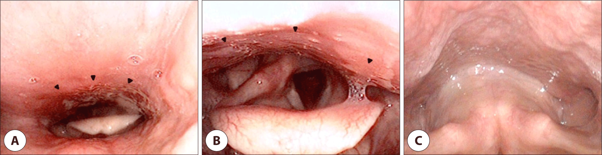
이상의 임상 양상 및 영상학적 소견 등을 종합하여 경장근의 급성 석회성 건염과 동반된 후인두 농양으로 진단하고, amoxicillin/clavulanic acid를 하루 60 mg/kg으로 투여하였고, 비스테로이드성 소염진통제도 투약하였다. 균배양검사에서는 녹색연쇄상구균(Streptococcus viridans)이 동정되었고, 기존 사용하는 항생제에 감수성이 있었다. 치료 2일째부터 경부통증에 대한 시각아날로그척도가 8점에서 3점으로 호전되었다. 입원 3일째 일반혈액검사에서 백혈구는 8,500/μL, 적혈구 침강속도는 54 mm/hr 및 C-반응단백은 23 mg/L 등으로 호전되었다. 입원 6일째 시각아날로그척도가 1점이었고, 인후두 내시경 소견에서도 후인두벽에 특이 소견이 없어서, 퇴원하였다(Fig. 1C). 5개월이 지난 현재까지 재발 소견 없이 추적관찰 중이다.

고찰

경추부 경장근의 급성 석회성 건염은 칼슘 수산화인회석(calcium hydroxyapatite)이 경장근에 축적되면서 발생한 염증성 질환이다.1) 30–60대에 호발하며, 성별에 따른 발생빈도 차이는 없다.6) 정확한 원인과 유병률 등은 밝혀지지 않았으나, 문헌 검색에서 2003년 이후 국내에 15건 정도 보고된 드물게 발생하는 질환이다.1,2,7) 경추 전방에 위치하는 굴곡근인 경장근은 상사부, 하사부 및 수직부 등으로 구성되며, 석회화는 주로 1–2번 경추의 전방에 있는 상사부에서 발생한다.8) 석회화의 진행 기전은 불명확하지만, 반복적인 외상 및 손상 등이 주변 조직에 허혈 및 괴사 등을 일으켜 발생하는 것으로 생각된다.8) 흔한 임상 양상은 경부 경직, 경부 통증, 연하통 등이며, 미열이 동반되고 드물게 등, 견관절 및 상지 부위 등에 동통 및 두통, 어지럼증 등도 나타날 수 있다.3,7) 경부 근육의 강직 또는 연축 등이 나타날 수 있으며, 경추부의 굴곡 및 신전 시 통증에 의한 심한 운동범위의 제한이 있고, 인후두 내시경 소견에서 후인두벽의 발적 및 부종 등이 관찰될 수 있다.1,5,9,10) 이러한 양상 때문에 후인두 농양, 감염성 척추염 및 뇌수막염 등과 감별하기 어렵지만, 다른 질환들과 비교해서 경과가 양호하므로 조기에 감별 진단하는 것이 매우 중요하다.5)

급성 경장근 석회성 건염의 진단은 혈액검사와 영상학적 검사를 통하여 이루어진다.4) 일반혈액검사에서 백혈구 및 C-반응단백 등의 수치가 상승한다.5) 영상검사는 단순 경부촬영, 경부 전산화단층촬영 및 자기공명영상 등을 시행할 수 있다.5) 단순 경부촬영에서 후인두 공간의 음영이 증가된 소견을 보이며, 전산화단층촬영은 경장근의 무정형 석회화 침착 및 척추체 전방의 연부조직 병변을 확인할 수 있어 가장 정확한 진단법이다.1,11) 특히 후인두 농양과 감별을 위해서는 척추 전강의 부종 및 병변 주변의 조영 증강 유무를 확인해야 하고, 조영 증강이 있는 경우는 후인두 농양을 고려해야 한다.4,8) 본 증례에서도 전산화단층촬영에서 저음영 병변 주변이 조영 증강되고, 경구강 천자에서 농성 분비물이 흡인되고, 백혈구 및 C-반응 단백질 등의 수치가 다른 증례보다 크게 증가되어, 염증보다는 농양에 더 합당한 소견으로 보인다. 자기공명영상은 척추 전방의 액체저류를 확인과 경추부의 염증을 감별하는 데는 도움이 되지만 석회화 침착을 발견할 확률이 낮으며, 필수적인 검사는 아니다.1) 후인두 농양은 편도염, 인후두에 저명한 염증, 이물 매복 및 치성 염증 등에 의해 유발될 수 있다.12) 본 증례에서는 이러한 명확한 유발 요인이 없어서, 인과관계를 단정할 수는 없지만, 석회성 건염에 의해 농양이 발생하였을 가능성이 있다고 생각된다.

급성 인두후 건염은 치료의 여부와 관계없이 1–2주 이내로 증상이 소실되는 것으로 알려져 있고, 증상이 있는 경우에는 소염진통제를 사용할 수 있다.1,8) 본 증례에서는 다른 임상과에서 소염진통제가 투여되었지만 증상의 호전이 적고, 전산화단층촬영에서 경장근의 석회성 건염 소견과 후인두 농양이 동반된 것으로 판단되어 흡입천자 후 바로 항생제 치료를 진행하였다. 저자들의 문헌 고찰에서는 급성 경장근 석회성 건염이 후인두 농양으로 오인되어 보고된 경우는 있었지만, 저명한 후인두 농양이 동반되어 보고된 경우는 국내에서는 없었던 것으로 사료된다.1,5,12) 저자들은 본 증례를 통하여 경부 강직, 경부통, 경부 운동 제한 및 후인두 벽의 종창 등의 소견을 보이는 환자에서 경장근에 발생한 급성 인후두 건염을 감별해야 하며, 드물지만 후인두 농양과 동반될 수 있으므로 정확한 이학적 및 영상 검사 등을 통한 조기 진단과 적절한 치료가 필수적이라는 교훈을 얻었다.

Acknowledgements

Not applicable.

Funding Information

Not applicable.

Conflicts of Interest

No potential conflict of interest relevant to this article was reported.

Author Contribution

Conceptualization: Kim SW, Beag MS.

Writing - original draft: Moon SK, Beag MS.

Writing - review & editing: Moon SK, Beag MS, Ko SY, Kim SW.

Ethics Approval

Not applicable.

References

1.

Suk SH, Jung TY, Kim S, Choi SH. A case of acute calcific tendinitis of the longus colli muscle in the cervical spine misconceived as acute retropharyngeal Abscess. J Clin Otolaryngol Head Neck Surg 2016;27(1):166-70.

2.

Yoon HJ, Son CN, Lee SH, Joo KB, Kim TH. A case of retropharyngeal calcific tendinitis in a patient with ankylosing spondylitis. J Rheum Dis 2013;20(6):385-8.

3.

Shawky A, Elnady B, El Morshidy E, Gad W, Ezzati A. Longus colli tendinitis. a review of literature and case series. SICOT J 2017;3:48.

4.

Raggio BS, Ficenec SC, Pou J, Moore B. Acute calcific tendonitis of the longus colli. Ochsner J 2018;18(1):98-100.

5.

Lee CH, Huh SH, Jin SM, Lee SH. 3 Cases of acute retropharyngeal calcific tendinitis misconceived as acute retropharyngeal abscess. Korean J Otorhinolaryngol-Head Neck Surg 2011;54(6):411-4.

6.

Kim YJ, Park JY, Choi KY, Moon BJ, Lee JK. Case reports about an overlooked cause of neck pain: calcific tendinitis of the longus colli. Medicine(Baltimore) 2017;96(46):e8343.

7.

Lyo IU, Kim Y, Yu HK, Ahn SK. Acute calcific retropharyngeal tendinitis: case report. J Korean Neurosurg Soc 2003;33(3):317-9.

8.

Kang CH, Son ES, Kim DH, Jang HG. Acute longus colli tendinitis without calcification. J Korean Orthop Assoc 2015;50(3):264-7.

9.

Kim JH. Acute calcific prevertebral tendinitis without differentiated by simple X-ray. Osteoporosis 2015; 13(2):117-21.

10.

Park R, Halpert DE, Baer A, Kunar D, Holt PA. Retropharyngeal calcific tendinitis: case report and review of the literature. Semin Arthritis Rheum 2010;39(6):504-9.

11.

Zibis AH, Giannis D, Malizos KN, Kitsioulis P, Arvanitis DL. Acute calcific tendinitis of the longus colli muscle: case report and review of the literature. Eur Spine J 2013;22(Suppl 3):434-8.

12.

Jeong JS, Lee DW, Yeon JY. Adequate management of retropharyngeal abscess. Korean J Otorhinolaryngol-Head Neck Surg 2004;47(9):899-903.