Journal of Clinical Otolaryngology Head and Neck Surgery
The Busan, Ulsan, Gyeoungnam Branch of Korean Society of Otolaryngology-Head and Neck Surgery
Case Report

안와 외향 골절 후 시력 저하를 동반한 지연성 안와기종

김정석1https://orcid.org/0000-0002-4799-3172, 이승주1https://orcid.org/0000-0001-8276-2567, 권재환2https://orcid.org/0000-0002-3260-6969, 박태정1,*https://orcid.org/0000-0002-2622-0138
Jungsuk Kim1https://orcid.org/0000-0002-4799-3172, Seungjoo Lee1https://orcid.org/0000-0001-8276-2567, Jaehwan Kwon2https://orcid.org/0000-0002-3260-6969, Taijung Park1,*https://orcid.org/0000-0002-2622-0138
1메리놀병원 이비인후과
2고신대학교 의과대학 이비인후과학교실
1Department of Otorhinolaryngology-Head and Neck Surgery, Maryknoll Hospital, Busan, Korea
2Department of Otorhinolaryngology, Kosin University College of Medicine, Busan, Korea
*Corresponding author: Taijung Park, Department of Otorhinolaryngology-Head and Neck Surgery, Maryknoll Hospital, Busan 48972, Korea Tel: +82-51-461-2692, Fax: +82-51-462-9419, E-mail: P791216@naver.com

© Copyright 2022 xxxxxxxxxxxxxxx. This is an Open-Access article distributed under the terms of the Creative Commons Attribution Non-Commercial License (http://creativecommons.org/licenses/by-nc/4.0/) which permits unrestricted non-commercial use, distribution, and reproduction in any medium, provided the original work is properly cited.

Received: Sep 15, 2022; Revised: Oct 04, 2022; Accepted: Oct 25, 2022

Published Online: Dec 31, 2022

ABSTRACT

Orbital emphysema is commonly associated with fractures of the orbital floor or medial wall. The air often dissipates spontaneously, but rarely can cause increased intraocular pressure and even loss of vision. It predominantly occurs with medial orbital wall fractures and is exacerbated by sneezing and nose blowing. We present a case of orbital emphysema with serious complications in which vision is affected in a 51-year-old male.

Keywords: Emphysema; Orbital fractures; Ocular vision

서론

안와기종은 안와 하벽 및 내벽 골절 시에 흔하게 동반된다. 대부분의 경우 시간이 흐르면서 저절로 흡수되나 드물게 안와내압을 상승시켜 시력을 저하시키기도 한다. 이러한 경우는 대부분 안와 내벽 골절에서 나타나며 재채기와 코풀기에 의해 더욱 악화된다.1) 저자들은 안와 외향 골절 후 발생하여 심각한 시력 저하를 초래한 안와기종 1예를 조기에 진단하고 수술적 치료로 교정하였기에 이를 문헌 고찰과 함께 보고하는 바이다.

증례

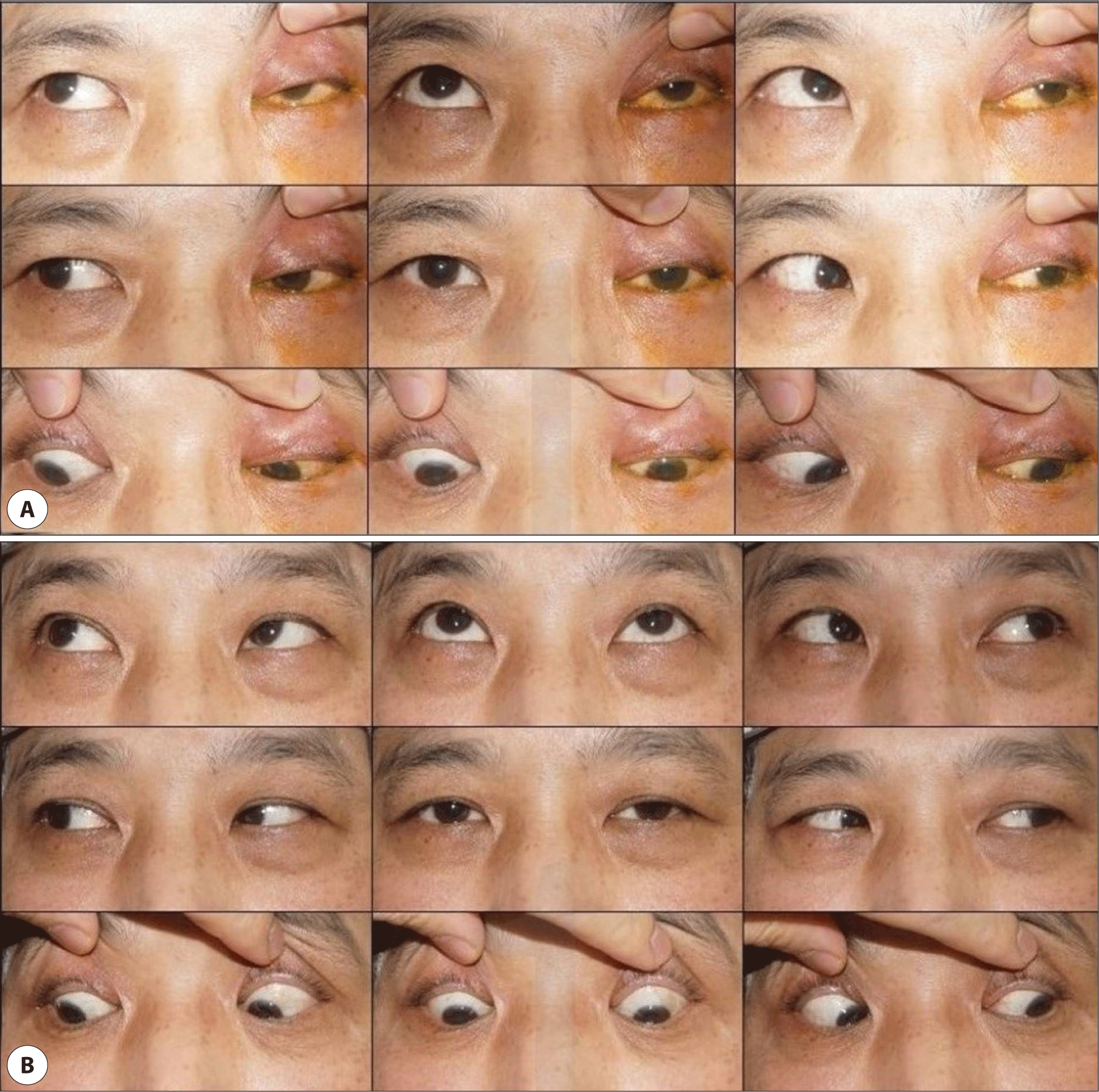
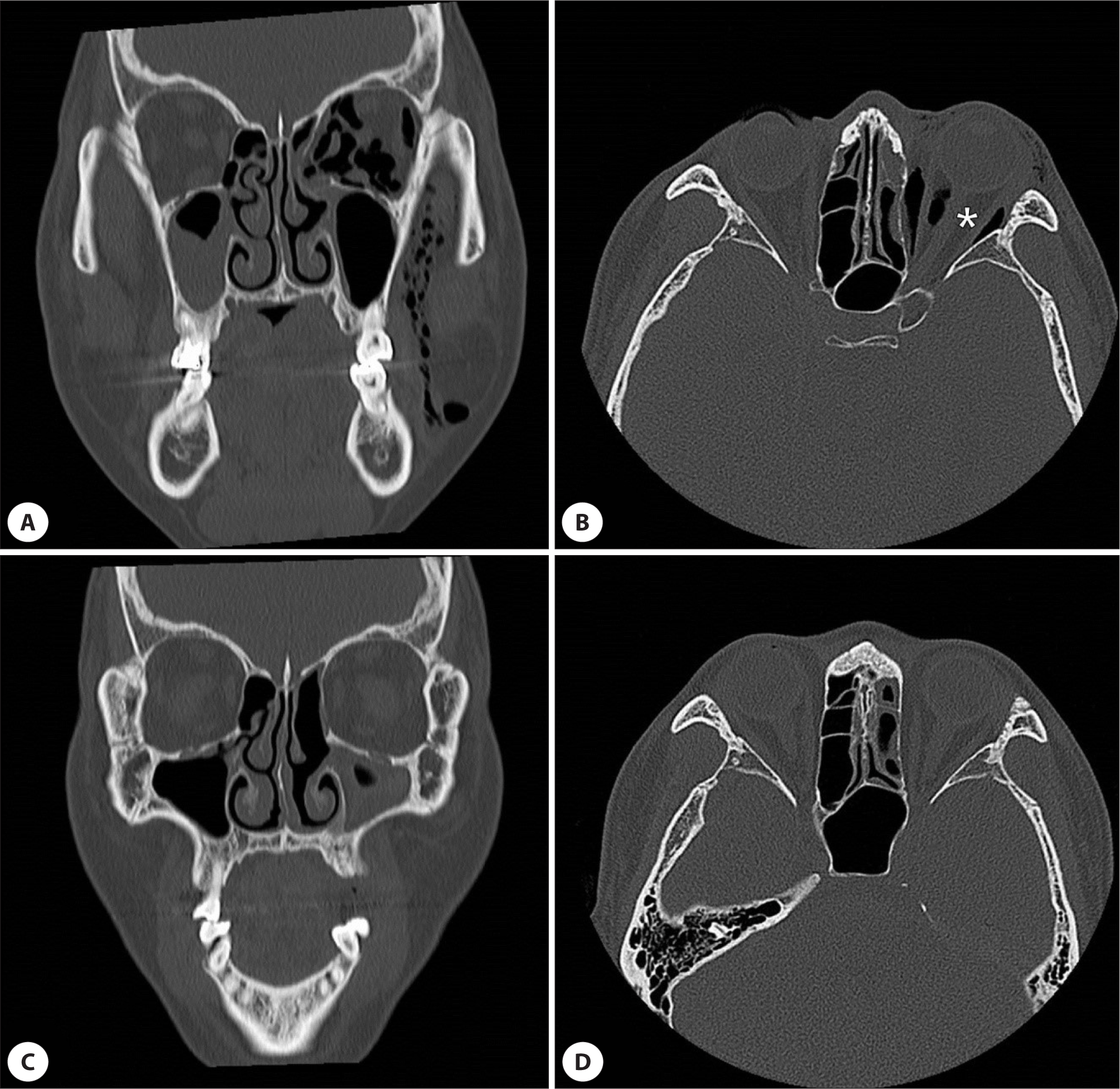
51세 남자가 내원 3일 전 집에서 넘어지면서 방문 손잡이에 좌측 안면부를 부딪힌 후 좌측 안구 주변 통증 및 안면부 부종 그리고 시력 저하를 주소로 본원 외래에 내원하였다. 환자는 수상 직후에는 코피 등 별다른 증상이 없었으며 수상 이후 특별히 코를 풀거나 재채기 등은 하지 않았다고 하였으나, 시간이 경과할수록 서서히 좌측 안구 주변 및 뺨이 붓고 내원 당일에는 부종으로 좌측 눈을 뜰 수 없었다. 이학적 검사상 좌측 안와 주위로 경미한 타박상 및 점상 출혈을 보였으며, 부종이 심해 손으로 눈꺼풀을 벌려야만 겨우 눈동자를 확인할 수 있었고 촉진 시 열발음이 있었다. 또한 통증을 동반한 좌안의 전방향 안구 운동 제한을 보였다(Fig. 1A). 안와 골절 유무 판단을 위해 시행한 안면부 전산화단층촬영에서 좌측 안와 하벽의 골절 소견은 경미하였으나 동측 안와 내벽의 들창문형(trapdoor type) 골절이 관찰되었고, 안와 내부 및 뺨 주변 연부조직에 다량의 공기 음영이 관찰되었다(Fig. 2A, B). 안과적 검사에서 안압은 우안 15 mmHg, 좌안 24 mmHg이었고, 안구돌출검사(hertel exophthalmometry)에서 좌안 3 mm 안구 돌출 소견을 보였다. 양안 대광반사 및 안저검사는 정상이었고 상대적 입력 동공 반사 이상(relative afferent pupillary defect) 역시 없었으나, 소수 시력은 우안 0.8, 좌안 0.15로 좌안 시력 저하가 관찰되었다.

jcohns-33-4-218-g1
Fig. 1. Preoperative and postoperative 9 cardinal view of gaze. A: Extraocular movements of left eye were completely disturbed in all directions and ptosis is observed. B: Three days postoperatively, normal extraocular movements of left eye were observed.
Download Original Figure
jcohns-33-4-218-g2
Fig. 2. A preoperative coronal (A) and axial CT scan (B). Blow out fracture of left medial orbital wall with left pneumophthalmos and emphysema at left infratemporal fossa. The stretched optic nerve (asterisk) is observed. A postoperative coronal (C) and axial CT scan (D) of same patient show well-corrected left medial orbital wall and decompressed orbit. CT: computed tomography.
Download Original Figure

저자들은 안와 내벽의 들창문형 골절로 발생한 안와 기종에 의해 발생한 안와구획증후군으로 진단하고, 지연된 정복술로 인한 영구적인 시력저하가 초래될 수 있는 점을 고려하여 응급 시험적 절개술 및 정복술을 계획하였다. 수술은 전신마취하 부비동 내시경 수술을 통해 전 사골동 절제술을 시행하여 안와 내벽의 들창문형 골절 부위를 확인한 후, 골편을 골절 부위에서 빼낸 다음 안구 마사지를 시행하여 안와 내 유입된 다량의 공기를 배출하였다. 안와 기종이 제거된 후 강제견인검사를 시행하여 기계적인 운동제한이 사라진 것을 확인한 다음, 안와 격막에 붙은 골편을 정렬한 상태에서 실라스틱 시트를 재단하여 역U자형으로 사골동에 삽입한 후 Merocel®을 충전하여 고정하고 수술을 마쳤다.

수술 직후 좌측 안구 주변 및 뺨의 부종과 시력은 크게 호전되었으며 안구운동 제한 역시 수술 당일 거의 소실되었다. 외상성 시신경염 발생 가능성에 대비하여 전신적 스테로이드를 술 후 1일째부터 1 mg/kg/day를 3일간 사용한 후, 0.2 mg/kg/day씩 3일에 걸쳐 감량하였다. 수술 후 1일째 좌안 안압은 11.8 mmHg로 감소하였고, 수술 후 3일째 안구 돌출 소견 및 안구운동장해가 완전히 소실되었음이 확인되었고(Fig. 1B), 소수 시력은 좌안 0.6으로 수술 전과 비교하여 현저히 회복되었다. 수술 2주째 시행한 안면부 전산화단층촬영에서 좌측 안와 내벽의 들창문형 골절은 잘 교정되었고 내원 당시 촬영 영상에서 좌측 안와와 뺨 등에서 보이던 비정상적인 공기음영은 모두 소실된 것을 확인할 수 있었으며(Fig. 2C, D), 술 후 12개월이 지난 현재까지 특별한 합병증 없이 경과 관찰 중이다.

고찰

안와기종은 대부분 외상으로 인하여 안면골 골절이나 안와 외상 등에 의한 합병증으로 발생한다.2) 기종이 발생하는 기전은 직접적인 안와 외상이 안와 외벽의 골절을 유발해 사골동 및 상악동과 안와 사이에 비정상적인 통로를 만들게 되고, 갑자기 높아진 비강 내 압력이 비강 내의 공기를 안와로 이동시켜 발생하는 것으로 알려져 있다.3) 대부분의 안와기종 환자들은 피하기종과 안검 부종 이외에 특별한 동반 증상이 없으나, 안와기종이 안와 내에서 공간을 차지하면 안구를 앞으로 밀어내어 안구 돌출을 유발하기도 하며, 심하면 외안근의 움직임을 제한할 수 있다.4,5) 안와기종이 시력에 손상을 주는 경우는 드물지만 안와 첨부까지 침투된 공기방울들에 의해 안압상승, 망막중심동맥 폐쇄, 시신경 위축, 공기색전에 의한 안정맥의 열상 등이 발생하여 시력저하가 올 수 있어 주의가 필요하다.6,7) 본 증례에서도 안와 외상 후 발생한 안와 내벽의 들창문형 골절로 인해 안와기종이 지연 발생하여 서서히 커지면서 안구 운동 제한은 물론 안압 상승과 시신경 위축에 의해 시력 저하까지 나타났다.

안와기종은 안구 돌출, 복시 및 안구운동장애 등의 증상이 동반되더라도 시신경 압박이나 시력 저하의 소견이 없다면 일반적으로 7일에서 10일 이내에 별다른 후유증을 남기지 않고 자연 호전된다.8) 그러나 예기치 않게 안와기종이 지속적으로 커져 안압이 일정 수준 이상 상승하여 시력 저하 및 안근육마비 증상이 생기게 되면 즉시적인 안와 내 감압을 위한 처치 및 수술을 고려해야 하고, 외상성 시신경염이 동반되는 경우 고용량 스테로이드 용법을 사용할 수 있다.79) 저자들은 본 증례에서 안와기종에 의한 안압 상승으로 시력 저하와 안구 운동 제한이 있음을 확인하고, 감압을 목적으로 한 응급 시험적 절개술 및 정복술을 시행하였다. 수술 후 빠르게 안구 운동 제한이 회복되었으며 시력 역시 빠른 회복세를 보였으나, 외상성 시신경염 발생을 우려하여 3일간 고용량 스테로이드 치료를 시행하였다.

안와 외상 후 발생한 안와 내벽의 들창문형 골절이 체크 밸브 형태로 작용하여 시간이 지남에 따라 안와기종의 크기를 증가시킨 것은 두개내기종의 발생과 많은 유사점이 있는데, 구조적으로 연조직을 싸고 있는 경질막 바깥의 얇은 골구조의 비교적 작은 골절이 원인이라는 점이 같으며 원리적으로 볼 밸브 매커니즘으로10) 설명이 가능하다는 것이 유사하다.

결론

안와 외상 후 발생한 안와 내벽 및 하벽의 경미한 골절은 특별한 치료를 요하지 않는 경우가 많다. 하지만 재채기나 기침, 그리고 코를 풀어 발생하는 안와기종으로 환자가 여러 불편을 겪을 수 있으므로 비강 내 압력이 높아지는 행위를 못하도록 교육하는 것이 필요하며, 앞서 본 증례와 같이 매우 드물게 정상 호흡 과정을 통해서도 안와기종이 발생하여 안구운동장애와 시력 저하 및 소실 등 치명적인 합병증을 일으킬 가능성이 있으므로, 안와 외상 후 내원한 환자를 진료할 때 좀 더 세심한 경과 관찰이 필요하다는 점을 임상의들은 항상 견지하여야 할 것이다.

Acknowledgements

Not applicable.

Funding Information

Not applicable.

Conflicts of Interest

No potential conflict of interest relevant to this article was reported.

Author Contribution

Conceptualization: Kim J, Lee S, Kwon J, Park T.

Data curation: Kim J, Lee S.

Formal analysis: Kwon J, Park T.

Methodology: Kim J, Park T.

Software: Lee S.

Validation: Kim J, Lee S, Kwon J, Park T.

Investigation: Kim J, Lee S, Park T.

Writing - original draft: Kim J.

Writing - review & editing: Kim J, Lee S, Kwon J, Park T.

Ethics Approval

Informed consent for publication of the images was obtained from the patient

References

1.

Jordan DR, White GL Jr, Anderson RL, Thiese SM. Orbital emphysema: a potentially blinding complication following orbital fractures. Ann Emerg Med 1988; 17(8):853-5.

2.

Brown SM, Lissner G. Orbital emphysema following remote skull trauma. Ophthal Plast Reconstr Surg 1995;11(2):142-3.

3.

Caesar R, Gajus M, Davies R. Compressed air injury of the orbit in the absence of external trauma. Eye 2003;17(5):661-2.

4.

Yuksel M, Zafer Yuksel K, Ozdemir G, Ugur T. Bilateral orbital emphysema and pneumocephalus as a result of accidental compressed air exposure. Emerg Radiol 2007;13(4):195-8.

5.

Mathew S, Vasu U, Francis F, Nazareth C. Transconjunctival orbital emphysema caused by compressed air injury: a case report. Indian J Ophthalmol 2008; 56(3):247-9.

6.

Burt B, Jamieson M, Sloan B. Medial wall fracture- induced pneumo-orbita mimicking inferior rectus entrapment. Am J Emerg Med 2010;28(1):119.e1-3.

7.

Papadimitriou P, Ntomouchtsis A, Antoniades K. Delayed traumatic ocular emphysema: a case report. Oral Surg Oral Med Oral Pathol Oral Radiol Endod 2006;102(6):e18-20.

8.

van Issum C, Courvoisier DS, Scolozzi P. Posttraumatic orbital emphysema: incidence, topographic classification and possible pathophysiologic mechanisms. A retrospective study of 137 patients. Oral Surg Oral Med Oral Pathol Oral Radiol 2013;115(6):737-42.

9.

Silver HS, Fucci MJ, Flanagan JC, Lowry LD. Severe orbital infection as a complication of orbital fracture. Arch Otolaryngol Head Neck Surg 1992;118(8):845-8.

10.

Shameer A, Pushker N, Lokdarshi G, Basheer S, Bajaj MS. Emergency decompression of orbital emphysema with elevated intraorbital pressure. J Emerg Med 2017;53(3):405-7.