
Introduction
Obstructive sleep apnea syndrome (OSAS) is the most common form of sleep-disordered breathing in adults, reported in 22% of men and 17% of women.1) The etiology is turbulent airflow caused by partial or complete narrowing of the upper airway.2) When considering surgery, the most important issue is assessment of the site of narrowing. Upper airway anatomical assessments based on computed tomography, magnetic resonance imaging, and cephalometry are often static and performed while patients are awake, and thus may not represent the dynamic upper airway behavior during sleep. In contrast, drug-induced sleep endoscopy (DISE) can be used to evaluate the upper airway using a flexible fiberoptic endoscope with subjects under pharmacological sedation that simulates natural sleep.3) DISE was first described by Croft and Pringle in 1991, and it is commonly used to assess the cause of sleep apnea and guide surgical management of OSAS.4–6) Although DISE has certain limitations (sleep is artificially induced and the observation time is limited), DISE data are more physiologically relevant than those obtained using other diagnostic tools. DISE evidences good test-retest reliability, especially when used to evaluate the hypopharyngeal airway.7)
Prior to surgery, it is essential to know which part of the upper airway collapses, as well as the pattern of airway obstruction. DISE often identifies several airway obstructions in the upper airways of patients with OSAS. Palatine tonsil hypertrophy is a frequently encountered potential source of upper airway obstruction in adults.8) Jara et al. found a positive correlation between palatine tonsil size and the apnea-hypopnea index (AHI).8) Cahali et al. reported a positive correlation between palatine tonsil volume and the AHI.9) Holmlund et al. suggested that tonsillectomy might effectively treat OSAS adults with large tonsils.10) However, few studies have explored the correlations between palatine tonsil size and obstructive upper airway patterns in adults. The palatine tonsil is easy to evaluate via simple physical examination. If obstructive airway patterns can be elucidated by viewing the tonsil, this would aid planning of initial OSAS treatment. Here, we explored whether the size of the palatine tonsil could be used to predict the extent and pattern of adult upper airway obstruction during snoring and apneic events.
Materials and Methods
We retrospectively evaluated 268 adult patients (mean age 42.80±12.34 years, range 17–73 years) who underwent polysomnography and were diagnosed with OSAS (AHI score>5) in a single medical center from 2013 to 2020. We recorded age, sex, body mass index (BMI), and AHI via chart review. We excluded patients with obvious facial deformities such as mandibular dysplasia, craniosynostosis, and cleft palate, as well as those who had undergone prior surgery of the soft palate or palatine tonsil. The research protocol was reviewed and approved by Busan Saint Mary’s Hospital Institutional Review Board (IRB approval number: BSM 2021-11).
Brodsky standardized system was used to evaluate palatine tonsil size via physical examination: grade 0=tonsils limited to the bilateral tonsillar fossa; grade I=tonsils occupy less than 25% of the space between the anterior pillars of the oropharynx; grade II=tonsils occupy 25%-50% of the space; grade III=tonsils occupy 50%-75% of the space; grade IV=tonsils occupy 75%-100% of the space.11)
DISE was performed by otolaryngologists in a dim and quiet operating room, with patients supine under pulse-oximetry and cardiac rhythm monitoring. Sleep was induced by intravenous midazolam. The anesthesiologist slowly titrated the drug to 0.07 mg/kg per patient; boluses of 1-2.5 mg were given (maximum 7.5 mg/patient) using a target-controlled infusion system. All DISE procedures employed a flexible 4-mm-diameter videolaryngoscope and were performed by the same otolaryngologist, who recorded upper airway obstruction patterns. We divided the upper airway into two parts using the Koo’s DISE classification system based on the Fujita scoring method: retropalatal (the region posterior to the soft palate) and retrolingual (the region of the pharynx posterior to the vertical portion of the tongue).12) The retropalatal level was subdivided into the palate (the anteroposterior [AP] diameter) and upper lateral pharyngeal wall (the lateral diameter). The retrolingual level was subdivided into the tongue base (AP diameter) and the lower lateral pharyngeal wall (lateral diameter). The extent of airway obstruction was classified as none (grade 0), partial (grade 1; 50%-75% obstruction), and complete (grade 2; >75% obstruction).
All statistical analyses were performed using SPSS for Windows ver. 18.0 (SPSS, Chicago, IL, USA). Simple linear regression was used to explore relationships between the Brodsky score and the extent of upper airway obstruction. A p<0.05 was considered to indicate statistical significance.
Results
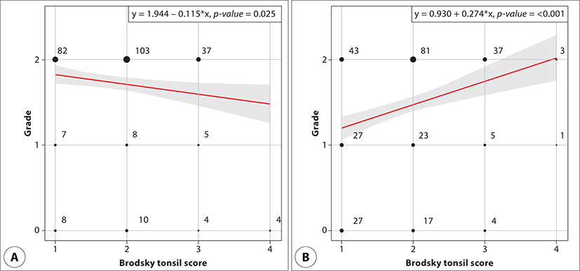
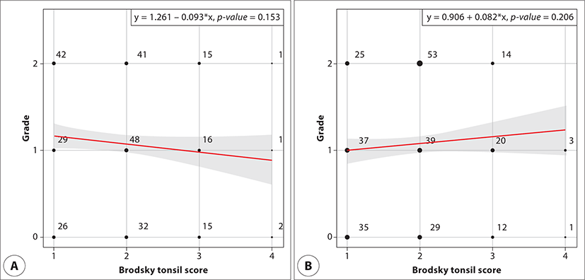
The study included 268 patients (230 males and 38 females); Table 1 lists the demographic characteristics. The mean age was 42.81±12.34 years and the mean BMI was 26.40±3.69 kg/m2. The mean AHI was 32.15±22.74. Table 2 lists the Brodsky tonsil scores: 36% (97) of patients were classified as grade I; 45% (121) as grade II; 17% (46) as grade III, and 1% (4) as grade IV. Upper airway obstruction was more common at the retropalatal than the retrolingual level. Table 2 also lists the extents of obstruction. At the retrolingual level, 75 patients had no AP obstruction (grade 0) and 77 had no lateral obstruction. At the retropalatal level, 26 patients had no AP and 48 had no lateral obstruction. Simple linear regression revealed a relationship between the Brodsky score and the extents of obstruction at the retropalatal level. A significantly negative relationship was evident between the score and AP collapse (slope coefficient=-0.115, p<0.05) and a significantly positive relationship was observed between the score and lateral collapse (slope coefficient=0.274, p<0.05) (Fig. 1). However, no significant relationship was observed at the retrolingual level (both p>0.05) (Fig. 2).
| Characteristics | Value (n=268) |
|---|---|
| Sex | |
| Male | 230 (84.87%) |
| Female | 38 (15.13%) |
| Age (y) | 42.81±12.34 |
| Body mass index (kg/m2) | 26.40±3.69 |
| Apnea-hypopnea index (event) | 32.15±22.74 |
Discussion
To date, correlation between palatine tonsil size and upper airway obstruction in OSAS patients remains unclear. In children with OSAS, the palatine tonsil is the most frequent source of obstruction.13) Miller et al. found a positive correlation between the Brodsky tonsil score and DISE lateral pharyngeal wall collapse in children with OSAS.14) They found that the majority of children with sleep-disordered breathing with a Brodsky tonsil score of 1 did not demonstrate lateral pharyngeal wall obstruction.14) Adenotonsillectomy is often recommended for children with OSAS, but if the tonsils are small, it may be necessary to identify the site of obstruction prior to or in conjunction with adenotonsillectomy.14) However, in adults, the correlation between palatine tonsil size and upper airway obstruction has not been well established.
When considering treatment, it is still unclear which is more relevant: the real tonsil volume or the tonsil size observed during physical examination. Jara et al. found that the subjective palatine tonsil grade (rather than the objective tonsil volume) was more strongly associated with AHI in adults.8) The space occupied by the palatine tonsils within the upper airway, rather than the tonsil volume per se, may be more relevant in terms of OSAS severity. Therefore, we used the subjective Brodsky tonsil score rather than objective palatine tonsil volume in this study.
For evaluating the upper airway, we used DISE, which reflects the conditions of natural physiological sleep, unlike other diagnostic tools. Koo et al. recently reported that the phonetic characteristics of snoring during drug-induced sleep are similar to those during natural sleep, so DISE screening appears to reflect the characteristics of natural sleep in terms of the occlusion site.15) DISE requires a scoring system to evaluate the results. The Kezirian VOTE (Velum, Oropharynx, Tongue base, Epiglottis) classification focuses on structures involved in obstruction.3) However, this classification tends to oversimplify the upper airway, rendering surgical decision-making difficult. We used the Koo’s classification system based on the Fujita scoring system; this divides the upper airway into retropalatal and retrolingual levels.11) The Koo’s classification is meaningful in terms of function and allows a more intuitive description of upper airway obstruction from the surgeon’s point of view.
We found a higher incidence of upper airway obstruction at the retropalatal than the retrolingual level, which is consistent with previous reports. Vanderveken et al. found that most upper airway obstructive patterns were AP collapses at the level of the palate.16) In a previous study, multilevel obstructions were more common than unilevel obstructions, but unilevel obstructions at the retropalatal level were about nine-fold more common than those at the retrolingual level.17) The most frequent anatomical sites were the palate and tonsils; lateral pharyngeal wall obstruction predominated.17) Ravesloot et al. reported that most (83%) of patients who underwent DISE had palatal obstructions.18)
Lin et al. assessed 55 OSAS patients using DISE and found a significant relationship between the Brodsky classification and oropharyngeal collapse.19) They applied the VOTE classification and revealed the location of the upper airway collapse but did not reveal the collapse patterns or their directions. In contrast, we found that at the upper airway retropalatal level, the obstruction of the lateral pharyngeal wall was more severe in the group with large palatine tonsils, and the obstruction of the AP direction was rather decreased with large palatine tonsils. Perhaps as the size of the tonsils increases, it acts as a factor that interferes obstruction with AP direction of the upper airway. We suggest that in patients with large tonsils, any decrease in the AP diameter of the upper airway is limited by the tonsil size.
As Brodsky tonsil scores increased, airway collapse at the retropalatal level during DISE tended to be lateral rather than AP or concentric. Based on these findings, the axial plane of the airway at the retropalatal level with large palatine tonsils forms an ellipse, the major axis of which is the AP diameter and the minor axis the lateral diameter.
We did not observe a significant relationship between the Brodsky tonsil score and retrolingual obstruction. Based on DISE assessment, Miller et al. found that the most common sites of obstruction in children with small tonsils were the base of the tongue and the adenoid region.14) However, we found that the obstruction pattern at the retrolingual level was not affected by palatine tonsil size. Isono et al. proposed a box model, suggesting that at the retrolingual level, change in the size of a bony structure (such as the mandible) rather than soft tissue determines airway patency.20) In such a case, palatine tonsil size would not affect the obstruction pattern at the retrolingual level. Thus, to determine the overall, upper airway pattern, especially in patients with small tonsils, DISE should be performed prior to OSAS surgery.
A strength of our study is that we evaluated 268 DISE video images. However, this large-scale study had several limitations. First, its design was retrospective. Second, although DISE can reflect conditions of natural sleep, it may not accurately reflect the locations and characteristics of airway obstructions occurring during natural sleep. Also, because Brodsky tonsil scores are discrete variables, the extent of airway obstruction may differ among subjects with the same scores. Third, consideration of chronic underlying diseases that could affect OSAS severity was insufficient. More prospective research is needed.
Conclusion
As the palatine tonsil size increased, lateral pharyngeal wall obstruction at the retropalatal level became more severe, and airway collapse at that level tended to be lateral rather than AP or concentric. We found no significant relationship between Brodsky tonsil score and retrolingual obstruction. Even in adults with small tonsils, DISE should be performed prior to OSAS treatment to assess the overall obstructive pattern of the upper airway.