Journal of Clinical Otolaryngology Head and Neck Surgery
The Busan, Ulsan, Gyeoungnam Branch of Korean Society of Otolaryngology-Head and Neck Surgery
증례

Ho:YAG 레이저를 이용하여 제거한 하비갑개에 발생한 동정맥기형 1례

조기헌1https://orcid.org/0000-0001-9629-9286, 이동우1https://orcid.org/0000-0002-4481-9339, 천봉권2https://orcid.org/0000-0003-3565-9609, 이환호1,*https://orcid.org/0000-0002-6533-8454
Ki Hun Jo1https://orcid.org/0000-0001-9629-9286, Dong Woo Lee1https://orcid.org/0000-0002-4481-9339, Bong Kwon Chun2https://orcid.org/0000-0003-3565-9609, Hwan Ho Lee1,*https://orcid.org/0000-0002-6533-8454
1고신대학교 의과대학 이비인후과학교실
2고신대학교 의과대학 병리과학교실
1Department of Otolaryngology-Head and Neck Surgery, Kosin University College of Medicine, Busan, Korea
2Department of Pathology, Kosin University College of Medicine, Busan, Korea
*Corresponding author: Hwan Ho Lee, Department of Otolaryngology-Head and Neck Surgery, Kosin University College of Medicine, Busan 49267, Korea, Tel: +82-51-990-6247·Fax: +82-51-245-8539·E-mail: hornet999@hanmail.net

© Copyright 2021 The Busan, Ulsan, Gyeoungnam Branch of Korean Society of Otolaryngology-Head and Neck Surgery. This is an Open-Access article distributed under the terms of the Creative Commons Attribution Non-Commercial License (http://creativecommons.org/licenses/by-nc/4.0/) which permits unrestricted non-commercial use, distribution, and reproduction in any medium, provided the original work is properly cited.

Received: Oct 05, 2021; Revised: Nov 12, 2021; Accepted: Dec 03, 2021

Published Online: Dec 31, 2021

ABSTRACT

Vascular malformations are rare vascular diseases composed of improperly connected vasculature. Arteriovenous malformations (AVMs), havening direct communication between artery and vein without capillary beds, are rare among all vascular malformations. Most AVMs are found intracranially, extracranial AVMs mostly affect the head and neck area. We report an unusual case of a 52-year-old female who was presenting nasal obstruction and diagnosed with AVM originated from the inferior turbinate as a final pathology. Preoperative selective embolization and complete excision of the tumor is the gold standard to treat AVM due to its rich vascular formation. As Holmium:yttrium-aluminium-garnet laser (Ho:YAG laser) is well-known for photocoagulation, hemostasis, and precise excision of tissue, we performed partial inferior turbinectomy with Ho:YAG laser without preoperative selective embolization. The patient was followed-up for four years and did not presented any other nasal symptoms. By this case, we propose that surgical excision with Ho:YAG laser without preoperative selective embolization is feasible for treatment of small AVM without invasion of adjacent tissue. (J Clinical Otolaryngol 2021;32:281-285)

Keywords: Nasal cavity; Arteriovenous malformations; Turbinates; Ho:YAG Laser

서 론

혈관 기형은 부적절하게 연결된 혈관계에서 기인하는 드문 질환으로 그 중에서도 동정맥기형은 동맥이 모세혈관을 거치지 않고 곧바로 정맥과 연결되는 기형으로 전체 혈관 기형 중 단 1.5%만을 차지하는 매우 드문 질환이다.1) 대부분의 동정맥기형은 두개 내(90%)에 발생하여 이비인후과 영역에서 흔히 관찰되는 질환이 아니지만, 두개 외에 발생하는 동정맥기형은 대부분 두경부 영역(47.4%) 또는 사지(28.5%)에 발생한다.1) 현재까지 증상이 있는 동정맥기형의 표준적인 치료 방법은 술 전 혈관경화요법(sclerotherapy) 혹은 혈관 내 색전술(intravascular embolization)과 외과적인 완전 절제의 병합 요법이다.2)

본 저자들은 좌측 코막힘을 주소로 내원한 52세 여자 환자의 비강 내 종양을 Holmium:yttrium-aluminium-garnet(Ho:YAG) 레이저와 절단 겸자(cutting forceps)를 사용하여 제거하였고, 수술 후 동정맥기형으로 진단하였다. 수술 후 특별한 합병증은 경험하지 못하였고, 이에 하비갑개에서 발생한 동정맥기형에 대한 증례 보고와 함께 관련된 문헌을 고찰해 보고자 한다.

증 례

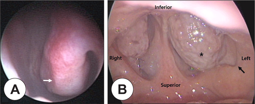
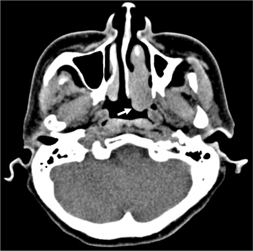
52세 여자 환자가 수개월 전부터 반복되는 재발성 중이염으로 내원하였으며, 내원 당시에는 고막 비후 외에 중이염 소견은 없었다. 코막힘 등 다른 비과적 증상은 호소하지 않았다. 과거에 외상이나 비과적 수술력은 없었으며, 가족력의 특이사항은 없었다. 고막운동성검사 상 양측 A형의 고실도 소견, 순음청력검사(6분법) 결과, 우측 20 dB, 좌측 23 dB의 정상 청력, 측두골 전산화 단층촬영 상 좌측 유양동 부분의 음영과 좌측 하비갑개 후방 비대가 관찰되었다. 비강을 통해 0도 내시경으로 관찰한 좌측 하비갑개에는 특별한 이상 소견이 없었으나(Fig. 1A), 구강을 통해 90도 내시경으로 관찰한 비인두 내시경 검사에서 좌측 하비갑개에서 후방의 표면이 불규칙한 종양이 비인두로 돌출되어 좌측 후비공(posterior choana)을 막고 있는 모습이 관찰되었으며, 좌측 이관 폐쇄는 관찰되지 않았다(Fig. 1B). 우측 하비갑개는 비대 소견은 없었지만, 후방 점막은 표면이 불규칙한 소견을 보였다. 부비동 전산화 단층촬영(non-enhanced paranasal sinuses(PNS) computed tomography(CT))에서는 좌측 하비갑개의 후방부위가 미만성으로 커져 있었으며, 주변 골 침범 소견은 없었고, 경계가 불명확한 종양이 좌측 비인두로 돌출되어 있었다(Fig. 2). 추가로 조영 증강 부비동 전산화 단층촬영(enhanced PNS CT)을 계획하였으나, 환자가 추가 검사를 원치 않아 좌측 비강 내의 양성 종양으로 추정하고, 진단적 치료를 위해 수술을 계획하였다. 수술 전 시행한 검사로는 종양의 혈행 공급에 대해 정확히 평가하는 데에 한계가 있었지만, 종양의 색상과 국소 소견으로 볼 때 혈행공급이 적다고 판단하였다. 그러나 술 중 예기치 못한 출혈에 대비하기 위하여 Ho:YAG 레이저를 준비하였다.

jcohns-32-3-281-g1
Fig. 1. Preoperative endoscopic image at the first visit. A : There is no definite mass-like lesion at the anterior aspect of left inferior turbinate (white arrow). B : The pinkish and irregular mass (asterisk) is protruded into the left nasopharyngeal cavity obstructing the left posterior choana. Pharyngeal orifice of the left Eustachian tube is not obstructed (black arrow).
Download Original Figure
jcohns-32-3-281-g2
Fig. 2. Preoperative CT image. Nonenhanced PNS CT shows diffusely enlarged inferior turbinate with protruding mass (white arrow) into the nasopharyngeal cavity. There is definite erosion of the surrounding bone. PNS: paranasal sinuses, CT: computed tomography.
Download Original Figure

종양은 전신마취 하에 비내시경으로 관찰하면서 제거하였다. 우측 하비갑개는 술 중 관찰 소견에서 특이 소견이 관찰되지 않았다. 술 중 출혈을 대비하여 Ho:YAG 레이저(OmniPulseTM, Trimedyne, Irvine, CA, USA)를 사용한 경비강 하비갑개 부분절제술을 시행하였다. Ho:YAG 레이저는 유리 섬유에서 조사되고, 18 gauge의 혈관카테터(angio catheter)를 씌워 사용하여 비강 내에서의 내시경과 같이 사용하기가 용이하였다. 수술 소견 상 좌측 하비갑개의 중후방이 불규칙한 표면을 가지면서 비대해져 3.0×1.5×1.0 cm 크기의 종양이 후비강까지 일부 막혀 있었다. 불규칙한 표면을 가진 부위를 종양 부위로 판단하고, 수술 시야 확보를 위해 하비갑개 전방의 정상 부위의 일부를 절단 겸자(cutting forceps)를 사용하여 제거하였다. 하비갑개 골부와 상악동 내벽에 부착하는 기시부는 정상 조직과의 충분한 변연을 확보한 후에 절단 겸자와 레이저를 사용하면서 분할절제술(piecemeal resection)로 제거하였다. 출혈이 있는 부위는 레이저로 지혈을 하면서 수술을 진행하였다. Ho:YAG 레이저가 조사되는 혈관카테터는 구부러질 수 있으며, 충분히 길어 후비공(posterior choana)까지 도달하는 데에 어려움이 없었다. 수술 중 대량 출혈은 없었고, 지혈을 위해 절제한 부위에 Greenplast®(Greencross, Seoul, Korea)를 뿌려주고, Merocel®(Medtronic Xomed, Jacksonville, FL, USA)로 패킹 시행 후 수술을 종료하였다. 수술 후 2일째 패킹을 제거하였고, 수술 후 3일째 출혈 소견이 없어 환자는 퇴원하였다.

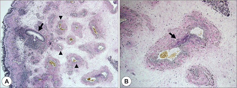
수술 후 확인된 병리조직 검사에서, 두꺼워진 혈관벽을 가진 큰 사행성 동맥과 다양한 크기의 얇은 혈관벽을 가진 정맥 등이 섬유점액성 바탕 내에서 관찰되었다. 이러한 동맥과 정맥이 직접 문합되는 혈관성 구조가 관찰되어 최종적으로 동정맥기형으로 진단되었다(Fig. 3).

jcohns-32-3-281-g3
Fig. 3. Histopathologic findings. A : It reveals a mixture of large thick-walled vessels (black arrow) which are occasionally tortous and thin-walled vessels (black arrowhead) in fibromyxoid background (Verhoeff-Van Gieson elastic stain, ×40). B : There is an anastomosing vessel (black arrow) showing fragmented elastic lamina in thick-walled portion (Verhoeff-Van Gieson elastic stain, ×100).
Download Original Figure

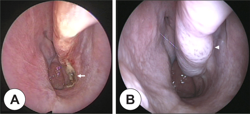
수술 후 1달째 외래 내원 시 환자가 호소하는 증상은 없었으며, 비내시경 검사 상 좌측 하비갑개 후방의 종양 절제 부위에 형성된 가피 외 특이소견은 없었다(Fig. 4A). 환자에게 추적관찰을 권유하였고, 이후 1달 간격으로 6번 추적관찰을 하다가 1년 간격으로 추적관찰을 하였다. 수술 후 2년째 좌측 하비갑개의 경한 비후가 보였으나 경과관찰하였고, 수술 후 4년째 호소하는 증상은 없었으나, 비내시경 검사에서 하비갑개의 비후가 여전히 보여서(Fig. 4B) 환자에게 추가적인 조직 검사를 권하였다. 그러나 환자는 증상이 없다고 거절하였고, 이후 병원에 내원하지 않았다.

jcohns-32-3-281-g4
Fig. 4. Postoperative endoscopic image. A : POD 1 month. Enlarged protruding mass from inferior turbinate is resected. No other specific findings are seen without some crusts at the operation site (white arrow). B : POD 4 years. Small mass with irregular-colored mucosa, which is suspicious of recurred AVM, is seen at the operation site of the inferior turbinate (white arrowhead). POD: postoperative day, AVM: arteriovenous malformations.
Download Original Figure

고 찰

혈관 이상(vascular anomaly)의 분류는 1982년에 Mulliken과 Glowacki가 제시한 조직학적, 생물학적, 임상적 특징을 기반으로 한 분류 방법에 따른다.3) 이는 혈관 내피 세포의 과형성(hyperplasia)이 원인인 혈관종(hemangioma)과 비정상적인 혈관 형성이 원인인 혈관 기형(vascular malformation)으로 두 가지 주요한 카테고리로 분류된다.3,4) 혈관 기형(vascular malformation)은 다시 혈류의 속도에 따라 저유속(slow-flow)과 고유속(fast-flow)으로 나눌 수 있고, 저유속 혈관 기형으로는 모세혈관 기형(capillary malformation), 정맥 기형(venous malformation), 림프관 기형(lymphatic malformation)이 있으며, 동정맥기형(arterivenous malformation)은 고유속 혈관 기형에 속한다.1,5)

동정맥기형은 태생 4-6주경 혈관 발달의 이상으로 발생하는 것으로 생각되며, 주로 소아기 때보다 청소년기와 성인기에 크기가 증가하는 양상을 보여 대부분 10-20대에 발견된다.6) 두개 외에 발생한 두경부 영역의 동정맥기형은 매우 드문 질환으로 국외에서는 Mulliken 등3), Jackson 등,7) Chen 등8)이 두개 외 동정맥기형 환자를 분석 및 보고한 바 있다. 국내에서는 Suh 등9)이 3예, Kim 등10)이 2예, Koh 등11)이 1예, Ahn 등12)이 1예의 두개 외 동정맥기형 환자 증례를 보고하였고, Noh 등13)은 1예의 비첨부에 발생한 동정맥기형 환자 증례를 보고하였다. 또한 Jeong 등14)은 19예의 두개 외 동정맥기형 환자를 후향적으로 분석 및 보고한 바 있다. 그들은 총 19예 중 13예에서 외과적 절제술을 시행하였는데, 절제술 단독 초치료로서 1예, 색전술과 함께 병합 초치료로서 7예, 에탄올 경화요법에 반응하지 않은 환자 중 5예가 있었다고 보고하였다.14) 또한 치료 후 재발 및 병변의 크기 증가는 평균 27.3개월의 관찰 기간 동안 19예 모두에서 발견되지 않았다고 보고하였다.14) 하지만 비강 내에서 발생한 동정맥기형은 국내에서 보고된 바가 없고, Ho:YAG 레이저를 사용하여 절제한 동정맥기형의 증례 또한 보고된 바가 없다.

진단적인 검사로는 혈관조영술, 혈류 초음파촬영술, 전산화 단층촬영(CT), 자기공명영상(MRI, magnetic resonance imaging) 등이 사용된다.1) 혈관조영술은 동정맥기형의 핵(nidus)과 공급 혈관의 상태를 확인하여 치료 방침을 정할 수 있는 중요한 검사이다.1) 증례의 경우, 병변 크기가 작고 환자가 침습적인 진단을 원하지 않아 시행하지 않았다. 전산화 단층촬영과 자기공명영상은 병변의 연부 조직의 침범 정도와 혈류의 특성을 파악하는데 도움을 준다.1) 증례의 경우는 저자들의 착오로 시행한 조영 증강을 하지 않은 전산화 단층촬영에서 병변의 크기가 크지 않았고, 골 침범 소견이 없어 자기공명영상은 시행하지 않았다.

치료 방법은 명확한 영양 혈관이 있고, 골 침범이 없는 작은 동정맥기형에 대해서는 수술적 치료를 단독으로 시행해볼 수 있지만, 진행된 병변에 대해서는 술 전 혈관경화요법 또는 혈관 내 색전술을 시행한 후 수술적으로 병변을 완전히 절제하는 치료의 단독 또는 병합 요법이 표준적인 치료 방법이다.1,2) 혈관경화요법은 종양 내로 경화제를 주입하는 방법으로 혈액 내 단백질 변성, 혈관내벽 손상으로 혈전을 형성시켜 기형을 폐쇄시키는 원리이며, 에탄올을 경화제로 널리 사용한다.1) 본 증례에서는 전산화 단층촬영 상 골 침범이 없는 작은 종양이어서 혈관경화요법, 혈관 내 색전술 등의 전 처치 없이 수술을 계획하였다. 그러나 술 중 출혈에 대비하기 위해 전통적인 절제술보다는 지혈 효과가 큰 Ho:YAG 레이저를 사용한 절제술을 시행하였다. Ho:YAG 레이저는 2,080 nm 파장의 빛을 방출하는 고출력 고체 레이저로, 연조직과 뼈를 함께 절단할 수 있고, 절제와 광응고를 동시에 할 수 있어 다른 레이저들보다 조직의 응고, 지혈 및 정교한 절제에 유리하다는 장점이 있다.15) 또한 레이저 섬유는 구부러질 수 있는 특징이 있어 연성 내시경에도 활용될 수 있고, 좁은 부위에서도 효과적으로 사용할 수 있다. 본 증례에서는 이러한 장점을 가진 Ho:YAG 레이저를 사용하여 술 중, 그리고 술 후 출혈을 비롯한 특별한 합병증 없이 손쉽게 동정맥기형을 절제할 수 있었다.

저자들은 본 증례를 통하여 반복적인 비출혈과 같은 전형적인 증상 없이도 비강 내 혈관성 병변이 발생할 수 있음을 경험하였다. 본 증례에 따르면 주변 조직 침범이 없는 동정맥기형의 경우에서는 술 전 처치 없이 Ho:YAG 레이저를 사용한 수술 단독 치료로도 특별한 합병증 없이 손쉽게 절제할 수 있으나, 정상 조직과의 경계에 대한 세심한 확인이 필요하다. 이에 관련된 문헌 고찰과 함께 Ho:YAG 레이저를 사용하여 동정맥기형을 절제한 증례를 보고하는 바이다.

Acknowledgements

Not applicable.

Funding Information

Not applicable.

Conflicts of Interest

No potential conflict of interest relevant to this article was reported.

Author Contribution

Conceptualization: Lee HH.

Data curation: Jo KH, Lee HH.

Formal analysis: Jo KH, Lee DW.

Methodology: Lee HH.

Software: Jo KH, Lee HH.

Validation: .Jo KH, Lee HH.

Investigation: Lee HH, Chun BK.

Writing - original draft: .Jo KH.

Writing - review & editing: Jo KH, Lee HH, Chun BK, Lee DW.

Ethics Approval

Not applicable.

References

1).

Fernández-Alvarez V, Suárez C, de Bree R, Nixon IJ, Mäkitie AA, Rinaldo A, et al. Management of extracranial arteriovenous malformations of the head and neck. Auris Nasus Larynx 2020;47(2):181-90.

2).

Pandhare MN, Jyoti DB, Mandale MS, Suresh RB. Acquired arteriovenous malformation of lip occurring as an occupational hazard: a case report with review of literature. J Oral Maxillofac Pathol 2018;22(2):287.

3).

Mulliken JB, Glowacki J. Hemangiomas and vascular malformations in infants and children: a classification based on endothelial characteristics. Plast Reconstr Surg 1982;69(3): 412-22.

4).

Richter GT, Friedman AB. Hemangiomas and vascular malformations: current theory and management. Int J Pediatr 2012;2012:645678.

5).

Kaban LB, Mulliken JB. Vascular anomalies of the maxillofacial region. J Oral Maxillofac Surg 1986;44(3):203-13.

6).

Fowell C, Jones R, Nishikawa H, Monaghan A. Arteriovenous malformations of the head and neck: current concepts in management. Br J Oral Maxillofac Surg 2016;54(5):482-7.

7).

Jackson IT, Carreño R, Potparic Z, Hussain K. Hemangiomas, vascular malformations, and lymphovenous malformations: classification and methods of treatment. Plast Reconstr Surg 1993;91(7):1216-30.

8).

Chen MT, Horng SY, Yeong EK, Pan QD. Treatment of high-flow vascular malformations in the head and neck with arterial ligation followed by sclerotherapy. Ann Plast Surg 1996; 36(2):147-53.

9).

Suh JC, Kim JW, Goh EK, Chon KM. Three cases of arteriovenous malformation of the auricle. J Clin Otolaryngol Head Neck Surg 1993;4(1):141-8.

10).

Kim DI, Rho YS, Kim HJ, Lim HJ, Jung CH. Two cases of arteriovenous malformation arising from head and neck. Korean J Otorhinolaryngol Head Neck Surg 1994;37(6):1335-41.

11).

Koh JW, Oh JH, Shin JC, Kim SY. Case report: intramasseteric vascular anomaly-misdiagnosed to parotid sialolithiasis. Korean J Otorhinolaryngol Head Neck Surg 1999;42(3):380-5.

12).

Ahn SK, Kim JG, Kim SH, Yu HK. A case of arteriovenous malformation of the parotid gland. Korean J Otorhinolaryngol Head Neck Surg 2000;43(10):1134-6.

13).

Noh Y, Ryu G, Kim HY. A case of arteriovenous malformation of the nasal tip. Korean J Otorhinolaryngol Head Neck Surg 2020;63(3):123-8.

14).

Jeong HS, Choi JY, Lee HJ, Kim TW, Kim MB, So YK, et al. Diagnosis and treatment of extracranial arteriovenous malformations in the head and neck region. Korean J Otorhinolaryngol Head Neck Surg 2005;48(9):1136-42.

15).

Leunig A, Janda P, Sroka R, Baumgartner R, Grevers G. Ho:YAG laser treatment of hyperplastic inferior nasal turbinates. Laryngoscope 1999;109(10):1690-5.