Journal of Clinical Otolaryngology Head and Neck Surgery
The Busan, Ulsan, Gyeoungnam Branch of Korean Society of Otolaryngology-Head and Neck Surgery
증례

중이염을 주소로 내원한 ANCA 음성 제한성 육아종증 다발 혈관염 1례

문지원1, 이진환2, 채성원1, 송재준1,*
Jee Won Moon1, Jin Hwan Lee2, Sung Won Chae1, Jae Jun Song1,*
1고려대학교 의과대학 구로병원 이비인후과학교실
2병리과학교실
1Department of Otorhinolaryngology-Head and Neck Surgery, Seoul, Korea
2Pathology, Korea University College of Medicine, Seoul, Korea
*교신저자: 송재준, 08308 서울시 구로구 구로동로 148, 고려대학교 의과대학 구로병원 이비인후과학교실 전화: (02) 2626-1299·전송: (02) 868-0475·E-mail: jjsong23@gmail.com

© Copyright 2021 The Busan, Ulsan, Gyeoungnam Branch of Korean Society of Otolaryngology-Head and Neck Surgery. This is an Open-Access article distributed under the terms of the Creative Commons Attribution Non-Commercial License (http://creativecommons.org/licenses/by-nc/4.0/) which permits unrestricted non-commercial use, distribution, and reproduction in any medium, provided the original work is properly cited.

Received: Dec 31, 2020; Revised: Jan 19, 2021; Accepted: Mar 04, 2021

Published Online: Jun 30, 2021

ABSTRACT

Granulomatosis with polyangiitis(GPA) is a systemic disease that causes granulomatous inflammation and vasculitis to small to medium sized vessels. Although prompt diagnosis is vital for the successful treatment of the disease, it is often misdiagnosed as a simple inflammatory disease in ENT clinic. Anti-neutrophil cytoplasmic antibody(ANCA) tests positive in up to 90% of GPA, and it is a useful diagnostic marker for its strong association with the disease. However, we experienced ANCA-negative limited GPA presenting with intractable otitis media, thereby reporting this case with a brief review of the literature. (J Clinical Otolaryngol 2021;32:59-64)

Keywords: 중이염; 육아종증 다발 혈관염; 항중성구세 포질항체
Keywords: Otitis media; Granulomatosis with polyangiitis; ANCA

서 론

육아종증 다발 혈관염(Granulomatosis with polyangitis, GPA)은 전신의 육아종성 염증과 작은·중간크기 혈관에 혈관염을 일으키는 원인 불명의 전신 질환으로, 심한 전신 증상을 보이는 경우 연구에 따라 96%까지도 혈중 항중성구 세포질 항체(anti-neutrophil cytoplasmic antibody, ANCA)가 발견된다고 알려져 있지만, 국소적 증상만 있는 경우 양성율이 낮아 진단에 유의해야 한다.1-3) 저자들은 잘 치유되지 않는 중이염을 주소로 내원하여 안면신경 마비까지 진행된 환자가 추후 ANCA 음성인 제한성 GPA로 진단되어 스테로이드 및 면역억제제 치료를 통해 호전된 1예를 경험하여 고찰과 함께 보고하는 바이다.

증 례

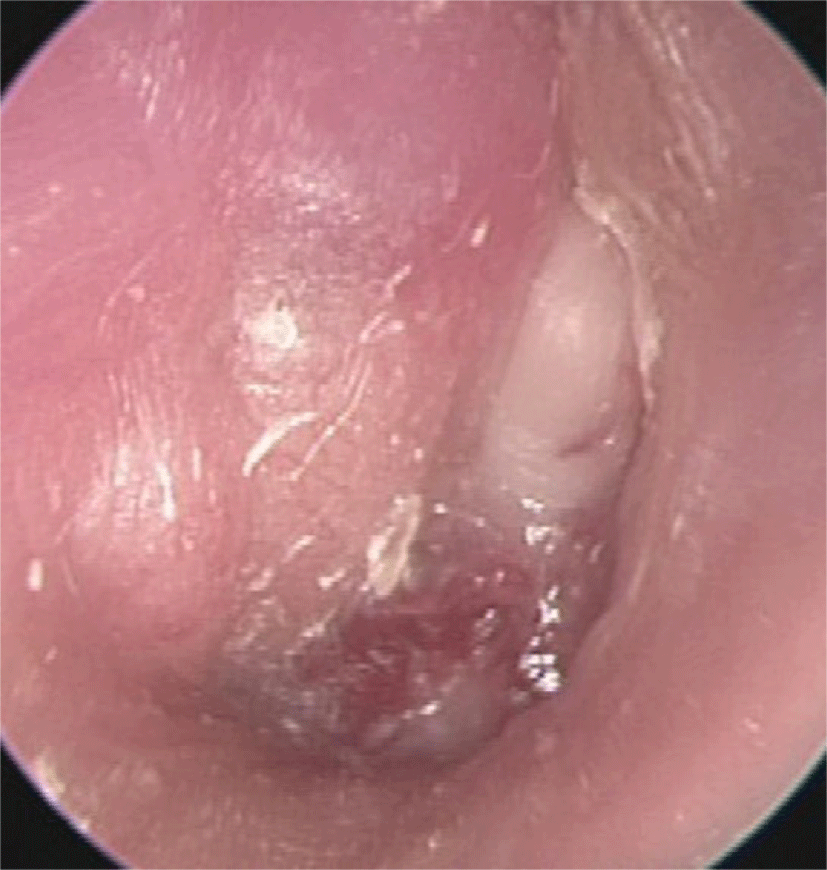
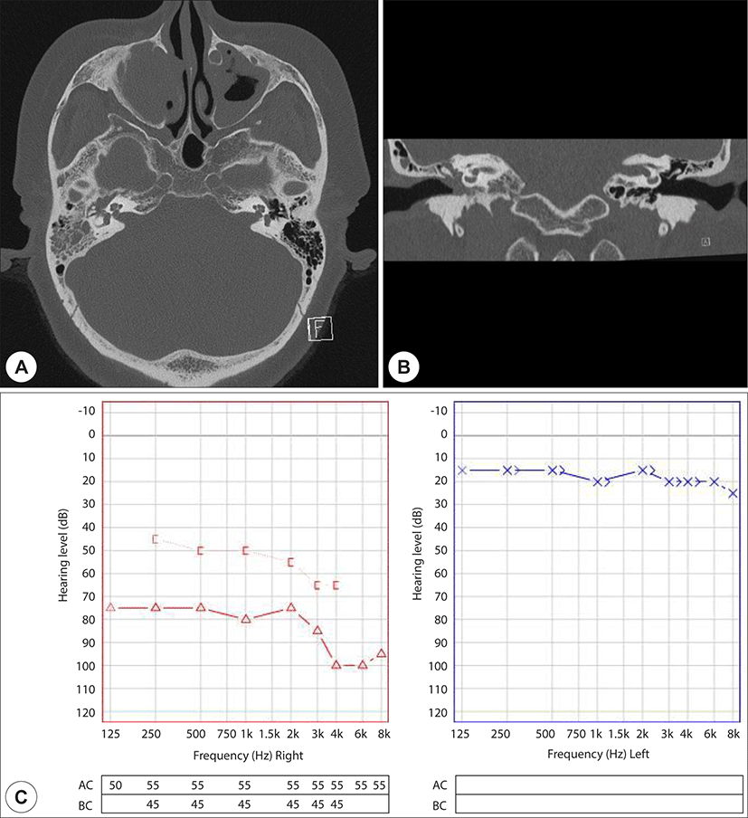
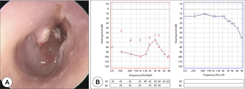
기저질환이 없는 57세 여자 환자가 내원 1개월 전부터 시작된 발열, 편두통을 동반한 우측 이통 및 이충만감으로 개인병원에서 치료받던 중 경과 호전이 없어 2020년 2월 본원 이비인후과 외래로 내원하였다. 이경 검사 상 좌측 고막은 정상이었으나, 우측 고막의 발적을 보여 우측 급성 중이염 의증 하 추가 검사를 시행하였고(Fig. 1), 고막운동성검사 상 우측은 C형의 고실도 소견, 순음청력검사(6분법) 상 우측 기도청력 81 dB, 골도청력 54 dB, 좌측 기도청력 및 골도청력 모두 18 dB, 측두골 전산화 단층촬영검사(computed tomography, CT) 상 우측 유양동 및 중이강 내 음영이 확인되었다(Fig. 2). 일주일 뒤 우측 이루 확인되어 미생물 검사 시행 후 보존적 치료를 지속하였으며, 열흘 뒤 우측 고막 발적은 다소 호전되었으나 이루는 지속되었고, 미생물 검사 상 균은 동정되지 않았다.

jcohns-32-2-59-g1
Fig. 1. Endoscopy of right tympanic membrane at initial outpatient clinic visit.
Download Original Figure
jcohns-32-2-59-g2
Fig. 2. Findings at initial outpatient clinic visit. (A),(B) Temporal bone CT showing soft tissue density in right mastoid and middle ear cavity(A: Axial view, B: Coronal view). (C) Pure tone audiometry.
Download Original Figure

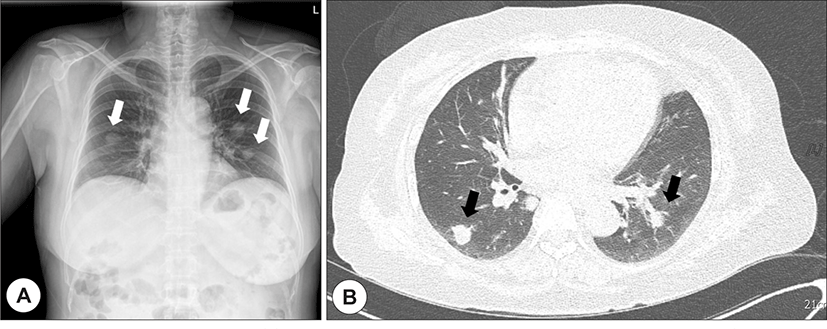
환자는 2019년 12월부터 지속되던 우측 유방염에 대한 치료를 위해 2020년 3월 본원 유방내분비내과에 입원하였으며, 입원 시 촬영한 흉부 X선 검사에서 폐 전이 암 의증, 추가 시행한 흉부 CT에서 양측 패혈성 폐렴 의증 소견으로 감염내과로 전과되었다(Fig. 3). 혈액검사 상 ANCA 음성, 혈중 요소 질소·크레아티닌 및 소변 검사는 정상이었고, C 반응 단백질은 150 mg/dL 이상, 적혈구 침강속도는 100 mm/hr 이상으로 상승되어 있었으며, 호전 없는 발열 및 두통으로 시행한 뇌척수액 검사 상 정상이었다. 입원 중 본과 협진을 통한 이경 검사 상 우측 박동성 고막 및 장액성 이루가 보여 국소마취 하에 우측 환기튜브 삽입술 및 단순 유양돌기절제술 시행 후 보존적 치료를 지속하였다(Fig. 4).

jcohns-32-2-59-g3
Fig. 3. Radiologic findings of chest. (A) Chest X-ray showing multiple lung nodules in both lung field (Possible lung metastasis, White arrow). (B) Chest CT showing variable-sized nodule with or without cavitation in both lungs (Possible septic emboli, Black arrow).
Download Original Figure
jcohns-32-2-59-g4
Fig. 4. Intra-operative findings and pathology of middle ear. (A) Granulations in middle ear (White arrow), (B) Acute inflammation with abscess (H&E, x200), (C) No definite vascular structure or vasculitis (Elastic, x200).
Download Original Figure

2020년 4월 House-Brackmann grade(H-B grade) 2인 우측 안면근육마비가 발생하였는데, 뇌 자기공명영상검사(magnetic resonance imaging, MRI) 상 정상, 신경학적 검사 상 이상소견 없어 우측 Bell씨 마비가 의심되었다. 이전 유방염에 대해 경구 스테로이드 투여 시 부작용이 발생한 기왕력 및 현재 감염으로 치료 중인 점을 고려하여 경구 항바이러스제만 추가 투여하며 경과 관찰하였고, 이후 시행한 측두골 MRI에서 안면신경의 특이 병변은 없었으며, 안면신경마비는 H-B grade 4-5까지 악화되었다.

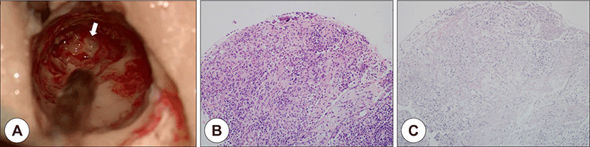
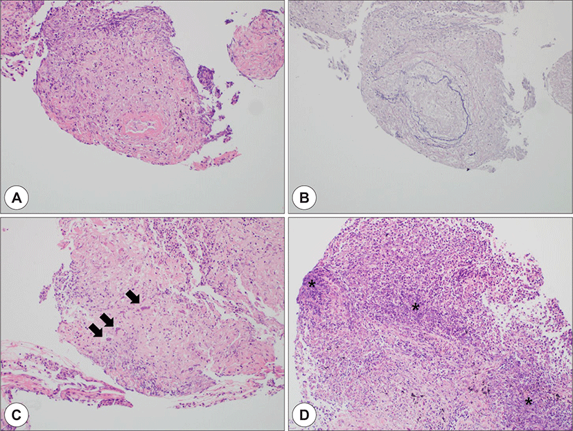
지속되는 발열로 재시행한 흉부 CT 상 기관지 주위로 염증이 증가되는 소견을 보여 기관지 내시경을 시행하였고, 우측 기관지의 결절성 병변에 대해 시행한 조직검사 결과, 괴사조직을 보이는 혈관염 및 육아종이 확인되어 ANCA 음성인 제한성 GPA로 진단되었다(Fig. 5). 정맥 스테로이드 투여 후(3일 간 Methylprednisolone 30 mg/일) 두통 및 발열이 호전되었으며, 환자 개인 사정으로 자의 퇴원한 뒤 류마티스 내과 외래를 통해 고용량 경구 스테로이드 및 면역억제제 병합치료를 시행하였다(경구 Prednisolone(PDS) 0.8 mg/kg/일 및 경구 Azathioprine(AZA) 0.4 mg/kg/일로 관해 유도 시작하여 경구 PDS 0.1 mg/kg 및 경구 AZA 2.5 mg/kg까지 6개월 간 단계적 증·감량, 정맥 내 Rituximab 1,000 mg 2주 간격으로 2회 사용, 관해 유지 위해 경구 Methotrexate 15 mg/주). 현재까지 청력은 호전되지 않았으나 안면마비는 H-B grade 3로 호전, 귀의 염증 또한 호전되어 외래 추적 관찰 중이다(Fig. 6).

jcohns-32-2-59-g5
Fig. 5. Representative pathologic findings of Wegener’s granulomatosis. (A),(B) Inflammatory cell infiltrations and vasculitis involving small to medium-sized vessels (A: H&E, x200, B: Elastic, x200), (C) Multinucleated giant cells (Center, Black arrow)(H&E, x200), (D) Geographic and basophilic necrosis composed of cell debris and neutrophils (Asterisk)(H&E, x200).
Download Original Figure
jcohns-32-2-59-g6
Fig. 6. 6 month follow-up evaluation. (A) Endoscopy of right tympanic membrane, (B) Pure tone audiometry.
Download Original Figure

고 찰

미국 류마티스 학회에서는 1)신장염을 시사하는 소변 침전물, 2)단순 흉부촬영 상 결절 또는 공동, 3)구강 또는 비강 염증, 4)조직검사 상 육아종성 염증의 4가지 기준 중 2가지를 만족할 때 88%의 민감도와 92%의 특이도로 GPA를 진단할 수 있다고 하였다.4) 혈중 ANCA는 진단 기준에는 포함되어 있지 않으나, ANCA 연관 혈관염에서 질병 활성도, 치료 반응도 등과 상관관계를 가지며, 질병의 활성도 및 병기에 따라 양성을 보이는 비율에 차이가 있는 것으로 알려져 있다.2,5-6)

GPA는 호흡기계의 침범이 가장 흔하지만 모든 장기의 침범이 가능하며, 정확한 발병기전은 불확실하나, ANCA가 육아종의 형성 및 혈관 손상을 일으키는 데에 중요한 역할을 하는 것으로 추정된다.5,7) 제한성 GPA의 경우, ANCA 양성률이 35%로 낮으며, GPA가 의심될 경우 최소 2번 이상의 ANCA 검사가 권고되는데, 증례의 환자는 이후 재검에서도 지속적으로 ANCA 음성으로 나타났다.8) 하부 호흡기를 침범하는 GPA의 경우, 영상 소견만으로는 폐렴과 같은 염증성 질환, 악성 종양 등과 감별진단이 어렵다고 알려져 있는데, 본 증례에서도 흉부 X선 검사 및 흉부 CT 상 전이성 폐암, 패혈 색전증으로 의심되는 소견이 보였다.9)

환자 대부분은 초기에 비강과 부비동에 증상이 나타나고, 약 20%에서 본 증례와 같이 귀를 침범하여 장액성·만성 중이염, 감각신경성 난청, 드물게는 안면신경마비도 발생할 수 있지만, 이는 단순 염증 질환들과 비슷하여 오진되는 경우가 많다.10) 증례의 환자의 경우, 양측 상악동 부비동염 및 부비동 수술 과거력이 있었지만 입원 당시 호소하는 증상은 없었고, 비강 내시경 상 특이 소견은 없었다. 이후 환자가 간헐적인 비강 내 통증을 호소하였으나, 이에 대한 진료를 거부하여 추가적인 평가는 시행하지 못하였다.

GPA 환자에서의 중이염은 대부분 일측성, 33%에서 양측성으로 나타난다고 보고된 바 있으며, 난청의 경우 치료를 통해 호전되기도 하지만, 다른 장기들이 치료에 반응하더라도 지속적으로 남아 있는 경우가 있는 것으로 알려졌다.11-13) 증례의 환자는 내원 당시 이미 난청이 상당히 진행된 상태로, 적절한 치료에도 불구하고 청력이 회복되지 않았던 것으로 생각된다. 또한 Nishino 등의 연구에 따르면 324명의 GPA환자 중 총 21명에서 뇌신경병증이 나타났고, 그 중 8명이 안면신경병증을 보였는데, 안면신경마비의 경우 스테로이드 및 면역억제제 병합 요법을 통한 표준치료 시 경과가 좋은 편이나, 수술적 치료 시 추가적인 신경 손상 가능성이 높다고 보고되었다.14-17)

33명의 GPA 환자를 대상으로 한 Park 등의 연구에서 육아종성 염증의 전형적인 소견인 괴사성 염증, 혈관염, 만성 육아종성 염증을 모두 보인 경우는 재검을 포함하여 27.2%에 불과하였고, 33.3%에서는 한 가지 이하의 소견을 보여 조직검사에서는 양성 판정을 받지 못했다.8) 증례의 환자도 유양돌기 검체에서는 괴사성 골조직 소견만 보였기 때문에 GPA와 별개로 발생한 중이염의 가능성을 배제할 수는 없으나, GPA에서 비강을 제외한 상기도의 다른 부위는 진단율이 떨어지기 때문에 의심되는 환자에게는 비강 및 부비동 진찰을 우선 시행하고, 병변이 없는 경우 폐, 신장 등 다른 장기에서 검사를 고려해야 한다.8,19)

GPA는 빠른 진단 및 치료가 이루어지지 않으면 장기적인 예후가 나쁜 질환으로, 초기증상이 이비인후과에서 흔하게 접하게 되는 염증성 질환과 유사하여 임상에서 이를 염두에 두고 의심하는 것이 가장 중요하다. 질환의 특성을 고려하여 전신 증상에 대한 문진과 함께 흉부 X선 검사, 소변검사 등 다장기 침범을 고려한 검사를 시행하고, ANCA가 음성이더라도 종합적으로 판단하여 의심이 된다면 확진을 위한 조직검사를 적극적으로 시행하는 것이 바람직하다.

REFERENCES

1).

Ozaki S. ANCA-associated vasculitis: diagnostic therapeutic strategy. Allergol Int 2007;56(2):87-96.

2).

Finkielman JD, Lee AS, Hummel AM, Viss MA, Jacob GL, Homburger HA, et al. ANCA are detectable in nearly all patients with active severe Wegener’s granulomatosis. Am J Med 2007;120(7):643. e9-14.

3).

Lamprecht P, Gross WL. Wegener’s granulomatosis. Herz 2004;29(1):47-56.

4).

Leavitt RY, Fauci AS, Bloch DA, Michel BA, Hunder GG, Arend WP, et al. The American College of Rheumatology 1990 criteria for the classification of Wegener’s granulomatosis. Arthritis Rheum 1990;33(8):1101-7.

5).

De Groot K, Gross W. Wegener’s granulomatosis: disease course, assessment of activity and extent and treatment. Lupus 1998;7(4):285-91.

6).

Schönermarck U, Lamprecht P, Csernok E, Gross W. Prevalence and spectrum of rheumatic diseases associated with proteinase 3-antineutrophil cytoplasmic antibodies (ANCA) and myeloperoxidase-ANCA. Rheumatology 2001;40(2):178-84.

7).

Bosch X, Guilabert A, Espinosa G, Mirapeix E. Treatment of antineutrophil cytoplasmic antibody–associated vasculitis: a systematic review. JAMA 2007;298(6):655-69.

8).

Park KT, Kong IG, Han DH, Kim DW, Kim SH, Rhee CS, et al. Clinical experiences of diagnosis and treatment for Wegener’s granulomatosis. Korean Journal of Otorhinolaryngology-Head and Neck Surgery 2008;51(12):1109-18.

9).

Ananthakrishnan L, Sharma N, Kanne JP. Wegener’s granulomatosis in the chest: high-resolution CT findings. AJR Am J Roentgenol 2009;192(3):676-82.

10).

Gottschlich S, Ambrosch P, Gross W, Hellmich B. Wegener’s granulomatosis in the head and neck region. HNO 2004;52(10):935-45.

11).

Bakthavachalam S, Driver MS, Cox C, Spiegel JH, Grundfast KM, Merkel PA. Hearing loss in Wegener’s granulomatosis. Otol Neurotol 2004;25(5):833-7.

12).

Joglekar S, Deroee AF, Morita N, Cureoglu S, Schachern PA, Paparella M. Polyarteritis nodosa: a human temporal bone study. Am J Otolaryngol 2010;31(4):221-5.

13).

Lidar M, Carmel E, Kronenberg Y, Langevitz P. Hearing loss as the presenting feature of systemic vasculitis. Ann N Y Acad Sci 2007;1107(1):136-41.

14).

Nishino H, Rubino FA, DeRemee RA, Swanson JW, Parisi JE. Neurological involvement in Wegener’s granulomatosis: an analysis of 324 consecutive patients at the Mayo Clinic. Ann Neurol 1993;33(1):4-9.

15).

Verma N, Gupta A. Wegener’s granulomatosis: an unusual presentation case report and review of the literature. The Internet Journal of Otorhinolaryngology 2012;14(1).

16).

Maranhão ASdA, Chen VG, Rossini BAA, Testa JRG, Penido NdO. Mastoiditis and facial paralysis as initial manifestations of Wegener’s granulomatosis. Braz J Otorhinolaryngol 2012;78(2):80-6.

17).

Roszkowska A, Morawska-Kochman M, Temporale H, Sikorska-Żuk M, Kręcicki T. Bilateral facial palsy in rapidly progressive course of Wegener’s granulomatosis: a case report. Case Reports in Otolaryngology 2013.

18).

Devaney KO, Travis WD, Hoffman G, Leavitt R, Lebovics R, Fauci AS. Interpretation of head and neck biopsies in Wegener’s granulomatosis. A pathologic study of 126 biopsies in 70 patients. Am J Surg Pathol 1990;14(6):555-64.