증례
구강 설부에 발생한 신경초종 1례
천용일1, 신성찬1, 이병주1, 권현근2,*
Schwannoma in the Oral Tongue : A Case Report with Review of Literature
Yong-Il Cheon1, Sung-Chan Shin1, Byung-Joo Lee1, Hyun-Keun Kwon2,*
1Department of Otorhinolaryngology-Head and Neck Surgery, Pusan National University Hospital, Busan, Korea
2Department of Otorhinolaryngology-Head and Neck Surgery, Pusan National University Yangsan Hospital, Yangsan, Korea
*교신저자: 권현근, 50612 경상남도 양산시 물금읍 금오로 20, 양산부산대학교병원 이비인후과 전화: (055) 360-2132·전송: (055) 360-2162·E-mail:
kwon-h-g@hanmail.net
© Copyright 2020 The Busan, Ulsan, Gyeoungnam Branch of Korean Society of Otolaryngology-Head and Neck Surgery. This is an Open-Access article distributed under the terms of the
Creative Commons Attribution Non-Commercial License (http://creativecommons.org/licenses/by-nc/4.0/) which permits
unrestricted non-commercial use, distribution, and reproduction in any
medium, provided the original work is properly cited.
Received: Oct 07, 2020; Revised: Nov 09, 2020; Accepted: Nov 27, 2020
Published Online: Dec 31, 2020
ABSTRACT
Schwannomas are benign, encapsulated and solitary masses originating from Schwann cells in the peripheral nerve sheath. Tumors grow slowly and develop about 25 to 40% of schwannomas in the head and neck area. Oral schwannoma occurs mostly on the tongue and can also occur in palate, buccal, mouth floor, gingiva and other oral mucosa. Schwannomas are usually treated by complete excision. A 21-year-old female patient with asymptomatic swelling on the tip of the tongue visited our hospital. Here, we report a case of schwannoma of the oral tongue with the review of literature. (J Clinical Otolaryngol 2020;31:281-285)
Keywords: 신경초종; 구강; 혀
Keywords: Schwannoma; Oral cavity; Tongue
서론
신경초종(schwannoma)은 단발성으로 잘 생기는 피막화된(encapsulated) 신경초(nerve sheath)의 Schwann 세포(Schwann cell)에서 기원하는 양성 종양이다. 정확한 유병률은 알려져 있지 않지만, 전체 신경초종의 약 25~45%가 두경부 영역에서 발생하며, 이 중 1~12%는 구강 내에서 발생한다고 보고되었다.1) 구강에서는 혀에서 가장 흔하게 생기며, 입천장, 구강저, 협측 점막, 잇몸, 입술 등에도 발생할 수 있다.2) 신경초종은 일반적으로 30~40대 사이에 잘 발생하며, 성별이나 인종에 따른 발생률의 차이는 없는 것으로 알려져 있다.3)
종양은 단독으로 느리게 성장하며, 표면이 매끄러운 덩어리로 나타나는데, 대부분 증상이 없지만 통증과 불편을 유발할 수도 있으며, 증상의 강도는 종양의 위치와 크기에 따라 결정된다. 일반적인 치료는 외과적 절제이며, 수술 후 재발률은 낮고 악성 변화는 드문 편이다.
이에 저자들은 21세 여성에서 혀의 앞부분에 발생한 무통성 신경초종을 외과적 절제로 치료한 증례를 경험하여 문헌고찰과 함께 보고자 한다.
증례
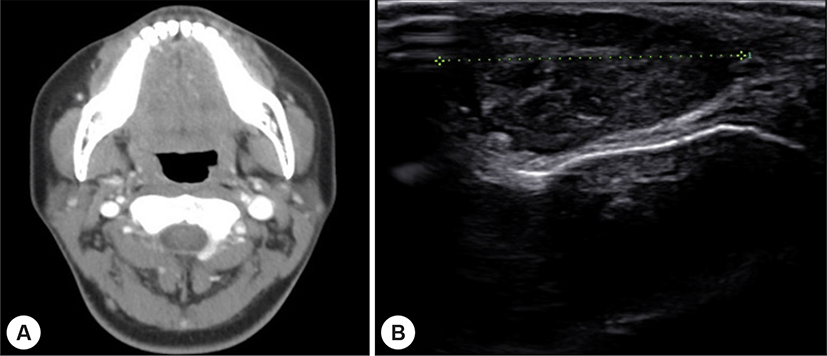
21세 여자 환자로 약 1년 전부터 발생한 구강 혀의 부종으로 외부 병원에서 컴퓨터 전산화 단층 촬영(computed tomography, CT) 검사 및 초음파 검사(ultrasonography)를 시행 후 의뢰되었다. 과거력상 특이 사항은 없었으며, 부종으로 인한 이물감 이외에 통증이나 미각 이상 등 불편감은 없다고 하였다. 처음 발견 당시와 비교하였을 때 크기 변화는 없었다고 하며, 구강 진찰상 구강 혀의 끝에서 약간 우측으로 치우쳐 있는 약 1.5 cm 크기의 종물이 관찰되었다. 촉지하였을 때 압통은 없었고, 경계는 비교적 명확한 형태의 종물이었다. 외부에서 촬영한 CT 검사에서는 특이 소견이 관찰되지 않았으며(Fig. 1A 초음파 검사상 경계가 좋으며, 내부는 비균질한 저에코성 음영에 후방 음영이 강조된 타원형 종물이 확인되었다(Fig. 1B). 임상적으로 양성 종물이 의심되었고, 혀의 표면에서 깊은 부위에 위치하여 조직검사를 하기 어려운 상태로 판단하여 추가적인 자기공명영상(magnetic resonance imaging, MRI)이나 조직검사없이 진단 및 치료 목적으로 수술하기로 결정하였다.
Fig. 1.
Preoperative radiologic findings.
A : Preoperative contrast enhanced neck CT axial view. No definite mass was observed at the tip of the tongue on CT. B : Ultrasonographic image. Encapsulated and several lobulated hypoechoic mass was visible. Posterior acoustic enhancement is also observed.
Download Original Figure
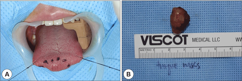
수술은 전신마취로 진행하였으며, 수술전 국소 소견은 다음과 같다(Fig. 2A). 종물이 혀의 끝에 위치하였기 때문에 양쪽 측방향으로 봉합(stay suture)하여 구강 외부로 견인하였으며 단극성 소작기(monopolar cautery) 및 sharp mosquito 등 기구를 이용하여 조심스럽게 박리하였다. 종물은 주위 조직과는 유착이 심하지 않고 잘 분리되어 종물만 적출(enucleation) 하였다. 수술장에서 확인된 종물의 크기는 1.8×1.4 cm 이었고, 표면은 매끈하였으나 형태는 불규칙한 모양이었다(Fig. 2B). 잔여 조직이 있는지를 확인하고, 지혈한 후 봉합하고 수술을 종료하였다.
Fig. 2.
Intraoperative imaging.
A : Preoperative local finding. A relatively rounded mass was identified at the tip of the tongue. B : Postoperative surgical specimen. The mass was completely removed and showed irregular lobulated shape.
Download Original Figure
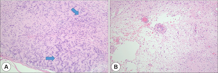
병리과에 요청하여 시행한 H & E 염색(Haemotoxylin & Eosin staining)에서 세포의 밀집도가 높고 늘어진 슈반 세포로 구성된 Antoni A형과 상대적으로 밀집도가 낮고 느슨한 형태의 Antoni B형이 관찰되어 신경초종으로 진단할 수 있었다(Fig. 3).
Fig. 3.
Histologic findings.
A : Antoni A area. Highly cellular dense and palisading nuclear arrangement was seen. (blue arrow) B: Antoni B area. Loosely arranged hypocellular region. (H&E stain, ×100).
Download Original Figure
술후 감각이상이나 부종 등 합병증은 없었으며, 술후 1개월째 혀의 점막은 정상화되어 수술의 흔적이 관찰되지 않을 정도로 완치되었고, 술후 3년째 재발없이 외래 경과 관찰 중이다.
고찰
신경초종의 병인은 명확하지 않지만, 뇌신경, 말초신경, 교감신경을 둘러싸고 있는 신경막의 Schwann 세포에서 기원하는 것으로 알려져 있다.4,5) 두경부 영역에서 흔하게 발생하는데, 이 중 구강내 신경초종은 혀에서 가장 흔하며, 구개, 구강저, 협부 점막, 잇몸, 입술 등에서도 발생할 수 있다.6) 종양은 서서히 자라기 때문에 발견되기 오래전부터 생겼을 가능성이 높으며, 혀의 신경초종의 발생률은 성별과는 무관하지만 30~40 대에 호발한다는 보고도 있고, 나이에 따른 차이를 보이지 않는다는 문헌도 있다.7,8) 일반적으로 평균 2.4 cm 크기의 무통성 종물로 혀의 어느 부위에서나 별견되며, 크기가 3.0 cm보다 커지면 연하장애, 통증, 발성장애, 음성변화를 호소할 수 있다.8) 본 증례에서는 조직검사 결과, 1.8 cm 크기로 측정되었고, 환자는 혀의 끝에 발생하였기에 이물감 이외에 다른 증상없이 내원하였다. 혀의 심부나 기저부에 발생하였다면 더 커진 상태에서 병원을 방문했을 가능성이 높다고 볼 수 있다.
CT 검사에서는 경계가 명확한 균질성 병변으로 나타나며, 만일 비균질성 병변이 관찰된다면 악성 변화를 의심해 볼 수 있다.8,9) MR 검사는 CT 검사보다 혀의 신경초종 진단에 도움이 된다. T1 강조영상(T1 weighted image, T1WI)에서는 근육과 비슷한 강도의 신호를 보이고, T2 강조영상(T2 weighted image, T2WI)에서는 고신호강도로 나타나는데,10,11) MR 검사를 통해 병변의 크기를 더 정확하게 알 수 있고, 주변 구조물과의 관계를 확인할 수 있다. 본 증례의 경우 CT 검사에서는 이상 소견이 관찰되지 않았고, 초음파 검사에서 종물이 확인되어 의뢰되었다. 초진 당시 혈관종이 의심되어 정밀한 평가 위해 의뢰되었는데, 신경초종의 경우 임상적으로 다른 피막으로 둘러싸인 양성 종물과 감별하기 어려우며, 조직검사를 통해 최종 진단이 내려질 수 있다. 일반적으로 신경초종과 감별해야 할 질환으로는 섬유종, 림프관종, 혈관종, 점액낭종, 지방종, 선종, 타액선 종양 및 설기저부에서 발생하였을 경우 갑상설 낭종 등이 있다.7,11)
두경부 영역의 신경초종에서 악성 변화는 8~10%로 보고되었다.12) 일반적으로 신경초종은 악성 변화를 하지 않지만, 두경부 영역에서는 보고된 적이 있으며, 혀에서는 1례가 보고되었다.1,2,13)
조직학적으로 신경초종은 피막화 되어 있으며, 피막 아래로 Antoni type A와 B 라는 두가지 특징적인 소견이 관찰된다. Antoni type A는 울타리 모양(palisading pattern)의 핵형을 가진 치밀한 슈반 세포가 치밀하게 배열된 것이 특징적이며, Antoni type B는 느슨하게 배열된 세망 조직에서 세포의 다 형태성이 표현된다. 이들 두 가지 형태는 대부분의 신경초종에서 발견되지만 재발이나 악성 변화와는 관계가 없다.14)
신경초종은 방사선치료에 반응하지 않으므로 일반적으로 침범한 신경과 함께 외과적으로 절제하는 것이 추천된다.14,15) 구강에 발생한 신경초종은 경구강 접근법이 가장 많이 사용되는 수술 방법이며, 최근에는 설기저부 신경초종의 치료를 위해 경구강 로봇수술(transoral robotic surgery)이나 CO2 레이저를 이용한 절제술을 이용하기도 한다.1,3) 설부에 발생한 신경초종 중 약 14.6%가 설기저부에 발생하며, 크기가 4.0 cm보다 크거나 설기저부 후측면에 위치한 경우 과거에는 하악 절개(mandibulotomy), 구순 절개(lip splitting)를 통한 접근 또는 경부 접근(cervical)을 통한 개방적 접근법이 사용되기도 하였으나, 앞서 언급한 바와 같이 크기가 크거나 접근이 어려운 경우 로봇수술이나 레이저 수술을 고려해볼 수 있다.16-19) 설기저부에 발생한 신경초종은 대부분 크기가 작을 때는 증상이 없다가 일정 이상 커지면 이물감, 코골이, 삼킴 불편감, 혀의 감각 이상, 구음장애 및 출혈, 통증 등을 유발할 수 있으나 수술의 적응증에 관하여는 아직 정립된 바가 없다.20) 종물이 큰 경우, 일반적인 전신마취가 어려워 기관지 내시경 유도하에 각성된 상태에서 비기관 삽관(nasotracheal intubation)을 시도하거나 기관절개술이 필요한 경우도 있으므로 술전 평가가 중요하다. 완전 절제술 후에 재발은 드물지만 불완전 절제시 재발할 수 있으므로 주의가 필요하다.
Acknowledgements
본 연구는 2019년도 양산부산대학교병원 임상연구비 지원으로 이루어졌음.
REFERENCES
Lira RB, Goncalves Filho J, Carvalho GB, Pinto CA, Kowalski LP. Lingual schwannoma: case report and review of the literature. Acta Otorhinolaryngol Ital 2013;33(2):137-40.

Arda HN, Akdogan O, Arda N, Sarikaya Y. An unusual site for an intraoral schwannoma: a case report. Am J Otolaryngol 2003;24(5):348-50.


Lopez JI, Ballestin C. Intraoral schwannoma. A clinicopathologic and immunohistochemical study of nine cases. Arch Anat Cytol Pathol 1993;41(1):18-23.

Weber AL, Montandon C, Robson CD. Neurogenic tumors of the neck. Radiol Clin North Am 2000;38(5):1077-90.


Dreher A, Gutmann R, Grevers G. Extracranial schwannoma of the ENT region. Review of the literature with a case report of benign schwannoma of the base of the tongue. HNO 1997;45(6):468-71.

Park SY, Min JH, Park SJ, Ryu JW. Two cases of neurilemmoma of the cervical vagus nerve including intracapsular enucleation of nerve preservation. Korean J Otorhinolaryngol-Head Neck Surg 2001;44(12):1350-4.

Kim MS, Kim YH, Jung HJ, Hong WP. A case of neurilemmoma of the posterior wall of the hypopharynx. Korean J Otorhinolaryngol-Head Neck Surg 1998;41(2):274-7.

George NA, Wagh M, Balagopal PG, Gupta S, Sukumaran R, Sebastian P. Schwannoma base tongue: case report and review of literature. Gulf J Oncolog 2014;1(16):94-100.

Park HS, Park BS, Myung NS, Koo SK. A case of schwannoma od the tongue tip. J Clinical Otolaryngol 2009;20(2):268-71.


