Journal of Clinical Otolaryngology Head and Neck Surgery
The Busan, Ulsan, Gyeoungnam Branch of Korean Society of Otolaryngology-Head and Neck Surgery
증례

신생아에서 공기가 찬 종괴로 발현한 이상와 누공 : 증례보고

김용우1, 한영미2, 이진춘3,*
Yong-Woo Kim1, Young Mi Han2, Jin-Choon Lee3,*
1부산대학교 의과대학 양산부산대학교병원 영상의학과학교실
2소아청소년과교실
3이비인후과학교실
1Department of Radiology, Pusan National University, Pusan National University Yangsan Hospital, Yangsan, Korea
2Department of Pediatics, Pusan National University, Pusan National University Yangsan Hospital, Yangsan, Korea
3Department of Otorhinolaryngology Head and Neck Surgery College of Medicine, Pusan National University, Pusan National University Yangsan Hospital, Yangsan, Korea
*교신저자: 이진춘, 50612 경상남도 양산시 물금읍 부산대학로 49, 부산대학교 의과대학 양산부산대학교병원 이비인후과학교실 전화: (055) 360-2652·전송: (055) 360-2162·E-mail: ljc0209@hanmail.net

© Copyright 2020 The Busan, Ulsan, Gyeoungnam Branch of Korean Society of Otolaryngology-Head and Neck Surgery. This is an Open-Access article distributed under the terms of the Creative Commons Attribution Non-Commercial License (http://creativecommons.org/licenses/by-nc/4.0/) which permits unrestricted non-commercial use, distribution, and reproduction in any medium, provided the original work is properly cited.

Received: Sep 17, 2020; Revised: Oct 08, 2020; Accepted: Oct 15, 2020

Published Online: Dec 31, 2020

ABSTRACT

Branchial cleft cysts are common causes of congenital neck masses in the pediatric population. However, neonatal presentation of branchial cleft cysts is uncommon, but recognizable secondary to acute respiratory distress from airway compression or complications secondary to infection. We report a 1-day-old infant presenting with an air-filled neck mass that enlarged with Valsalva and was not associated with respiratory distress. The infant was found to have a third branchial cleft cyst with an internal opening into the pyriform sinus. The cyst was conservatively managed with endoscopic surgical decompression and cauterization of the tract and opening. (J Clinical Otolaryngol 2020;31:276-280)

Keywords: 새열기형; 이상와 누공; 신생아; 초음파; 자기공명영상
Keywords: Branchial cleft anomalies; Pyriform sinus Fistula; Neonate; US; MRI

서론

새열낭종은 소아에서 두 번째로 흔한 목의 선천성 종괴이며, 전체 선천성 목 종괴의 20%를 차지한다. 태생학적으로 아가미기관은 태생 2주와 7주 사이에 발달하며, 아가미기관의 잔유물이 남으면 새열낭종(cyst), 누공(fistula), 동(sinus) 들이 형성되게 된다.1) 제2새열기형이 가장 흔하며, 전체 새열기형의 90% 이상을 차지하고 있다. 제3, 4새열기형은 그 빈도가 극히 드물어서, 대부분 증례보고 형식으로만 보고되고 있다. 그리고 대부분 늦은 나이에 발현된다.2-4) 제3, 4새열기형은 이상와에 내공을 가지는 새열기형이라 ‘이상와 누공(pyriform sinus fistula)’으로 통칭하여 명명되고 있다.5-6)

저자는 일시적인 빈호흡과 구토 증상을 가진 신생아에서 커지는 좌측 경부의 종괴가 영상 검사를 통해 진단되고, 후두경하 화학적 소작술 치료로 호전된 이상와 누공을 가진 제3새열기형의 증례를 경험하였기에 문헌고찰과 함께 보고하는 바이다.

증례

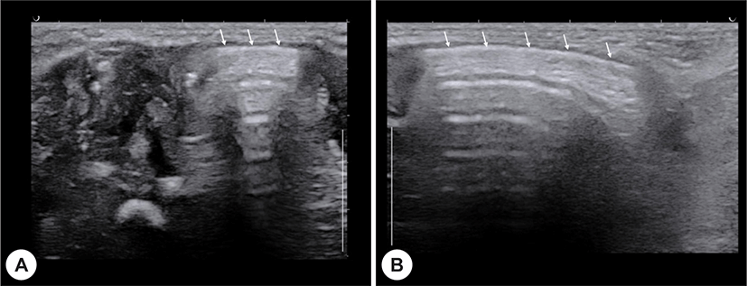
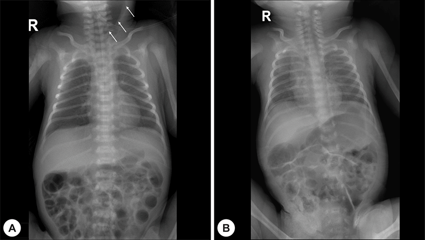
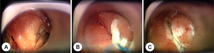
조기양막파수로 33주에 태어난 저체중(2,180 gram)의 미숙아가 입원 치료중 경증의 구토와 빈호흡을 보였다. 경과 관찰 중 환아의 빈호흡과 전신 상태는 호전되었으나, 신체 검사상 좌측 경부 종괴가 출생 8일경에 발견되었고, 종괴 부위에 누공이나 피부의 변화는 없었다. 종괴를 확인하기 위하여 경부 초음파를 시행하였다. 좌측 경부의 종괴는 공기가 찬 고에코의 낭종으로 초음파에서 확인되었다(Fig. 1). 인판토그램(infantogram)들을 검토하였을 때 좌측 경부에, 출생 직후에는 보이지 않던, 저음영의 공기가 차 있는 종괴가 커져 있는 소견을 발견하였다(Fig. 2A). 종괴의 정확한 위치와 주변 구조물과의 관계를 확인하기 위해서 경부 자기공명영상을 시행하였고, 2.8 cm 크기의 저신호강도의 낭종이 좌측 경부의 외측으로 위치하고 있으며, 작은 공기액체층(air-fluid level)을 동반하고 있었다. 수평면 T2 강조영상에서 이 낭종의 하부 끝단이 좌측 이상와에 연결이 의심되는 소견을 보였다(Fig. 3). 이상와 누공이 확인하고, 식도와 연결을 가지는 낭종을 감별하기 위해서 식도 조영술을 시행하였다. 식도 조영술상에 이상와 누공은 확인되지 않았으며, 식도와 연결되는 소견도 보이지 않았다. 소아 기관지 내시경상에서 좌측 이상와 누공이 의심되어서 이비인후과에 후두경하 화학적 소작술이 의뢰되었다. 수술장 후두경에서 좌측 이상와의 내측면 상부에 누공이 확인되었고, 25% trichloroacetic acid(TCA)를 이용한 화학적 소작술을 시행하였다. 치료후 좌측 이상와 누공은 폐쇄되었고, 좌측 경부 종괴도 점차 감소하였다(Fig. 4). 수술후 4주에 시행한 경부 초음파 상에 고에코의 공기가 찬 낭종의 크기는 감소하였고, 술후 6주에 시행한 인판토그램(infantogram)에서 더 이상 저음영의 공기가 찬 낭종은 보이지 않았다(Fig. 2B). 환아는 수술후 외래에서 시행한 신체검사상 종괴의 소실이 확인되었고, 특이한 증상의 발현없이 경과를 관찰하고 있다.

jcohns-31-2-276-g1
Fig. 1. Axial (A, B) high-resolution US images, performed with a linear 12 MHz transducer, show an air-filled, hyperechoic cyst (arrows) in left neck.
Download Original Figure
jcohns-31-2-276-g2
Fig. 2. Preoperative infantogram (A) reveals the low-attenuated left neck mass (arrows) on 7 days after birth and postoperative infantogram (B) shows complete resolution of air-filled left neck mass on 6 weeks after cauterization with 25% TCA.
Download Original Figure
jcohns-31-2-276-g3
Fig. 3. The coronal enhanced T1-weighted (A) and T2-weighted (B) MR images show a 2.8 cm sized, well-defined mass lesion with low signal intensity at left neck. Axial T2-weighted images at the level of pyriform sinus demonstrate round mass with small air-fluid level (C) and prominent left pyriform sinus with high signal intensity (C, D).
Download Original Figure
jcohns-31-2-276-g4
Fig. 4. Direct Laryngoscopy. (A) Intraoperative view of fistula opening at the lateral aspect of left pyriform sinus (arrow). Asterisk indicates left vocal cords. (B) It shows cottonoid pledget soaked in 25% TCA on the opening of left pyriform sinus fistula. (C) Postoperative view reveals ablated opening of fistula after cauterization with 25% TCA.
Download Original Figure

고찰

이상와 누공을 가지는 새열기형은 제3, 4새열기형으로 그 빈도가 극히 드물어 전체 새열기형의 1% 미만을 차지하는 것으로 알려져 있다. 따라서 우리 증례처럼 대부분 증례형식으로만 보고되고 있다.7-9) 제3, 4새열기형은 Liston의 분류에 따르면 인두 내 개구부가 이상와의 상부에 위치한 경우를 제3새열기형으로, 그리고 이상와의 첨부(apex)에 위치한 경우를 제4새열기형으로 구분한다.10,11) 빈도는 제3새열기형이 2.5%, 제4새열기형은 6.7%로 보고되고 있다.12) 본 증례는 후구경에서 죄측 이상와의 내측면 상부에 누공이 위치하는 소견을 보여 제3새열기형에 해당된다.

이상와 누공과 관련된 첫 증상은 거의 모든 환자에서 경부 종창을 동반한 갑상선 주위의 농양이 주된 증상이고, 평균 11.9세에 첫 증상이 나타났다. 갑작스러운 경부 종창으로 인해 심한 호흡곤란이 나타날 수 있는데, 이는 신생아에서 발생하였다.12-14) 제3새열기형이 평균 5세, 제4새열기형은 9세에 흔히 증상이 발현한다고 보고되고 있다.13,14)

이상와 누공의 진단을 위해서는 바륨 식도 조영술이나 후두경 검사에서 이상와의 누공을 확인하는 것이 필요하다. 하지만 식도 조영술을 통한 누공의 확인은 감염이 있는 경우 염증이나 부종에 의해 동로(fistula tract)가 막히거나, 경부 농양의 농이 이상와 누공쪽으로 역류되어 제대로 조영되지 않는 경우가 흔하기 때문에 실제로 임상적으로 경부 염증이 있는 환자에게 확진을 위한 검사로 사용하기는 힘들 것으로 생각된다. 기존 문헌들의 보고에서도 식도 조영술의 민감도는 50%에서 80%까지 다양하게 보고되고 있다.6,15-17) 또한 본 증례와 같이 이상와 누공이 주로 소아에서 발견된다는 점에서 환자의 적극적인 협조가 필요한 식도 조영술로 진단하는 것이 어려울 것으로 생각된다. 따라서 전산화단층촬영 검사에서 이상와 누공으로 인한 경부 염증의 가능성이 의심될 때에는 전신마취 하 후두경 검사를 통해 기저 새열기형의 존재 여부를 확인하는 것이 꼭 필요할 것이다.

신생아와 유아에서 목의 종괴나 천명 등이 있을 때, 진단을 위한 검사의 순서는 환자의 임상 상태에 따라 결정된다. 특히 심각한 기도 장애 증상이 있으면, 굴절성 섬유기관지경 검사를 첫 번째로 필요로 한다. 만약 종괴가 의심되면, 단순 방사선 촬영과 초음파가 진단에 도움이 된다.

경부 방사선 촬영에서 이상와 누공과 연관된 제3새열기형은 경부의 외측으로 돌출된 종괴로 보일 수 있다. 일부에서는 공기, 공기액체층, 그리고 액체로 채워진 종괴로 발견된다. 초음파는 쉽게 시행할 수 있고, 고해상도 탐촉자를 사용하면 목부위의 연부조직과 근육의 관찰에 유용하다.18,19) 초음파는 낭종의 위치 확인과 다른 연조직 종괴와 감별에 도움이 된다.20) 저자의 증례에서도 초음파를 사용하였고, 낭종의 위치와 다른 연조직 종괴와 감별을 할 수 있었다. Takahiro 등21)에 따르면 초음파에서 크기가 작고, 타원형의 낭종에서는 누공이 확인되나, 크기가 크거나, 염증 소견이 있는 경우, 공기가 찬 낭종일 때는 누공의 확인이 어렵다. 본 증례도 공기가 찬 낭종으로 초음파에서 누공의 확인이 되지 않았다.

추가적인 검사로는 신생아에서 환자와 비슷한 증상을 보일 수 있는 혈관기형 또는 전장중복낭종(foregut duplication cyst), 후두낭(laryngocele)등 다른 기형 질환을 배제하기 위해 전산화단층촬영이나 자기공명영상이 필요하다.15) 제3, 4새열기형과 연관된 이상와 누공의 확진은 기관지 내시경이나 후두경 검사로 할 수 있다. 본 증례에서도 기관지 내시경이나 후두경 검사로 이상와 누공이 확인되었다. 하지만 Noriko 등22)의 보고에 따르면 감염에 의해 크기가 커져서 호흡 장애의 증상을 보이는 경우, 후두경 검사로 이상와 누공의 확인이 어려웠고, 수술시에 누공을 확인할 수 있었다.

이상와 누공의 치료에는 외부 수술적 절제와 TCA를 이용한 내시경적 화학적 소작술이 있다. 누공 경로의 절제술은 그에 따르는 광범위절개와 반회후두신경 손상 등의 위험요소를 고려하면, TCA를 이용한 내시경적 화학적 소작술은 누공 입구의 폐쇄를 도모하는 매우 안전하고 효과적인 치료방법으로 사료된다.5,12-14) 저자의 증례에서는 TCA를 이용한 내시경적 화학적 소작술을 시행하였고, 치료한 후 다른 합병증은 보이지 않았다. 그러나 Noriko 등22)의 보고와 같이 감염에 의해 낭종이 커져서 호흡 장애의 소견을 보이고, 후두경 검사로 이상와 누공의 확인이 되지 않는 경우에는 수술적 절제술을 고려하여야 하겠다.

결론적으로 이상와 누공과 연관된 제3새열기형은 매우 드물지만, 목 외측의 종괴가 있을 경우, 수유 곤란과 호흡곤란 같은 상기도 폐쇄 증상이 없더라도 감별진단에 포함시켜야 된다. 초음파가 이상와 누공과 연관된 제3새열기형의 진단에 도움이 되며, 병변의 범위와 감별진단과 같은 추가적인 수술전 정보가 필요시에는 전산화단층촬영이나 자기공명영상이 도움이 된다. 신생아에서 치료는 수술보다는 TCA를 이용한 내시경적 화학적 소작술이 추천된다.

REFERENCES

1).

Wilson DB. Embryonic development of the head and neck: part 2, the branchial region. Head Neck Surg 1979;2(1):59-66.

2).

Agaton-Bonilla FC, Gay-Escoda C. Diagnosis and treatment of branchial cleft cysts and fistulae. A retrospective study of 183 patients. Int J Oral Maxillofac Surg 1996;25(6):449-52.

3).

Doi O, Hutson JM, Myers NA, McKelvie PA. Branchial remnants: a review of 58 cases. J Pediatr Surg 1988;23(9):789-92.

4).

Ford GR, Balakrishnan A, Evans JN, Bailey CM. Branchial cleft and pouch anomalies. J Laryngol Otol 1992;106(2):137-43.

5).

Kim KH, Sung MW, Koh TY, Oh SH, Kim IS. Pyriform sinus fistula: management with chemocauterization of the internal opening. Ann Otol Rhinol Laryngol 2000;109(5):452-6.

6).

Kim KH, Sung MW, Lee KJ, Roh JL, Kwon TK, Kim IS, et al. Management of pyriform sinus fistula with chemocauterization. Korean J Otolaryngol-Head Neck Surg 2002;45(9):906-10.

7).

Drumm AJ, Chow JM. Congenital neck masses. Am Fam Physician 1989;39(1):159-63.

8).

Finn DG, Buchalter IH, Sarti E, Romo T, Chodosh P. First branchial cleft cysts: clinical update. Laryngoscope 1987;97(2):136-40.

9).

Nicollas R, Ducroz V, Garabédian EN, Triglia JM. Fourth branchial pouch anomalies: a study of six cases and review of the literature. Int J Pediatr Otorhinolaryngol 1998;44(1):5-10.

10).

Liston SL. Fourth branchial fistula. Otolaryngol Head Neck Surg 1981;89(4):520-2.

11).

Liston SL, Siegel LG. Branchial cysts, sinuses, and fistulas. Ear Nose Throat J 1979;58(12):504-9.

12).

Seok JH, Ahn DB, Sohn JH, Choi JH, Jung YY, et al. Pyriform sinus fistula: a single center experience. Korean J Otorhinolaryngol-Head Neck Surg 2013;56(3):154-8.

13).

Nicoucar K, Giger R, Jaecklin T, Pope HG Jr, Dulguerov P. Management of congenital third branchial arch anomalies: a systematic review. Otolaryngol Head Neck Surg 2010;142(1):21-8.e2.

14).

Nicoucar K, Giger R, Pope HG Jr, Jaecklin T, Dulguerov P. Management of congenital fourth branchial arch anomalies: a review and analysis of published cases. J Pediatr Surg 2009;44(7):1432-9.

15).

Park SW, Han MH, Sung MH, Kim IO, Kim KH, Chang KH, et al. Neck infection associated with pyriform sinus fistula: imaging findings. AJNR Am J Neuroradiol 2000;21(5):817-22.

16).

Yang C, Cohen J, Everts E, Smith J, Caro J, Andersen P. Fourth branchial arch sinus: clinical presentation, diagnostic workup, and surgical treatment. Laryngoscope 1999;109(3):442-6.

17).

Pereira KD, Losh GG, Oliver D, Poole MD. Management of anomalies of the third and fourth branchial pouches. Int J Pediatr Otorhinolaryngol 2004;68(1):43-50.

18).

Garel C, Legrand I, Elmaleh M, Contencin P, Hassan M. Laryngeal ultrasonography in infants and children: anatomical correlation with fetal preparations. Pediatr Radiol 1990;20(4):241-4.

19).

Garel C, Hassan M, Legrand I, Elmaleh M, NarcyP. Laryngeal ultrasonography in infants and children: pathological findings. Pediatr Radiol 1991;21(3):164-7.

20).

Imhof H, Czerny C, Hörmann M, Krestan C. Tumors and tumor-like lesions of the neck: from childhood to adult. Eur Radiol 2004;14:L155-65.

21).

Hosokawa T, Yamada Y, Sato Y, Tanami Y, Amano H, Fujiogi M, et al. Five neonatal causes of pyriform sinus fistula with cervical cystic lesion: a comparison between sonography and other modalities. J Med Ultrason (2001) 2015;42(4):579-85.

22).

Hamaguchi N, Ishinaga H, Chiyonobu K, Morishita H, Takeuchi K. A case of Pyriform sinus fistula with respiratory distress in the neonatal period. Case Rep Otolaryngol 2018; 2018:1696875.