Journal of Clinical Otolaryngology Head and Neck Surgery
The Busan, Ulsan, Gyeoungnam Branch of Korean Society of Otolaryngology-Head and Neck Surgery
증례

전경부에 발생한 성숙 기형종 증례

김연지1, 임일계1, 조의주2, 한성준1,*
Yeon Ji Kim1, Il-Gye Lim1, Uiju Cho2, Sungjun Han1,*
1가톨릭대학교 의과대학 성빈센트병원 이비인후과학교실
2병리과학교실
1Department of Otolaryngology-Head and Neck Surgery, The Catholic University of Korea, Seoul, Korea
2Pathology, St. Vincent’s Hospital, College of Medicine, The Catholic University of Korea, Seoul, Korea
*교신저자: 한성준, 16247, 경기도 수원시 팔달구 중부대로 93, 가톨릭대학교 의과대학 성빈센트병원 이비인후과학교실 전화: (031) 881-8887·전송: (031) 257-3752·E-mail: ithilim@naver.com

© Copyright 2020 The Busan, Ulsan, Gyeoungnam Branch of Korean Society of Otolaryngology-Head and Neck Surgery. This is an Open-Access article distributed under the terms of the Creative Commons Attribution Non-Commercial License (http://creativecommons.org/licenses/by-nc/4.0/) which permits unrestricted non-commercial use, distribution, and reproduction in any medium, provided the original work is properly cited.

Received: May 25, 2020; Revised: Jun 08, 2020; Accepted: Jul 17, 2020

Published Online: Dec 31, 2020

ABSTRACT

A teratoma is a tumor which has cells derived from all three germ layers (endoderm, mesoderm, ectoderm). Frequent sites of teratoma are sacrococcyx and gonads, and cervical teratoma is relatively rare. The ninety percent of its cases occurs in children. Cervical teratomas are typically located in the midline of neck, extending to mediastinum. Here we report a case of cervical mature teratoma in 21-year old male which is successfully removed by surgery, with the review of literature. (J Clinical Otolaryngol 2020;31:257-262)

Keywords: 기형종; 선천성; 경부
Keywords: Teratoma; Neck; Congenital

서론

기형종은 3가지의 생식세포층(ectoderm, mesoderm, endoderm)이 모두 발생하는 종양으로 주로 난소, 고환, 꼬리뼈부위(sacrococcyx)에 호발한다.1) 그 중 경부의 기형종은 전체 기형종의 3~5%를 차지하며,2) 소아와 성인을 모두 포함해서 그 유병률은 20,000~40,000명 중 1명으로 알려져 있다.3) 경부 기형종은 90%에서 소아에서 발견되며, 성인에서 발견되는 경우는 비교적 드물다. 경부의 위치에 따라 대부분은 정중선에서 종격동으로 확장된 형태로 나타나고, 측경부는 비교적 드물게 나타난다.4-6) 경부 기형종은 영상학적으로 낭성과 고형이 비균일성적으로 섞인 종괴로 나타나며, 낭성 부분이 많은 기형종의 경우 낭림프관종 등의 림프관 기형, 갑상선 낭종, 아가미낭종 등의 질환과 감별이 필요하다. 저자는 21세의 젊은 남성에서 경부에서 발견된 성숙 기형종의 증례를 경험하여 보고하는 바이다.

증례

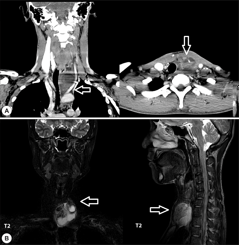
21세 남자 환자가 전경부의 종괴를 주소로 내원했다. 환자는 종괴가 5일 사이에 갑자기 커졌다고 기술했으며, 음성변화나 호흡곤란 등의 다른 증상은 호소하지 않았다. 촉진 시 전경부 중앙의 갑상선 부위에 3 cm 가량의 튀어나온 종괴가 만져졌으며, 부드러웠고 고정되어 있지 않았다. 피부 침범 소견 및 압통은 없었으며, 주변 경부 림프절의 비대는 촉지되지 않았다. 전경부 종괴에 대한 영상학적 검사와 세침흡인검사를 시행한 결과, 세침흡인검사에서는 탁한 액체가 흡인되었고, 슬라이드 도말검사에서 몇 개의 특징 없는 편평상피세포와 조직구만 관찰되었다. 경부 컴퓨터단층촬영검사에서 갑상선의 좌측 하방에 6.8 cm 크기의 미세 석회화와 지방조직을 동반한, 비균일적으로 낭성 부분과 고형 부분이 혼합된 종괴가 보였다. 경부 자기공명영상에서도 마찬가지로 비슷한 크기 (5 × 4.5 × 6 cm)의 비균일적이고 다중격성으로 낭성과 고형이 혼합된 종괴를 보였으며, 종괴로 인해 기관지는 우측으로 밀려 있었다. 다른 비정상적인 경부림프절 비대는 관찰되지 않았다(Fig. 1).

jcohns-31-2-257-g1
Fig. 1. Radiologic evaluation. A : Coronal and axial images of computed tomography show heterogenous cystic and solid mass at the left inferolateral aspect of thyroid gland. (arrows) Tracheal deviation is also shown. B : Coronal and sagittal T2 MRI images show heterogenous multiseptated cystic and solid mass with high signal intensity at anterior neck. (arrows).
Download Original Figure

영상학적 검사 결과를 바탕으로 고려하였을 때 경부 기형종이 의심되어 전신마취 하 경부 종괴의 제거를 계획했다. 종괴는 피대근 밑에서 노출되었으며, 위치는 좌측 갑상선엽과 흉선 사이에 위치하고 있었다. 갑상선 및 주변조직과의 유착은 없었으며, 손가락으로 무딘 박리를 이용해 갑상선과 주변 조직으로부터 쉽게 제거가 되었다(8.3 × 6 × 3 cm).

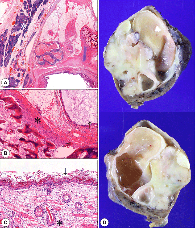
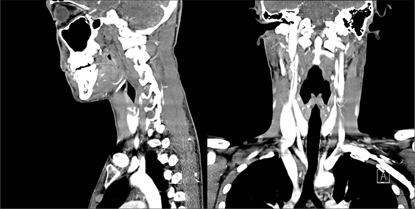
검체의 육안 검사상 조직의 단면은 부드러운 미색의 낭성 조직과 고형성 절편이 섞여 있었다. 낭성 부분은 털 및 치즈같은 피지 물질을 일부 포함하고 있었다. 현미경적 소견상 종괴의 가장자리는 흉선 조직으로 둘러싸여 있었고, 내부에는 연골, 뼈, 췌장조직, 피부, 장 점막, 지방 조직 등으로 구성되어 있었고, 그 외 미분화된 조직은 보이지 않았다(Fig. 2). 환자는 특별한 합병증 없이 수술 후 3일째에 퇴원하였다. 이후 수술 후 3개월째에 촬영한 컴퓨터 단층촬영에서 재발의 증거는 없었고, 기관지 편위는 정상으로 회복되었다. 수술 후 10개월간 재발 소견은 발견되지 않았다(Fig. 3).

jcohns-31-2-257-g2
Fig. 2. Microscopic (H&E) and gross pictures of specimen. A : Thymus tissue is seen on the left (white asterisk), and structures derived from other germ cell layers such as adipose tissue, intestinal mucosa, cartilage, and bone are observed on the right. (x8) B : Bone tissue (asterisk) and respiratory mucosa (arrow) are seen but there is no immature or malignant-differentiating portion. (x50) C : Some part of cystic portion is composed of skin tissue (asterisk) including skin appendages like epidermis and follicles (arrow) (x100) D : Gross view of specimen. The encapsulated mass is a mixture of cystic and solid portions. The cystic part contains hair and cheese-like sebum material.
Download Original Figure
jcohns-31-2-257-g3
Fig. 3. Postoperative evaluation. Sagittal (left) and coronal (right) images of computed tomography show no evidence of recurrence of tumor. Tracheal deviation is corrected.
Download Original Figure

고찰

기형종은 3가지의 생식세포층(ectoderm, mesoderm, endoderm)이 모두 발생하는 종양으로 조직내 신경상피 같은 미성숙 부분의 비율에 따라 성숙기형종과 미성숙기형종으로 분류된다.7) 미성숙 기형종이 항상 악성인 것은 아니지만 선천성 경부 기형종의 악성 비율은 5% 이내로 추정되며, 진단 시의 나이가 많을수록 악성의 확률이 증가한다. 1,7-9)

두경부 영역에서 기형종은 비교적 드물게 발생하며, 그 세부 부위는 비인두, 안구, 구인두, 전경부 등 다양하다. 발생 빈도에 있어 성별 간에 큰 차이를 보이지는 않는다.10) 본 증례에서는 해당되지 않지만, 전경부에서 발견된 경우 많은 기형종이 갑상선에서 유래하며, 갑상선의 한 엽(lobe)이 없거나 종괴가 밀고 있다면 그것은 기형종을 시사하는 소견이다.11) 만약에 수술 시 기형종이 갑상선에서 쉽게 박리되지 않는다면 갑상선엽절제술도 고려해야 한다.11)

대부분의 경부 기형종은 소아에서 많이 발견되며, 산전 영상검사의 발달로 출생 전후에 진단된다. 하지만 성인의 경부 기형종은 다른 증상 없이 대부분 만져지는 종괴로 병원에 내원한다.

경부의 기형종은 목의 정중부 또는 종격동으로 확장된 형태가 일반적이다. 초음파에서는 석회화를 동반한 비균일적으로 낭성과 고형이 혼합된 종괴로 보이고, 자기공명영상에서 전형적인 모습은 경계가 좋은 고형 부분 혹은 단방이나 다방성의 낭성 부분을 둘 다 가진 종괴의 모습이다.12) 석회화가 있으면 진단에 도움이 되지만, 없다고 해서 기형종의 진단을 배제할 수는 없다.

악성 기형종에서 종양세포가 융모상피암(choriocarcinoma)이나 난황난종양(yolk sac tumor)의 조직을 가지고 있다면 혈액검사에서 알파 태아단백질(alpha-fetoprotein, αFP)나 베타-인간 융모성 성선자극호르몬(beta-human chorionic gonadotrophin, β-hCG)의 혈중 수치가 증가할 수 있다. 하지만 αFP의 경우 기형종의 악성전환의 지표로 측정하기에는 민감도가 낮다.13)

치료는 수술적 제거가 원칙이다. 수술 후 양성인 경우 예후는 좋으며 재발은 드물다.14) 재발은 드물지만 불완전하게 절제가 된 경우 재발할 수 있다.7)

태아나 신생아에서 발견된 성숙 혹은 미성숙 기형종에 관한 증례들이 국내외 학회지에 여럿 보고되어 있다. 하지만 본 증례와 같이 청소년기 이후의 성인에서 발생한 양성 기형종은 증례가 그에 비해 적은 수가 보고되어 있다.15-17)

국내에 발표된, 젊은 연령에서 발견된 성숙 기형종의 증례들에서 종괴의 크기는 4.7~12.0 cm의 비교적 직경으로 크고 다양했으며, 갑상선의 종괴를 주소로 내원한 경우도 있었지만, 크기가 큰 경우 종격동으로 확장되어 흉부단순촬영 이상으로 내원한 경우도 있었다. 큰 크기로 인해 영상학적 평가에서 대부분 총경동맥 등의 대혈관이나 기도의 전위를 보였다. 본 증례에서는 실시하지 않았지만 αFP과 β-hCG의 혈중 수치를 수술 전 검사에서 시행한 경우 모든 증례에서 정상범위 안이었다. 종양의 크기와 위치에 따라 수술적 접근은 경경부(transcervical) 접근, 중위 흉골절개술, 후방 외측 흉벽절개술 등 다양했다. 또한 대부분의 증례에서 종괴와 주변 조직 사이 유착은 없었으며, 쉽게 박리되었다. 수술 후 예후는 모두 재발 없이 양호하였다.

결론

성인에서 발생하는 경부 기형종은 드물지만 술전 영상학적 평가를 통해 의심할 수 있으며, 악성이 아니라면 주변 조직과의 유착이 적기 때문에 크기가 크더라도 쉽게 수술로 절제가 가능하다. 저자들은 성인에서 짧은 시기에 커져 발현한 성숙 기형종을 수술로 치료한 증례를 경험하였기에 문헌 고찰과 함께 보고하는 바이다.

REFERENCES

1).

Byard RW, Jimenez CL, Carpenter BF, Smith CR. Congenital teratomas of the neck and nasopharynx: a clinical and pathological study of 18 cases. J Paediatr Child Health 1990; 26(1):12-6.

2).

Jordan RB, Gauderer MW. Cervical teratomas: an analysis. Literature review and proposed classification. J Pediatr Surg 1988;23(6):583-91.

3).

Berge SJ, von Lindern JJ, Appel T, Braumann B, Niederhagen B. Diagnosis and management of cervical teratomas. Br J Oral Maxillofac Surg 2004;42(1):41-5.

4).

Shah A, Latoo S, Ahmed I, Malik AH. Head and neck teratomas. J Maxillofac Oral Surg 2009;8(1):60-3.

5).

Gorur K, Talas DU, Ozcan C. An unusual presentation of neck dermoid cyst. Eur Arch Otorhinolaryngol 2005;262 (4):353-5.

6).

Alimehmeti M, Alimehmeti R, Ikonomi M, Saraci M, Petrela M. Cystic benign teratoma of the neck in adult. World J Clin Cases 2013;1(6):202-4.

7).

Carr MM, Thorner P, Phillips JH. Congenital teratomas of the head and neck. J Otolaryngol 1997;26(4):246-52.

8).

Abemayor E, Newman A, Bergstrom L, Dudley J, Magidson JG, Ljung BM. Teratomas of the head and neck in childhood. Laryngoscope 1984;94(11 Pt 1):1489-92.

9).

Batsakis JG, el-Naggar AK, Luna MA. Teratomas of the head and neck with emphasis on malignancy. Ann Otol Rhinol Laryngol 1995;104(6):496-500.

10).

Wolter NE, Siegele B, Cunningham MJ. Cystic cervical teratoma: a diagnostic and management challenge. Int J Pediatr Otorhinolaryngol 2017;95:97-100.

11).

Riedlinger WFJ, Lack EE, Robson CD, Rahbar R, Nose V. Primary thyroid teratomas in children: a report of 11 cases with a proposal of criteria for their diagnosis. Am J Surg Pathol 2005;29(5):700-6.

12).

Robson CD. Cysts and tumors of the oral cavity, oropharynx, and nasopharynx in children. Neuroimaging Clin N Am 2003;13(3):427-42, ix.

13).

Touran T, Applebaum H, Frost DB, Richardson R, Taber P, Rowland J. Congenital metastatic cervical teratoma: diagnostic and management considerations. J Pediatr Surg 1989;24(1):21-3.

14).

Alexander VR, Manjaly JG, Pepper CM, Ifeacho SN, Hewitt RJ, Hartley BE. Head and neck teratomas in children--a series of 23 cases at Great Ormond Street Hospital. Int J Pediatr Otorhinolaryngol 2015;79(12):2008-14.

15).

Lee SH, Hwangbo C, Kang YC, Shon KR. A case of cervical teratoma. Korean Journal of Otorhinolaryngology-Head and Neck Surgery 1996;39(5):892-6.

16).

Cho SW, Chee HK, Ann HS, Shin YC, Nam ES. Rapidly grown huge mediastinal benign teratoma; one case report. Korean Thorac Cardiovasc Surg 2000;33(6):521-4.

17).

Yoon JH, Nam KH, Jang HS, Park CS. 갑상선 종괴로 의심되었던 전종격동 성숙 낭성 기형종. Korean J Endocr Surg 2004;4(2):127-9.