Journal of Clinical Otolaryngology Head and Neck Surgery
The Busan, Ulsan, Gyeoungnam Branch of Korean Society of Otolaryngology-Head and Neck Surgery
증례

TnR Nasal Mesh®를 이용한 내시경적 안와내벽외향골절 정복술 2예

박근우1, 배교한1, 노웅재1, 정태영1,*
Geun Woo Park1, Gyo Han Bae1, Woong Jae Noh1, Tae Young Jung1,*
1메리놀병원 이비인후과
1Department of Otorhinolaryngology Head and Neck Surgery, Maryknoll Hospital, Busan, Korea
*교신저자: 정태영, 48972 부산광역시 중구 중구로 121, 메리놀병원 이비인후과 전화: (051) 461-2692·전송: (051) 462-9419·E-mail: handsjung@naver.com

© Copyright 2020 The Busan, Ulsan, Gyeoungnam Branch of Korean Society of Otolaryngology-Head and Neck Surgery. This is an Open-Access article distributed under the terms of the Creative Commons Attribution Non-Commercial License (http://creativecommons.org/licenses/by-nc/4.0/) which permits unrestricted non-commercial use, distribution, and reproduction in any medium, provided the original work is properly cited.

Published Online: Dec 31, 2020

ABSTRACT

Orbital blow out fractures can cause functional and cosmetic problems by prolapse or entrapment of the orbital tissue. Surgical intervention is performed to prevent limitation of gaze, diplopia and cosmetic disturbance. Implant material is used to support prolapsed orbital tissue. Implant material should be selected in consideration of multiple conditions. In this study the experiences with a new surgical material for reduction of orbital blow out fracture are presented. This report suggests TNR Nasal Mesh® can be new option for reduction of orbital blow out fracture. (J Clinical Otolaryngol 2020;31:243-247)

Keywords: 안와골절; 내시경
Keywords: Blow out fracture; Absorbable implants

서론

안와 외향 골절(Orbital blow out fracture)은 산업화, 교통 수단의 증가 및 고속화, 폭력 사고의 증가 등으로 인해 발생이 증가하고 있으며, 기능적, 미용적 문제가 발생할 수 있어 임상적 중요성이 강조되고 있다.1,2)

안와 외향 골절 정복술은 1991년 Yamaguchi 등3)이 내시경을 이용한 비내 정복술을 보고한 이후 이비인후과 영역에서 상기 접근법이 확대되고 있다. 비내시경적 정복술 시 사용하는 안와 삽입물은 골절 부위의 결손을 막는 동시에 안와 내용물의 탈출 억제를 통해 안와 기능 및 해부학적 구조 회복에 중요한 역할을 한다. 안와 삽입물은 크게 자가삽입물과 인공삽입물로 나눌 수 있으며, 인공삽입물은 다시 비흡수성, 흡수성으로 나눌 수 있다. 그 중 흡수성 삽입물의 경우, gelatin film, polyglycolic acid membrane, polydioxanone, poly L-/DL-lactide 70/30 implant4) 등 다양한 물질들이 사용되고 있다.

TnR Nasal Mesh®는 polycaprolactone(PCL)으로 구성된 다공성의 흡수성 인공삽입물이다. 이물 반응이 적고 분해 속도가 느려 골 치유 시기 동안 신체 구조물을 충분히 지지할 수 있는 장점과 안전성5)은 현재 사용되는 poly L-/DL-lactide 70/30 implant와 유사할 뿐만 아니라, 다른 제품에 비해 유연성이 좋으며, 재단이 쉬워 술자가 다루기 편리한 이점이 있어 비재건술에 사용하기 적합하며, 현재 비중격 천공 및 외비성형술에 널리 사용되고 있다.

이에 본 저자는 TnR Nasal Mesh®를 사용하여 안와 내벽 골절을 성공적으로 치유한 사례가 있어 이를 보고하려 한다.

증례

증례 1

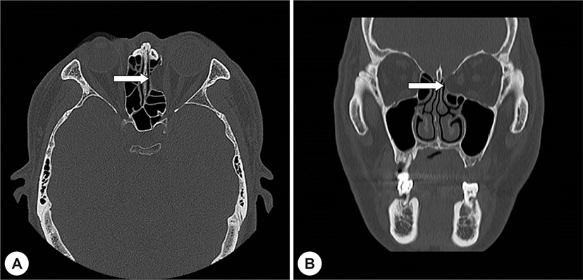
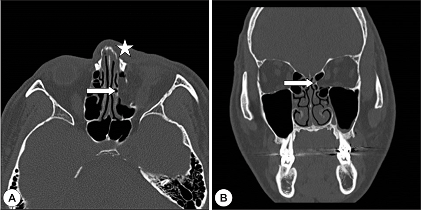
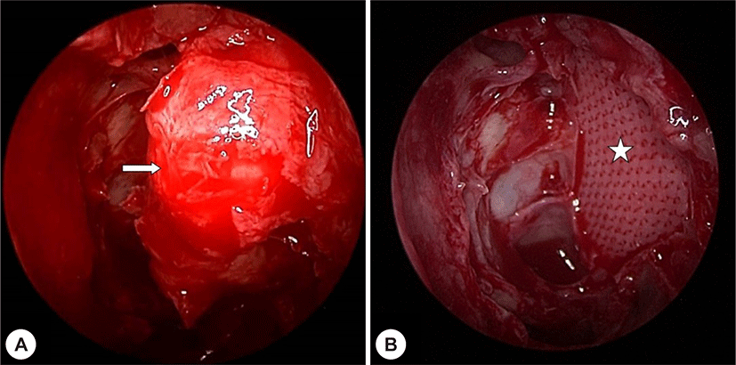
48세 남자 환자로, 8일 전에 튀어오른 에어 호스에 좌측 코와 내안각 부위를 맞아 타병원에서 전원되었다. 안와컴퓨터단층촬영 상 좌측 약 2 cm2 크기의 광범위한 안와 내벽 골절 및 비골 골절이 확인되었으나(Fig. 1), 안과적 검사에서 안구 함몰, 시야 이상, 안구 움직임 제한, 복시 등의 이상 소견은 없었다. 전신마취 하에 비관혈적 비골골절정복술을 먼저 시행하고, 비내시경을 이용하여 안와 내벽 골절 정복술을 시행하였다. 내시경으로 경상돌기, 사골포 등을 조심스럽게 제거하여 골절된 지판을 노출시킨 후, 탈출한 안와 조직을 정복하였으며, 골절의 크기를 재어 실제 골절 크기보다 크게 TnR Nasal Mesh®를 재단하여 골절부위에 속넣기 이식으로 고정하였다. 그리고 사골동에 반절개를 가한 inverted U형의 1 mm 두께를 가진 실라스틱 시트를 삽입 후 merocel®을 충전시킨 뒤 수술을 마쳤다(Fig. 2). 수술 후 1개월간 안와 조직의 탈출 없이 잘 유지되고 있으며(Fig. 3), 미용적, 안과적 이상 및 불편감 호소 없이 경과 관찰 중에 있다.
jcohns-31-2-243-g1
Fig. 1. Preoperative computed tomography findings. Axial (A) and coronal (B) image showed Lt. med. orbital wall fracture(arrow) & nasal bone fracture(star).
Download Original Figure
jcohns-31-2-243-g2
Fig. 2. Intraoperative finding. Intraorbital tissue (arrow) is herniated from original site (A). TnR Nasal Mesh® (star) is used for reduction of med. orbital wall fracture (B).
Download Original Figure
jcohns-31-2-243-g3
Fig. 3. Postoperative finding. TnR Nasal Mesh® insertion site (star) is covered with nasal mucosa without herniation of intraorbital tissue 1month after surgery.
Download Original Figure
증례 2

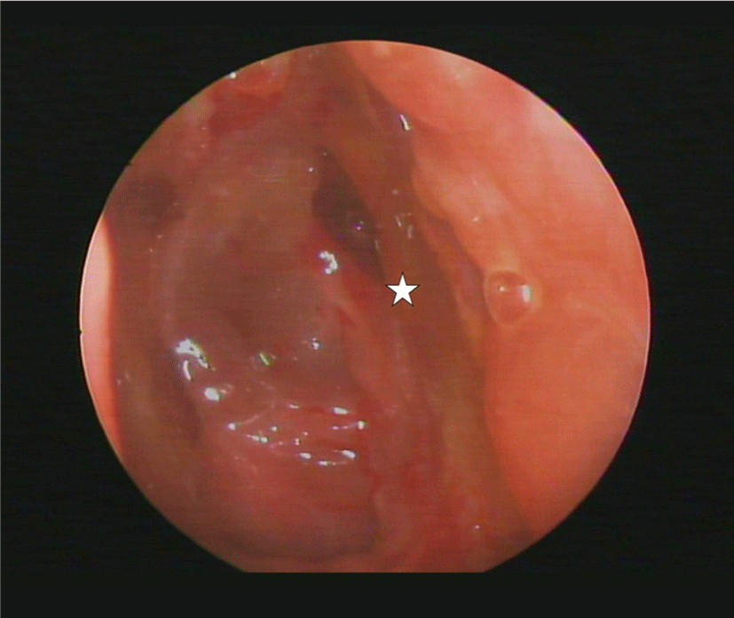
58세 남자 환자로, 7일 전 택시 조수석에서 사고가 나면서 좌측 안와부위를 부딪혀 내원하였다. 특이 내과적 과거력은 없었으며, 내원 당시 안구 함몰, 시야, 안구 움직임 제한, 복시 등 안과적 이상은 없었다. 외래에서 시행한 부비동컴퓨터단층좔영 상 약 2 cm2 크기의 광범위한 안와 내벽 골절이 확인되어 수술적 치료를 결정하였다(Fig. 4). 전신마취 하에 증례1의 경우와 같은 방법으로 비내시경적 정복술을 시행하였으나, 증례1의 경우와 다르게 안와 내벽 삽입물로 poly L-/DL-lactide 70/30 plate를 사용하였고, 이는 70℃ 이상의 온도에서 휘어지는 특성이 있기 때문에 가열된 생리 식염수에 재단된 고정판을 1초간 담근 후 골절 부위의 형태에 맞게 변형시켜 속넣기 이식 형태로 고정하였다(Fig. 5A, B). 수술 후 25일째 실라스틱 시트를 제거하였고, biosorb®의 이탈이 확인되었다(Fig. 5C). 수술 후 32일째 안구 함몰이 지속적으로 보여 재수술을 시행하였다. 재수술에서는 흡수성 인공 삽입물인 TnR Nasal Mesh®를 사용하였으며, 불완전하게 정복된 안와 조직을 안구 내로 정복하였다(Fig. 5D). 수술 후 3개월간 미용적, 안과적 이상 및 불편감 호소 없이 경과 관찰 중에 있다.
jcohns-31-2-243-g4
Fig. 4. Preoperative computed tomography findings. Preoperative axial (A) and coronal (B) image showed Lt. med. orbital wall fracture (arrow).
Download Original Figure
jcohns-31-2-243-g5
Fig. 5. Intraoperative imaging of first reduction (A, B). Intraorbital tissue (arrow) is herniated from original site (A). Poly L-/DL-lactide 70/30 plate (star) is used for reduction of med. orbital wall fracture (B). Postoperative finding (C). Poly L-/ DL-lactide 70/30 plate (star) escaped from insertion site. Intraoperative imaging of second reduction (D). TnR Nasal Mesh® (arrowhead) is used for reduction of med. orbital wall fracture.
Download Original Figure

고찰

안와 외향 골절(Orbital blowout fracture)은 둔한 안면부 외상에 의한 충격이 안와내 연부조직으로 전달되어 안와 내압의 급격한 상승으로 안와 외벽에 골절이 발생하는 것을 말한다. 안구운동의 기계적 제한이 있는 경우, 복시가 2주 이내 사라지지 않거나, 새로 발생할 경우, 2 mm 이상의 안구 함몰이 있는 경우, 방사선학적으로 광범위한 골절이 보일 경우, White-eyed 안와 외향 골절이 있을 경우 정복술을 시행하게 되며, 향후 안구 함몰이나 안구하수가 예상될 경우 정복술을 고려해볼 수 있다.

기존에 보고된 방법은 poly L-/DL-lactide 70/30 plate를 이용하여 골절의 경계보다 2 mm 정도 여유를 두고 재단한 후 70℃ 이상의 생리식염수에 판을 담궈 모양을 만든 후 골절 부위에 삽입하는 방법6)이나, 본원에서는 0.5 mm 두께의 TnR Nasal Mesh® 를 사용하여 안와 내벽 골절부위를 지지하였고, 실라스틱 시트를 반전된 U자형으로 사골동 내 골절부위에 위치시키고, Merocel®을 이용하여 고정하였다.7)

안와 재건에 있어서 이상적인 삽입물은 안와 결손부의 크기와 위치, 삽입물을 지지할 남아 있는 구조물에 따라 선택하고, 안와의 굴곡, 부비동의 상태, 수술자와 환자의 기호에 따라서도 선택할 수 있다.8-10)

삽입물은 크게 자가 조직과 인공 삽입물로 나눌 수 있다. 자가 조직은 생체적합성이 가장 우수하나 양이 제한되어 있으며, 이식물의 재흡수, 공여부의 합병증 발생 등의 단점이 있다.11,12) 인공 삽입물은 다루기 쉽고 3차원적인 재건이 가능하며, 수술시간이 짧다는 장점이 있어 널리 이용되나, 가격이 비싸고 생체 적합성이 낮은 단점이 있다.9,13-14)

기존에 사용되던 poly L-/DL-lactide 70/30 implant의 경우, 골절 부위에 맞게 재단하기 위해서 열을 가해야 하는 점, 짧은 시간 동안 결손 부위를 고려하여 재단하여 삽입해야 한다는 점, 일단 재단한 후에는 다시 단단해져 수술 중 모양 변형을 위해서는 재단한 고정판에 다시 열을 가해 모양을 바꾸거나 새로 재단해야 한다는 점에 불편함이 있어 초심자가 사용하기에는 다소 어려움이 있었다.

TnR Nasal Mesh® 는 미세다공성 polycaprolactone(PCL)으로 구성된 인공 삽입물로, 3D 프린팅 기술을 통해 제조되어 다양한 크기와 모양을 만들 수 있다. 정복술 시 신체 조직으로 이식된 후 주변 조직에 정상적으로 존재하는 다양한 세포들이 지지체 내부로 이동하는 것을 허용하여, 콜라겐 등과 같은 세포 외 기질의 생성을 유도하여 손상된 조직의 재건에 기여한다. 유연성이 매우 좋아, 특정 모양을 만들기 위해 기존의 다른 흡수성 인공물질과는 달리 열성 가열이 필요 없으며, 결손 부위에 맞추어 재단 후, 골절 부위에 삽입을 위한 조작도 용이하여 초심자들도 쉽게 이용할 수 있는 장점이 있다.

한계점으로 TnR Nasal Mesh®의 완전 흡수에 약 2년의 기간이 소요되며, 완전한 치료 결과 확인을 위해서는 정복술 후 2년 뒤 환자의 상태 확인이 필요하다. 이에 대해서는 이후 추가적인 연구가 필요하겠다.

이에 저자들은 두 증례에서 특이 합병증 없이 안와 내벽 외향 골절 정복술을 성공적으로 시행하였기에 이를 보고하는 바이다.

REFERENCES

1).

Burm JS, Oh SJ. Direct local approach through a W-shaped incision in moderate or severe blowout fracture of the mdial orbital wall. Plast Reconstr Surg 2001;107(4):920-8.

2).

Ilnuma T, Hirota Y, Ishio K. Orbital wall fractures. Conventional views and CT. Rhinology 1994;32(2):81-3.

3).

Yamaguchi N, Arai S, Mitani H, Uchida Y. Endoscopic endonasal technique of the blowout fracture of the medial orbital wall Oper Tech Otolayngol Head Neck Surg 1991;2(4):269-74.

4).

Han JU, Im JS, Suk SH, Kim JY. The surgical results of isolated orbital blowout fractures using bioresrobable Poly L-/DL-Lactide 70/30 implant. Korean J Otorhinolaryngol-Head Neck Surg 2014;57(11):759-65.

5).

Park SH, Yun BG, Won JY, Yun WS, Shim JH, Lim MH, et al. New application of three-dimensional printing biomaterial in nasal reconstruction. Laryngoscope 2017;127(5):1036-43.

6).

We JW, Kim YH, Jung TY, Bae KB, Cho JH, Kwon JH. Modified techinique for endoscopic endonasal reduction of medial orbital wall fracture using a resorbable panel. Ophthal Plast Reconstr Surg 2009;25(4):303-5.

7).

Kwon JH, Han CY, Bahn JM, Kim JG, Kim GC, Park GH, at el. Clinical study on blowout fractures of the inferior and medial orbital walls. Korean J Otorhinolaryngol-Head Neck Surg 1998;41(9):1152-5.

8).

Garibaldi DC, Illiff NT, Grant MP, Merbs SL. Use of porous polyethylene with embedded titanium in orbital reconstruction: a review of 106 patients. Ophthal Plast Reconstr Surg 2007;23(6):439-44.

9).

Jin HR, Shin SO, Choo MJ, Yoo IS. Reconstruction of traumatic orbital defects using porous polyethelene sheet. Korean J Otorhinolaryngol -Head Neck Surg 1998;41(10):1291-5.

10).

Clauser L, Galie M, Pagliaro F, Tieghi R. Posttraumatic enopthalmos: etiology, principles of reconstuction, and correction. J Craniofac Surg 2008;19(2):351-9.

11).

Fan X, Li J, Zhu J, Li H, Zhang D. Computer-assisted orbital volume measurement in the surgical correction of late enophthalmos caused by blow out fractures. Ophthal Plast Reconstr Surg 2003;19(3):207-11.

12).

Mass CS, Merwin GE, Wilson J, Frey MD, Maves MD. Comparison of biomaterials for facial bone augmentation. Arch Otolaryngol Head Neck Surg 1990;116(5):551-6.

13).

Zide MF. Late posttraumatic enophthalmos corrected by dense hydroxylapatite blocks. J Oral Maxillofac Surg 1986;44(10):804-6.

14).

Karesh JW, Horswell BB. Correction of late enophthalmos with polyethylene implant. J Craniomaxillofac Trauma 1996;2(2):18-23.