
Introduction
Low-grade nasopharyngeal papillary adenocarcinomas (LGNPPAs) are very rare; the tumor was first described and characterized by Wenig et al.1) in 1988. In the 2005 World Health Organization (WHO) classification of head-and-neck tumors, malignant epithelial neoplasms arising in the nasopharynx are divided into the following three entities: nasopharyngeal carcinomas, salivary gland-type carcinomas, and nasopharyngeal papillary adenocarcinomas of low-grade malignancy (LGNPPAs). The latter two tumor types are classified as adenocarcinomas or primary nasopharyngeal adenocarcinomas and are very rare, constituting only 0.7% of all malignant epithelial neoplasms in this region. In the nasopharynx, salivary gland-type carcinomas arise from submucosal seromucinous glands and are of various histological types, as are carcinomas of the major and minor salivary glands. In contrast, an LGNPPA is a region-specific tumor originating in the nasopharyngeal surface epithelium.2) Thyroid-like LGNPPAs (TL-LGNPPAs) constitute a small minority of LGNPPAs and are characterized by abnormal expression of thyroid transcription factor-1 (TTF-1), thus mimicking papillary thyroid carcinomas. A TL-LGNPPA was first described by Carrizo et al.3) in 2005. TL-LGNPPA predominantly occurs in the roof of the nasopharynx and at the posterior edge of the nasal septum.4) In a review of TL-LGNPPAs, only 9 of 25 cases arose from the nasal septum.5) Here we describe a TL-LGNPPA arising from the posterior nasal septum in a 25-year-old male, followed by a brief discussion.
Case Report
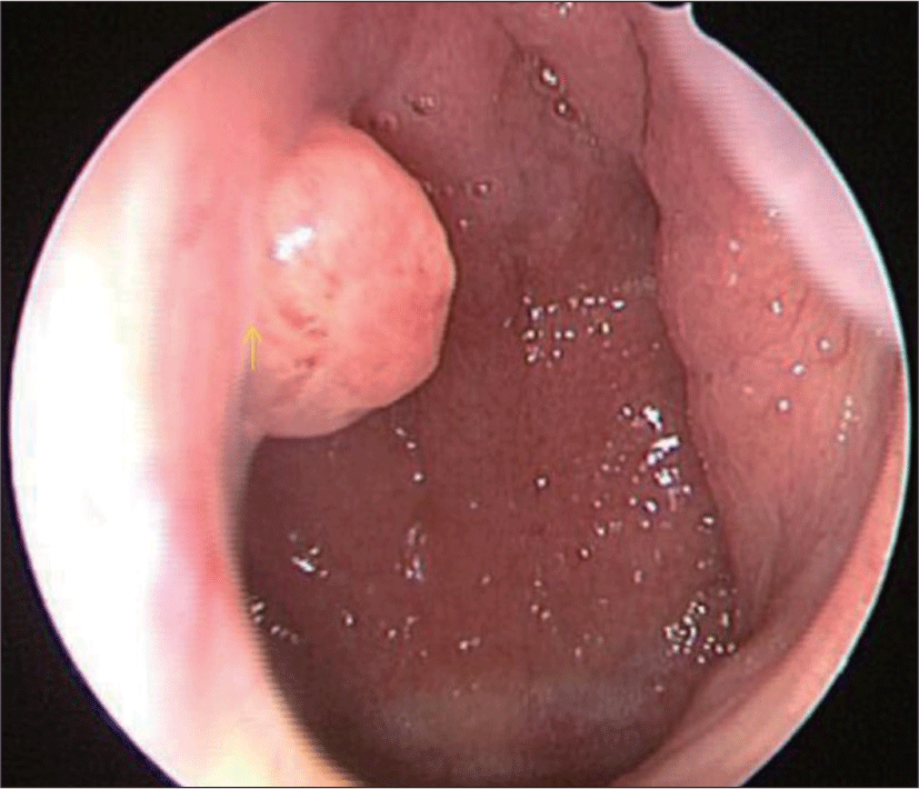
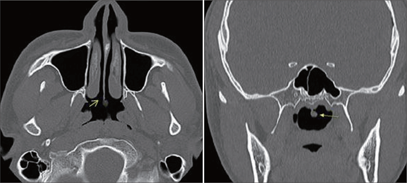
A 25-year-old Korean male discovered a mass on his nasal septum and was referred to the Department of Otolaryngology, Busan Saint Mary’s Hospital for further evaluation. A single, protruding solid mass about 4-5 mm in diameter was evident on nasopharyngoscopy of the left posterior septal wall (Fig. 1). Laboratory data were unremarkable; the C-reactive protein level was normal. Diagnostic computed tomography (CT) revealed a 0.4×0.3 cm-sized, solid nodular tumor confined to the left posterior end of the nasal septum (Fig. 2). Neither CT nor physical examination of the cervical lymph nodes revealed any abnormality. A regional excisional biopsy was performed under local anesthesia. We decided to perform transnasal endoscopic resection employing laryngeal cutting forceps to approach and resect the tumor. The tumor was completely removed, bleeding was minimal, and the patient did not complain of nasal pain, congestion or epistaxis. Microscopically, the tumor exhibited a papillary structure with a fibrovascular core. A psammoma body was evident, as in thyroid carcinomas. Most papillae were lined with epithelium, and exhibited round-to-ovoid nuclei, again similar to thyroid papillary carcinomas (Fig. 3).
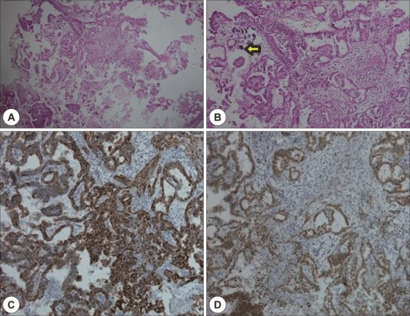
On immunohistochemical examination, the tumor cells were diffusely positive for TTF-1 but negative for other thyroid-related proteins including thyroglobulin (TG). The neoplastic cells were positive for cytokeratin (CK) 7 and negative for S-100 (Fig. 3). Thus, we diagnosed a TL-LGNPPA. Thyroid function tests performed after biopsy confirmed that the patient was euthyroid. Thyroid ultrasound revealed a normal gland lacking any focal lesion or obvious jugular lymph node enlargement. We scheduled systemic radiological imaging. TORSO F18 fluorodeoxyglucose (FDG)-positron emission tomography (PET) CT was performed 1 h after intravenous injection of 11.87 mCi F18 FDG. No abnormal FDG uptake was evident from the skull base to the upper thigh; we thus confirmed the absence of a metastatic lesion. No postoperative adjuvant treatment was administered. The patient remains well and free of disease 9 months after surgery.
Discussion
LGNPPA is an extremely rare neoplasm.6) A thyroid-like LGNPPA morphologically resembles a papillary thyroid carcinoma; the nuclei are immunoreactive for TTF-1.7) A TL-LGNPPA is a type of the principal papillary adenocarcinoma group of nasopharyngeal tumors. The tumor arises in the epithelium lining the nasopharynx, exhibits no gender bias, and can develop over a wide age range(11-64, mean 37 years).7) The lesion is usually a polypoid or pedunculated mass <1-3 cm in diameter, often in the posterosuperior wall of the nasopharynx.8) Only 9 of 25 cases of TLLGNPPAs arose from the nasal septum.5) In this case, the mass was confined to the posterior nasal septum; a TL-LGNPPA confined to the nasal septum have not been reported so far in Korea. Differential diagnosis requires microscopic evaluation and immunohisto-chemical staining. As an LGNPPA is a pedunculated nodular mass <3 cm in diameter, it can be misdiagnosed as a benign tumor such as a nasal polyp. It is difficult to distinguish an LGNPPA from a benign adenoma; pathological diagnosis is essential.
Malignant epithelial neoplasms arising in the nasopharynx are divided into the following three entities: nasopharyngeal carcinomas, salivary gland–type carcinomas, and nasopharyngeal papillary adenocarcinomas of low-grade malignancy (LGNPPA). LGNPPA and salivary gland-type carcinomas are histologically similar,9) but the latter carcinomas are surrounded by submucosal capsules; an LGNPPA exhibits a transitional zone below a normal epithelium. Immunohisto-chemical (IHC) staining is helpful; a salivary gland-type adenocarcinoma can be easily excluded, being S-100-negative.10) To distinguish an LGNPPA from other nasopharyngeal carcinomas, IHC staining for TTF and EMA is required. As an LGNPPA is morphologically similar to a thyroid papillary carcinoma, IHC staining for TG is required to confirm that the tumor is not of thyroid origin. The most common IHC staining pattern of TL-LGNPPA tumor cells is TTF-1-positive TG-negative; a papillary thyroid carcinoma is TTF-1-negative TG-positive.
To the best of our knowledge, a TL-LGNPPA is a benign tumor exhibiting indolent behavior with an excellent prognosis, and thus, without any angiolymphatic spread or metastasis. The treatment of choice is complete resection.11) If complete resection is impossible, adjuvant radiotherapy should be considered.1) Most patients with TL-LGNPPAs report nasal symptoms such as epistaxis or nasal obstruction; there are no specific symptoms. Our patient presented because of accidental discovery of a nasal mass, and he had no specific symptoms. TL-LGNPPAs are rare. Nasopharyngeal papillary adenocarcinoma is less common than epithelial or salivary gland carcinomas, and since it shows histological findings and expression of TTF-1 similar to thyroid papillary carcinoma, it is essential to exclude the possibility of metastatic cancer. Therefore, an accurate differential diagnosis and appropriate surgical resection should afford an excellent prognosis.