
Introduction
Engel used “pseudocyst” of the auricle (APC) to report the intracartilageneous cyst without epithelial lining.1,2) APC also known as endochondral APC and idiopathic cystic chondromalacia.3)
APC is characterized by an asymptomatic unilateral benign cystic lesion in the triangular or scaphoid fossa of the auricle.4-6) The APC is a very rare entity of the auricle and exclusively found in male.5,7,8)
As “idiopathic” implies, APC has an unknown etiology of cyst formation in the auricular cartilage so far. However, it may be related to an embryological or developmental malformation.9) Minor repetitive trauma from various sources such as wood pillows,10,11) motorcycle helmets11) or headphones,11) and other causes of ear folding or repeated rubbing, all have been reported to trigger the cyst formation.5,10) The presence of hemosiderin supports the trauma theory and it implies preceding leakage of blood from the neighboring vessels as a result of trauma.11) Those insults have been reported to lead to the release of lysosomal enzymes resulting in cartilaginous autolysis and degeneration with separation, fibrosis, progressive dilatation, and subsequent cyst formation.1) However, lysozyme has not been isolated in later studies.6,10) This intracartilaginous cavity without epithelial lining, is filled with serous fluid.7) The concentrations of protein, albumin, glucose, and cholesterol was similar to those of them in serum.6,9)
Repeated trauma can initiate a leakage of glycosaminoglycans, which, forming as intracartilagenous microcysts, coalesce to join a larger cyst.10) However, there are many reports without apparent preceding trauma history. Detailed questioning and thoroughly investigation should be applied to uncover it.10)
Various nonsurgical treatments for ACPs are described in the literature, including aspiration and intralesional injection of steroids and other sclerosing agents such as trichloroacetic acid with the aim of inducing fibrosis.12) Variable surgical procedures have also been used to reduce the high recurrence rate after performance of less invasive treatments.4)
Case Report
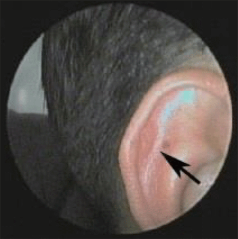
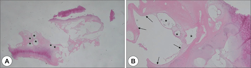
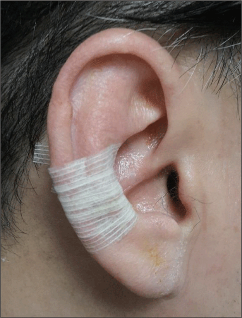
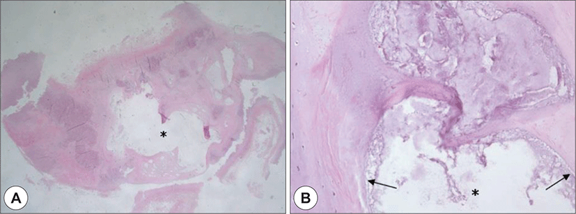
A 26-year-old man was referred to our clinic for the right auricular mass in several years. He denied any history of preceding trauma. Transfer report said trials of several aspirations were unsuccessful. A 0.5 cm-diameter firm and slightly fluctuating mass was palpated at the scapha of the right auricle. It was palpated at anterior and posterior surfaces of the auricle (Fig. 1). The helix also slightly protruded outward by the mass. The needle aspiration was tried, but until reaching the hard part, only a dry tap was obtained. The impression of organized subpericondrial hematoma, APC, or chondroma was made. After obtaining the consent of possible floopy ear, auricular mass excision via posterior approach was planned. The entire auricle was sterilized with betadine and drapped for surgery. After local injection with 2% lidocaine and 1 : 80,000 epinephrine around mass including anterior and posterior auricular skin. A helical incision of the posterior auricular skin was made. Then, the skin flap was elevated beyond the boundary of the mass. The mass was located within the cartilage. When the cut through the posterior cartilage was performed, serous fluid was released. The posterior cartilage leaf of the cyst was excised along the margin and the borders were smooth-ened. However, anterior and helical bulging were still remained. So, remaining anterior cartilage leaf was also excised. The skin was repositioned to its original site and closed with simple 5-0 Nylone sutures. To prevent hematoma, we applied several adhesive surgical tape strips (ASTs) (Nexcare Steri-Strips, 3M, MN, USA) applied to compress the raised skin flap. No additional external dressing was required. The sutures were removed after one week. At that time, ASTs were changed new one and reapplied firmly. After 2 weeks, remaining strips were gently removed. Histological findings were compatible with an APC without an epithelial lining (Fig. 2). There was no recurrence with fine cosmesis at 8-month of follow-up after excision (Fig. 3).
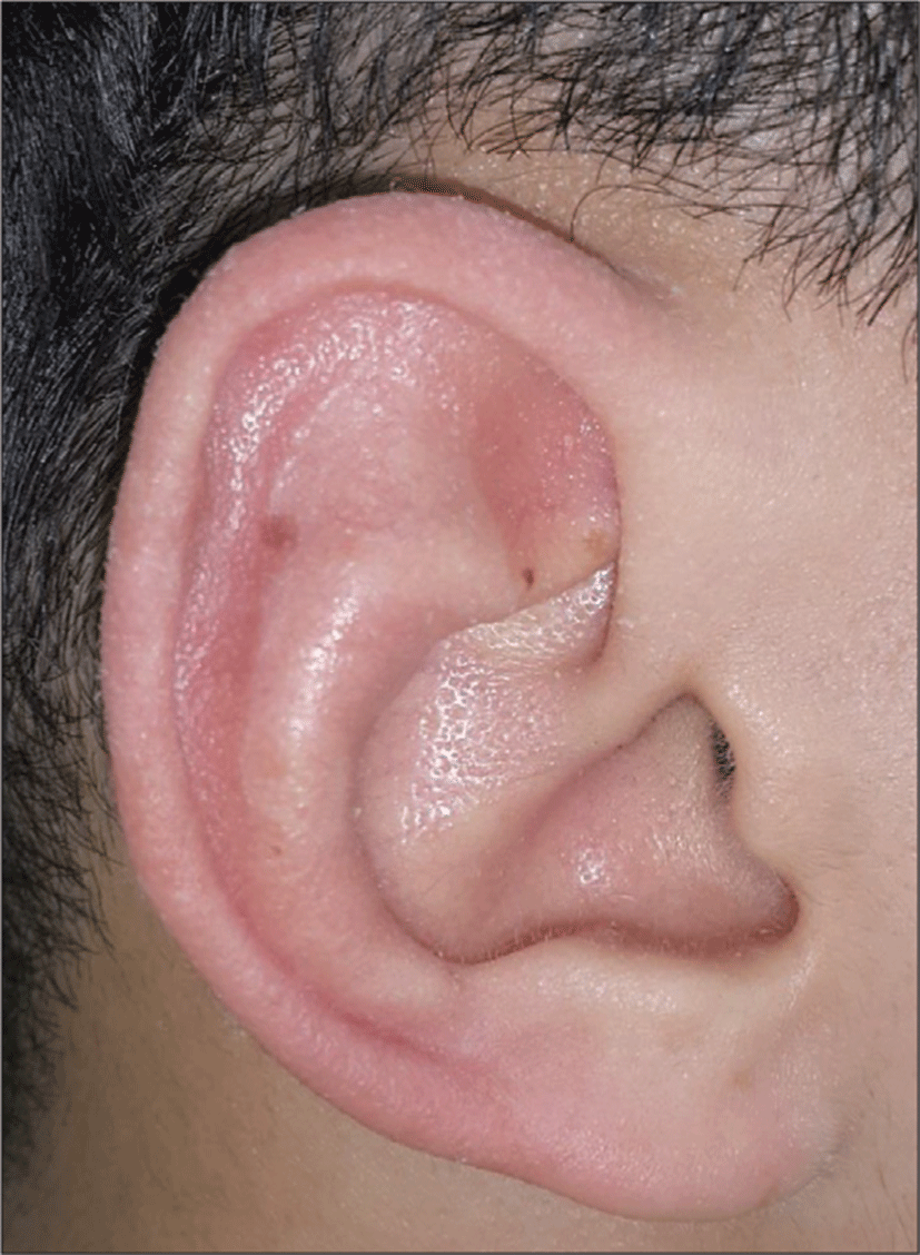
A 60-year-old man presented with a 4-year history of a slow growing mass of the right ear. He could not recall any preceding trauma. Physical examination revealed a 1.4×1 cm sized firm mass in the scaphoid fossa. The overlying skin was unremarkable (Fig. 4). The trial of needle aspiration was unsuccessful. The differential diagnosis included subperichondrial organized hematoma, APC, and relapsing polychondritis.
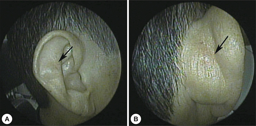
The patient wanted to excision for diagnosis. The entire auricle was sterilized with betadine and drapped for surgery. After injection with 2% lidocaine and 1 : 80,000 epinephrine around mass, a linear incision was made just above the mass. Then, the skin flap was elevated beyond the boundary of the mass. A tangential incision was made around mass through the anterior leaf of cartilage and perichondrium. The release of the straw-colored fluid was observed. The anterior wall of perichondrium and cartilage leaf of the cyst were excised along the margin and the borders were smooth-ened. Thorough curettage of the granulation above posterior cartilagenous leaf of the cyst was performed. Meticulous hemostasis was followed to prevent hematoma formation. The skin and anterior pericondrial flap was repositioned above the remaining posterior cartilageneous leaf and closed with simple 5-0 Nylone sutures. Postoperative compression was done as same as previous case one (Fig. 5). Histological findings were compatible with an APC without an epithelial lining. There also were focal destruction of the cartilage and fibrosis in surround tissue (Fig. 6). There was no recurrence of the lesion with good cosmesis over a follow-up period of one year.
Discussion
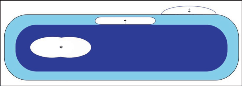
The differential diagnosis of soft swellings of the auricle includes benign cysts such as epidermal or dermoid cysts, auricular seroma, subperichondrial hematoma, and APC, or ; inflammatory lesions such as relapsing polychondritis, and chondrodermatitis nodularis helicis; and benign tumors such as chondromas, trichilemmoma, and rarely angiomas.13) Interestingly many reports of APC might misdiagnosed seroma.13) Many reports did not specify which plane the fluid was.
Characteristic histological positions and features are main points of differentiation. The lesion within the cartilage is considered as APC. On the other hand, epidermal or dermoid cyst are located at the subcutenous layer above the perichondrium. Otohematoma and auricular seroma are located subperichondrial and above the cartilage (Fig. 7).9) There are characteristic clinical features such as painful chondrodermatitis nodularis helicis, diffuse reddish relapsing polychondritis, and well aspirated subperichondrial hematoma.
Managements aim at removal the cyst without relapsing and restoring the shape of the auricle. Despite variable treatment modalities has been designed, the treatment of choice cannot be decided so far because of the high recurrence rate, which made APC is very annoying for the clinician.
Simple observation has not been recommended, untreated APCs used to induce progressive hardening or subsequently destruction or deformity of cartilage.11)
Simple aspiration of cystic fluid tends to reaccumulation.14) Therefore, aspiration was considered a diagnostic procedure rather than a therapeutic one.15) Aspiration followed by compression has been designed to decrease the recurrence rate.
Aspiration and additional intracystic injection of corticosteroid16) or sclerosing agent such as minocycline, 50% trichloro acetic acid,12) bleomycin, fibrin glue,17) or iodine tincture also have been applied with reports of variable success rate.
Intralesional steroid injections have the risk of some complications such as a skin pigmentation, atrophy, auricular deformity, and a high recurrence rate.11) Systemic corticosteroids also failed to subside.
The cystic fluid is gradually originated from the perichondrium in the anterior wall.18) Therefore, simple aspiration or inducing sclerosis cannot prevent reaccumulation, since the remained perichondrium can reproduce the fluid. The treatment should focus to remove the anterior perichondrium.
Variable surgical approaches have been applied for treating the APC. Incision and drainage,19) insertion the drain5) and punching the inferior border of the APC2) were reported. To reduce recurrence, following compressive dressing has been proposed. Besides traditional contour dressing, variable compression material such as bolstered pressure sutures,20) plaster of Paris casts,8) clip,4,16) and button15) have been applied with variable success rates. However, problems of APC recurrences, prolonged application and deformed cartilage still remained.
To overcome this issue, more invasive procedures of deroofing of the APC was introduced.10) After removing the anterior cartilagenous leaf, curettage of the posterior wall is performed, followed by the compression dressing. Recurrence could be reduced if the anterior degenerated cartilage of the APC can be removed completely. However, the posterior leaf should be preserved to prevent floppy ear deformity.1,10) But extirpation of cyst is inevitable in some case like our second case to restore original shape of auricle. Surgical deroofing of the APC was reported with no recurrence and good cosmesis in most cases.10) Original contour dressings10) was modified to use buttoning as a compression method without recurrence.15) button bolsters14) were not convenient to be resized. Therefore, the use of AST is very convenient to apply variable length. Customized AST with suitable lengths or widths could have definite effects on the auricular compression.
We use the adhesive surgical tape strips in the postoperative compression of APC. We prefer excision of the anterior leaf of the APC, leaving intact posterior leaf, followed by AST compression of the wound.
Conclusion
For the clinician, APC could be very annoying in terms of recurrence. We prefer excision of the anterior leaf of the APC, leaving intact posterior leaf as far as possible, followed by AST compression of the wound. This method is better for cosmesis and patient’s comfort without prolonged bulky pressure compression.