증례
비인두강내 식물성 괴사조직 1예
진호경1, 노양섭1, 홍상덕1,*
A Case of Necrotic Vegetable in Nasopharynx
Hokyung Jin1, Yangseop Noh1, Sang Duk Hong1,*
1성균관대학교 의과대학 삼성서울병원 이비인후과학교실
1Department of Otorhinolaryngology-Head and Neck Surgery, Samsung Medical Center, Sungkyunkwan University School of Medicine, Seoul, Korea
*교신저자: 홍상덕, 06351 서울 강남구 일원로 81 성균관대학교 의과대학 삼성서울병원 이비인후과학교실 전화 :(02) 3410-3579·전송:(02) 3410-3879 E-mail:
kkam97@gmail.com
© Copyright 2019 The Busan, Ulsan, Gyeoungnam Branch of Korean Society of Otolaryngology-Head and Neck Surgery. This is an Open-Access article distributed under the terms of the Creative Commons Attribution Non-Commercial License (http://creativecommons.org/licenses/by-nc/4.0/) which permits unrestricted non-commercial use, distribution, and reproduction in any medium, provided the original work is properly cited.
Received: Feb 19, 2019; Revised: Mar 28, 2019; Accepted: Apr 25, 2019
Published Online: Jun 30, 2019
ABSTRACT
Nasopharyngeal foreign bodies are rare and usually asymptomatic. Although there have been some reports about nasopharyngeal foreign body with sponges, inset or metallic bolt, there was no nasopharyngeal vegetable foreign body. We report a case of 56 year-old female patient with unusual nasopharyngeal foreign body (vegetable) presenting with postnasal drip. The foreign body was detected at right rosenmuller fossa in sinonasal computed tomography. After foreign body was removed by endoscopic endonasal approach, her postnasal drip was gone. In our knowledge, this is the first report about nasaopharyngeal foreign body with vegetable tissue. (J Clinical Otolaryngol 2019;30:92-96)
Keywords: 비인두강내 이물질; 식물성 괴사 조직
Keywords: Nasopharyngeal foreign body; Necrotic vegetable tissue
서 론
비강 혹은 인두강내 이물질은 비교적 흔하게 발생하 는 것으로 알려져 있으나, 비인두강내 이물질은 굉장히 드물게 보고되고 있다. 비인두강내 이물질로서 기존에 보고된 증례를 살펴보면 솔방울, 거머리, 대리석, 금속 철판 등 다양한 이물질이 있으나,1-5) 야채나 채소와 같은 식물성 조직이 이물질로 발견된 증례는 없어, 이에 대해 증례 보고를 하는 바이다.
증 례
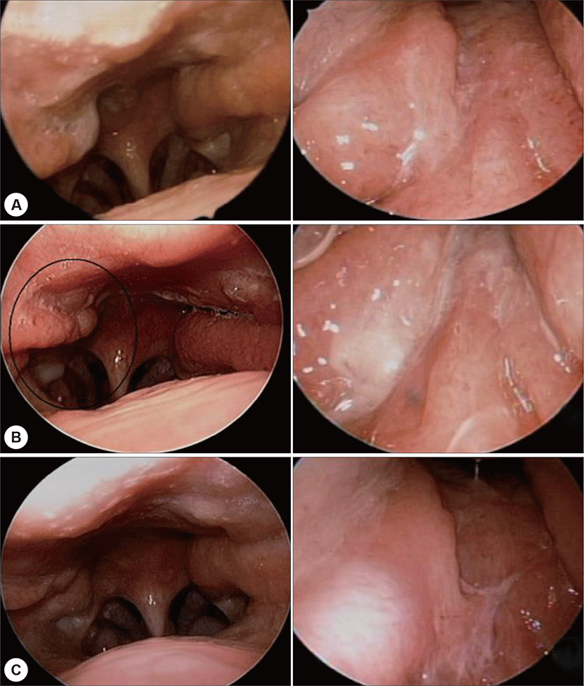
56세 여자가 10년 전부터 발생한 코골이와 3주 전부 터 발생한 후비루를 주소로 2017년도 1월 16일 본원 외 래를 방문하였다. 계통 문진 상 코골이와 후비루 외에 목 에 이물감, 악취 등의 다른 증상은 호소하지 않았다. 외 래에서 시행한 내시경 검진 상 우측으로 휘어진 비중격 만곡증 소견 및 우측 로젠뮬러 와(Rosenmuller fossa)의 부종 및 협착 소견이 관찰되었다(Fig. 1A).
Fig. 1.
A : This image showed a bulging of right rosenmuller fossa taken at the first outpatient visit. Left image was transoral endoscopic (90 degree) imaging of nasopharynx and right image was transnasal endoscopic (30 degree) imaging of nasopharynx. B : This image showed a bulging of right rosenmuller fossa still being observed after three months from the first outpatient visit. Left image was transoral endoscopic (90 degree) imaging of nasopharynx and right image was transnasal endoscopic (30 degree) imaging of nasopharynx. C : This image showed the disappearance of a bulging of right rosenmuller fossa at three months after surgery. Left image was transoral endoscopic (90 degree) imaging of nasopharynx and right image was transnasal endoscopic (30 degree) imaging of nasopharynx.
Download Original Figure
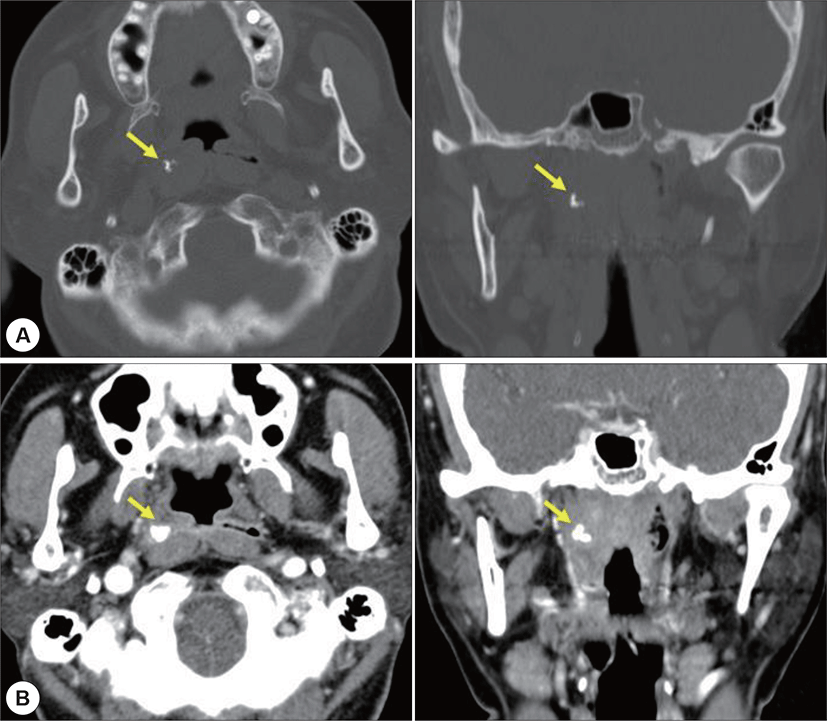
내원 한 달 후 수면다원검사를 시행하였으며, 종합적 으로 중등도 수면 무호흡증에 해당하였다. 외래에서 수 술 전 영상 확인을 위해 안면골의 컴퓨터단층촬영(facial bone CT)를 시행하였으며, 우측 비인두강에 석회화를 포 함한 이관을 앞으로 밀고 있는 병변이 우연히 발견되었 다(Fig. 2A). 당시 문진 상 코골이나 후비루의 양상은 첫 외래 때와 비교하여 큰 변화가 없었다. 상기 비인두강 병 변은 환자가 2개월 전 두통으로 타 병원 방문 후 시행했 던 뇌 자기공명영상에서는 관찰되지 않는 병변으로 2개 월 이내에 새로 발생한 병변으로 판단되었다.
Fig. 2.
A : This image showed a preoperative calcification lesion in right nasopharynx indicated by the yellow arrow. This image was taken at one month after the first outpatient visit. Left image was an axial imaging of facial bone CT and right image was a coronal imaging of facial bone CT. B : This image showed a right pharyngeal wall thickening and calcification lesion in right nasopharynx indicated by the yellow arrow. This image was taken at five months after the first outpatient visit. Left image was an axial imaging of enhance paranasal sinus CT and right image was a coronal imaging of enhance paranasal sinus CT.
Download Original Figure
우측 비인두강의 부종 부위에서 악성 종양을 배제하 기 위하여 조직검사를 시행하였으나 조직 검사상 림프 과다형성(lymphoid hyperplasia) 소견만 보였다. 조영 증 강 코곁굴 컴퓨터단층촬영(enhance paranasal sinus CT) 을 통해 우측 비인두강 병변을 추적관찰 하였으며, 석회 화 병변은 기존과 동일하게 관찰되었고, 병변 주위 점막 의 조영 증강 소견이 관찰되었다. 외래에서 다시 시행한 내시경 검진 상으로는 좌측에 비해 비대해진 우측 인후 두벽(pharyngeal wall) 비대가 관찰되었으며, 후비루도 지속되고 있었다(Fig. 1B). 5개월후, 다시 한 번 조영 증 강 코곁굴 컴퓨터단층촬영을 하였으며, 우측 비인두강 에 관찰되는 석회화 병변 크기는 동일하였고, 동측의 인 후두벽의 비후(ipsilateral pharyngeal wall thickening) 가 더 증가한 것으로 관찰되었다(Fig. 2B).
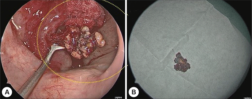
우측 비인두강 병변에 대한 절제술 및 조직검사를 위 해 전신마취하에 수술을 시행하였다. 네비게이션 시스 템을 이용하여 석회화 병변의 위치를 확인 후 곡선 흡인 기(curved suction)와 링 큐렛(ring curette)을 사용하여 로센뮬러 와(rosenmuller fossa)안에 묻혀 있는 석회화 병 변을 제거하였다. 이와 동시에 우측 로센뮬러 와(rosenmuller fossa)의 점막 조직에 대해 조직검사도 같이 시행 하였다. 수술장에서 관찰한 병변은 육안상 진균구(fungal ball)와 유사한 형태를 보였다(Fig. 3).
Fig. 3.
A : Intraoperative imaging. Using ring curette, the foreign body was removed from right lateral recess of nasopharynx. B : Gross morphology of foreign body.
Download Original Figure
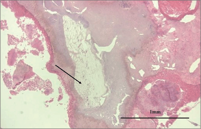
조직검사상 석회성 병변은 식물성 괴사조직(necrotic change of vegetable)이였으며(Fig. 4), 로센뮬러 와의 조 직은 괴사가 동반된 육아조직(granulation tissue with necrosis)이었다. 술 후에 음식물의 역류 등의 병력에 대하 여 확인하였으나, 특이 병력은 없었다.
Fig. 4.
Histology of foreign body. The black arrow showed the necrotic change of the vegetable’s spongy structure. The black line represented 1 mm as the size parameter.
Download Original Figure
수술 후 3개월 째 외래 추적관찰을 시행하였으며, 기 존에 관찰되던 비인구강내 비대 조직은 사라진 상태였 고, 내시경 검진 및 환자의 주관적 증상으로 존재하던 후비루 역시 사라졌음을 확인하였다(Fig. 1C).
고 찰
본 증례의 경우 환자의 주호소는 코골이와 후비루였 다. 외래 내원 시 비인두 내시경 사진을 보면 약간의 팽 창(bulging) 소견은 보였으나, 따로 의심하지 않으면 정 상으로 오인하기 쉬운 소견이었다. 따라서 본 환자에서 비인두강내 병변을 병력과 내시경 소견만으로 진단하는 것은 매우 어려웠다. 이물질은 수면무호흡증 및 코막힘 검사 중, 안면골의 컴퓨터단층촬영에서 우연히 발견되 었으며, 안면골의 컴퓨터단층촬영은 조영제를 사용하지 않는다는 점을 고려했을 때, 병변의 석회화가 병변의 존 재를 아는데 큰 역할을 하였다.
컴퓨터단층촬영에서 관찰되는 본 증례의 병변은 경계 가 불분명하였고, 미세 석회화가 동반되었으며, 종양의 형태보다는 염증성 병변에 가까웠다. 또한 본원 초진 2 개월 전 타원에서 촬영한 자기공명영상에서는 상기 병 변이 관찰되지 않았다. 부비동내 석회화가 동반된 병변 이 보이는 경우 진균 병변을 의심할 수 있기 때문에, 저 자들도 처음에는 비인두강내 진균 병변 가능성을 고려 해봤다.6,7) 현재까지 Dogan이 2004년도에 52세 여자환 자에서 비인두강내 진균구가 발견되었다고 보고한 문헌 이 유일한 증례였다.8) 한편 비인두강내 기형종의 경우 미세 석회화를 동반할 수 있다고 알려져 있지만, 본 증 례의 경우는 영상에서 종양의 형태를 보이지 않았기 때 문에 이 가능성이 낮다고 판단하였다.6)
여러 가지 가능성에 대해 고려하며, 수술적으로 조직 검사를 시행하였다. 최종 조직검사 결과는 울타리 조직 과 해면 조직이 관찰되는 식물성 괴사 조직으로 보고되 었다. 조직검사 결과가 보고 된 후 환자에게 병력 청취를 다시 시행하였으나, 채소나 야채 등의 음식물을 섭취 후 구토를 했던 기억이나, 목에 이물감을 느꼈던 적은 특별 히 없다고 하였다. 기존에 비인두강내 이물질로 보고된 문헌들을 살펴보면 거머리, 솔방울, 대리석, 금속 철판 등 다양한 종류의 이물질이 보고되었으나,1-5) 식물성 괴 사조직으로 보고된 증례는 없었으며, 이번 증례가 첫 증 례로 생각된다. 비인두강내 이물질이 발견되는 경우는 매우 드물지만, 후비루와 같은 흔한 증상이 동반될 수 있 으며, 수술적으로 특별한 합병증 없이 이물질을 제거할 수 있다는 점을 공유하고자 저자들은 본 증례를 보고하 게 되었다.


