
Introduction
Lemierre’s syndrome, which is known as postanginal septicemia or human necrobacillosis, is a rare disease of the head and neck that often affects healthy adolescents and young adults.1,2) It has been described as internal jugular vein thrombosis with recent oropharyngeal infection.
Clinical presentation of Lemierre’s syndrome was mainly sore throat, neck mass and neck pain. In addition, it could cause several fatal complications such as meningitis, brain abscess, pulmonary thromboembolism with mortality rate of 5%.2)
Because of its variety of clinical presentation, early suspicion and treatment of Lemierre’s disease has been considered as difficult. In some cases of Lemierrre’s syndrome, they revealed neurologic symptoms accompanied by encephalopathy and stroke.2-4) In this report, we demonstrated rare clinical presentation of Lemierre’s syndrome as a complex partial seizure.
Case report
A 17 year-old female was previously healthy and lost body weight about 10 kg for the purpose of diet during 2 months. Recently, she had cough, sputum and fever for 2 weeks, and symptoms were not relieved in spite of oral antibiotics therapy (amoxicillin/clavula-nate). She had complex partial seizure with upper eye ball deviation and loss of consciousness during 1 hour after agitation.
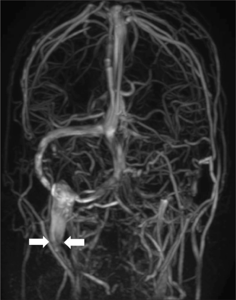
She was transferred to emergency room of our hospital, and seizure was stopped with antiepileptic drugs. Electoroencephalography was normal and brain magnetic resonance imaging (MRI) revealed no significant focal lesion in the brain parenchyme. However, diffuse inflammatory change adjacent to right internal jugular vein (Fig. 1A) was identified on neck contrast computed tomography (CT) and occluded right internal jugular vein with brain cortical veins enlargement was shown on MR angiography (Fig. 2). On laboratory exam, level of white blood cell (WBC) was 11730/μL, C-reactive protein (CRP) was 33.69 mg/dL and erythrocyte sedimentation rate (ESR) was 120 mm/hr on blood test. And there was no abnormal findings on cerebrospinal fluid (CSF).
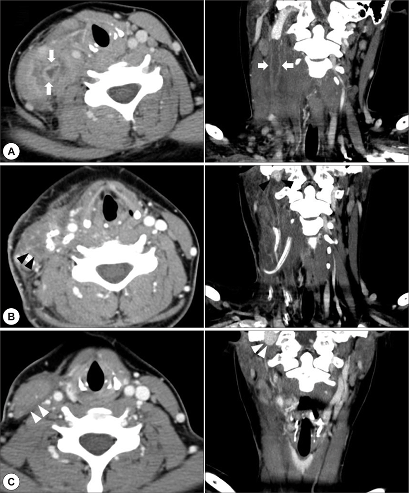
With the diagnosis of septic thrombophlebitis of right internal jugular vein (Lemierre’s syndrome), she underwent radical debridement of necrotic tissue and ligation of right internal jugular vein with intravenous antibiotics administration (piperacillin/tazobactam). The gram stain and culture of surgical specimen was negative. 1 week after the surgery, neck CT demonstrated improved inflammatory change of the right neck, but progression of thrombus was observed at the distal part of the ligated internal jugular vein from level II to level IV (Fig. 1B). Therefore, anticoagulation therapy has been started using intravenous heparin (50 sec≤target aPTT<70 sec) which was converted to warfarin (2 sec≤target PT<3 sec) in 5 days after the initiation of anticoagulation. After the surgery, antibiotics therapy was performed for 3 weeks until normalization of blood lab (WBC : 6820/μL, CRP : 0.44 mg/ dL, ESR : 9 mm/hr) and anticoagulation therapy lasted for 3 months. Three months after surgery, neck CT revealed no inflammation of right neck and no change of internal jugular vein thrombosis (Fig. 1C). She was followed-up until 6 months after the surgery without symptom including fever or seizure.
Discussion
Lemierre’s syndrome is diagnosed in patients who had history of upper respiratory tract infection and radiological evidence of internal jugular vein thrombosis.1,2) Recently, incidence of Lemierre’s syndrome has been decreased because of development of antibiotics and diagnostic tools. Otherwise, it demonstrated various clinical presentation such as otalgia, joint pain, and headache, and fatal complication.2,5) Initial presentation of Lemierre’s syndrome is mostly tonsillitis followed by pharyngitis and pneumonia.2) In this report, patient presented complex partial seizure with leukocytosis. In this situation, we usually suspect infectious disease of brain such as encephalitis and meningitis, but Lemierre’s syndrome could be a final diagnosis. Lemierre’s syndrome with seizure or complications of central nervous system (CNS) is rare. Twenty-two cases of CNS complications resulting from Lemierre’s syndrome were reported from 1980 to 2010.6 Because CNS complications could result in fatal outcomes including death, we should consider Lemierre’s syndrome diagnosing patients who have CNS symptoms. The diagnosis of Lemierre’s syndrome is based on clinical suspicion. The computed tomography (CT) scan is the best diagnostic method to evaluate internal jugular vein thrombosis and primary upper respiratory tract infection.6) In addition, Doppler ultrasonography could be used to check blood flow and echogenicity of internal jugular vein and adjacent structures.7) Magnetic resonance imaging (MRI) could increase the sensitivity of diagnosis, because it reflect intraluminal signal intensity change by the thrombosis.6) We used CT and MRI together to evaluate the disease extent of thrombosis, brain, other soft tissue status.
Surgical drainage and antibiotics therapy has been cornerstone of treatment of Lemierre’s syndrome. The first-line antibiotic therapy should involve penicillin/ β-lactamase inhibitor for β-lactamase producing pathogens and oropharyngeal oraganisms.2,6) In this case report of culture negative Lemierre’s syndrome, piper-acillin/tazobactam seemed to be effective medical treatment.
Most of Lemierre’s syndrome patients have been managed with surgery and antibiotics, and the effect of anticoagulant therapy remains controversial. In this case, we treated with anticoagulant because this could result in systemic septic emboli including lung, joint and brain. And we performed serial CT scan after the surgery, and detected improvement of thrombosis progression at distal stump of internal jugular vein. Because the possibility of CNS complications in Lemierre’s syndrome, we followed up for 6 months until confirmation of stabilization of thrombosis on radiologic examination.
In summary, Lemierre’s syndrome has been decreased but it could make fatal complications. Therefore we should have precise diagnosis with early suspicion, and perform the proper treatment including surgical drainage and antibiotics.